18
Role of radiographs in periodontal diagnosis
Figure 18.1 Ways of minimising radiation exposure when using radiographs.
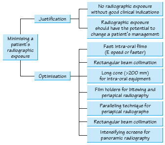
Figure 18.2 Key aspects of a radiographic periodontal assessment. CEJ, cement–enamel junction; TMJ, temporomandibular joint.
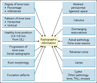
Figure 18.3 Panoramic radiograph showing bone loss.
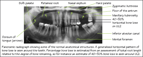
Figure 18.4 Furcation defects.
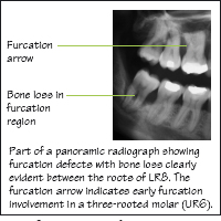
Figure 18.5 Calculus and deficient crown margin.
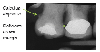
Figure 18.6 Plaque retention factors. Courtesy of Ms V. Yorke.
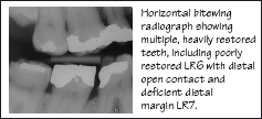
Figure 18.7 Overhanging restoration margins.
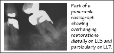
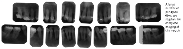
Figure 18.8 Full mouth periapical radiographs for a patient with advanced bone loss.
Radiographs are used as an adjunct to a thorough clinical examination in order to make a diagnosis and draw up a treatment plan. The details of national regulations governing the use of radiographs vary in different countries, but in principle they all seek to protect the workforce and general public and to limit the radiation dose to patients.
Patients are protected by application of the principles of justification and optimisation (Fig. 18.1):
Stay updated, free dental videos. Join our Telegram channel
VIDEdental - Online dental courses


