17: Problem Solving in the Management of Teeth With Subgingival Carious and Fractured Margins Including Radicular Defects

Chapter 17

Problem Solving in the Management of Teeth With Subgingival Carious and Fractured Margins Including Radicular Defects

Problem-Solving List

Problem-solving issues and challenges in managing compromises in sound tooth margins and radicular defects addressed in this chapter are:

Normal Periodontium in Relation to Surgical Treatment Planning
Indications for Crown Lengthening

    Convenience of tooth isolation
    Establishing increased clinical crown for the “ferrule effect”
    Preventing restorative failure
    Avoiding marginal problems with restorations
    Managing deep fractures
    Location of obscure root canal orifices
    Following orthodontic extrusion of teeth or roots
Potential Contraindications for Crown Lengthening

    Medical status
    Esthetic zone
    Minimal vertical dimension of the root trunk
Surgical Techniques for Crown Lengthening

    Surgical procedures
    Envelope tissue flap
    Thinning or undermining the tissue
    Recontour of alveolar bone
    Tissue closure
    Periodontal surgical dressing
    Healing
    Complications with surgical crown lengthening
Application of Crown Lengthening Techniques to Other Endodontic Problems

    Iatrogenic perforations in the marginal periodontium
    Resorption defects in the marginal periodontium
    Vertical root fracture repair

“Once the biologic width of the supporting periodontal attachment apparatus has been severely violated, more extensive procedures are often necessary to manage compromised root structure and supporting bone.”< ?xml:namespace prefix = "mbp" />20

J.L. Gutmann, 1991

Preserving the natural dentition in symptom-free clinical function is a major goal of dentistry and its specific disciplines. Prosthetic dentistry has provided fixed and removable substitutes for teeth that have been lost, and the advent of implant dentistry has brought about a new concept, replacement of the natural dentition. While the development of implantology has been a marvelous addition to the practice of dentistry and has enhanced patient well-being,9,35,42 the enthusiasm for this option has in some cases clouded the process of sound treatment planning.8,43 However, many teeth that could be preserved in normal function through reasonable and adjunctive procedures (nonsurgical endodontic revision, surgical endodontic revision, crown lengthening, orthodontic root extrusion, root resection, etc.) are removed in favor of replacement with implants. Data do not support the superiority of implants over these other procedures when treatment planned and executed properly.24,25 In many respects, implant dentistry is not an option for a great many people, and tooth retention is not only possible but in the patient’s best interest and the best choice.

In this chapter, surgical procedures will be presented that can be used effectively to retain teeth with a variety of complications in addition to pulpal disease. The most common problems encountered are clinical crowns with subgingival carious or fractured margins and insufficient tooth structure to retain a restoration. In many of these cases, crown lengthening is an ideal solution. Exposure of sound tooth structure can make restoring certain types of compromised teeth a routine procedure, enhancing not only the restoration of the tooth but the biologic widths of the supporting periodontium. The basic techniques of this surgical intervention can be extended to the management of other endodontic problems such as resorption and perforation defects.

Normal Periodontium in Relation to Surgical Treatment Planning

Prior to considering periodontal surgical procedures, a review of the normal anatomy of these tissues is essential. A discussion of average sulcus depth was included at the beginning of Chapter 4. The essential anatomic relationships between the tooth, bone, and soft tissues of the average healthy periodontal sulcus are depicted in Fig. 17-1. These dimensions have been described as the biologic width,* defined as the physiologic dimension of the junctional epithelium and connective tissue attachment.16,33

image

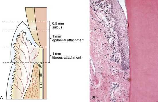
FIGURE 17-1 A, Normal periodontium. B, Histologic section through normal periodontium (H&E stain ×10).

The width of the attached gingiva varies from 1 to 9 mm,6 with an average of 3.7 mm in the anterior areas, 3.8 mm in the premolar regions, and 4.2 mm in the molar37 (Fig. 17-2). However, at least 5 mm of attached gingiva are recommended for restorative dentistry.32 Ideally, areas of attached gingiva with less than 5 mm should be considered for augmentation procedures if restorative margins are to be placed in the sulcus.4 The healthy gingival sulcus has been shown to have an average depth of 0.69 mm.33

image

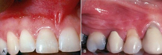
FIGURE 17-2 A, Attached gingiva of normal, ample width. B, Width of attached gingiva in this maxillary right premolar area is 1 mm or less.

The contours of the normal alveolar bone are also of great significance. Most often, surgical crown lengthening cannot be achieved without removing some crestal bone. Postoperatively, the measure of success is not only increased clinical crown length but also restoration of a normal periodontium circumferentially. In recontouring bone, the clinician should reproduce or create the contours of normal periodontium. Except in the mandibular second and third molar areas, the buccal and lingual plates are usually fairly thin throughout the dentition (Fig. 17-3). If the buccal plate appears excessively thick with a shelflike contour, it can be reduced (Fig. 17-4). Occasionally, a dehiscence or fenestration will be found after the flap is elevated (Fig. 17-5). The only significance of this finding is that the exposed areas of the root should be left untouched. Reattachment will invariably occur, providing that these root surfaces are not curetted.

image

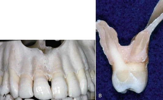
FIGURE 17-3 A, Labial plate of bone is normally thin. B, Cross-section of maxilla in the first molar area, showing buccal and palatal plates of bone.

image

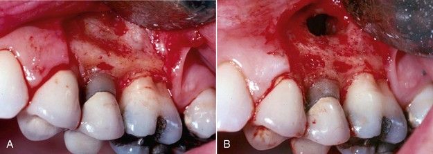
FIGURE 17-4 A, Excessive width of buccal plate of bone. B, Recontoured buccal plate during periapical surgery procedure.

image

FIGURE 17-5 Dehiscence of root surfaces after flap elevation. These areas should not be curetted.

image

CLINICAL PROBLEM

Problem

Can the interproximal bone throughout the mouth be recontoured using a standardized technique?

Solution

No, because there is a significant difference between the interproximal contours of the bone between the posterior teeth and the contours found between the anterior teeth. The normal contour between posterior teeth is flat buccolingually (Fig. 17-6). Crown lengthening will require an apical relocation of the bone while maintaining the identical form. Frequently, bony defects from chronic periodontitis are found interproximally, leaving intact ridges of the buccal and lingual or palatal plates. These ridges are normally reduced to restore as flat a buccal lingual contour as possible (Fig. 17-7). Conversely, in the anterior dentition, there is an interproximal crest (Fig. 17-8). In crown lengthening, the reproduction of these contours is necessary for optimal periodontal healing and also esthetics (Fig. 17-9). The interdental papillae are supported and shaped by the underlying bone. If the supporting bone is either lost by periodontal disease or failure to recreate the proper contour during surgery, the papilla will collapse (Fig. 17-10).

image

FIGURE 17-6 Normal interproximal anatomy of bone is flat in posterior dentition.

image

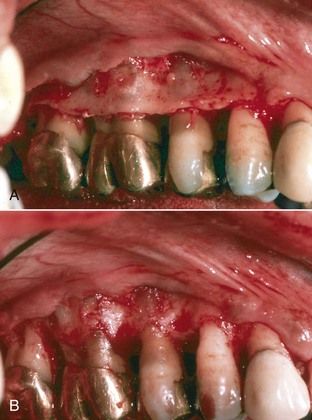
FIGURE 17-7 A, Periodontal surgery for severe chronic periodontitis. B, Following osseous recontouring and pocket elimination. Note ramping of buccal plate into interproximal spaces.

(Courtesy Dr. Curtis Wade.)

image

FIGURE 17-8 Normal interproximal anatomy of bone in the anterior dentition is characterized as a crest.

image

FIGURE 17-9 A, Crown lengthening on fractured maxillary central incisors. B, Restoration of interproximal bony crests which will support the papillae.

image

FIGURE 17-10 Loss of the interproximal crest of bone results in collapse of the interdental papilla and an unesthetic “black triangle.”

image

Indications for Crown Lengthening

There are multiple considerations in which crown lengthening can and should be considered in the treatment planning for tooth retention.

Convenience of Tooth Isolation

Before arriving at crown lengthening as the treatment of choice, a discussion of common alternatives is indicated. Most teeth with significant carious lesions can be isolated without crown lengthening. One simple solution is to place the dental dam clamp directly on the gingiva and complete the root canal treatment (Fig. 17-11). The difficulty of this approach is that it merely postpones the problem of treatment planning the final restoration. A second method of isolation is caulking the gaps around the dental dam with temporary filling materials or a specialized putty (Fig. 17-12). Once again, root canal treatment can usually be completed without leakage, but the restorability issues remain. Temporary filling materials can be used to seal large excavations within the tooth as well (Fig. 17-13). Unless the caries is thoroughly removed and a high quality restoration is placed, leakage can be a problem, especially if the root canal treatment requires multiple visits. Leakage between appointments is a very common cause of reinfection.

image

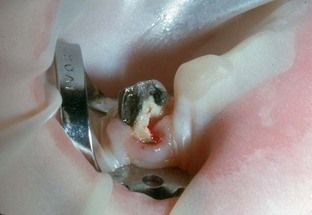
FIGURE 17-11 Placing dental dam clamp directly on gingival margin does not address subgingival caries or restorability following root canal treatment.

image

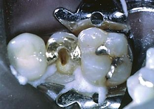
FIGURE 17-12 Dental dam sealers are effective, but restorability of the deep fracture in this case remains a problem.

image

FIGURE 17-13 Temporary restorations often leak.

Many clinicians favor complete restoration of the endodontically involved tooth. Access is then made through the new restoration (Fig. 17-14), which helps prevent leakage. After the root canal treatment is completed, however, the restoration often does not provide sufficient structural integrity to support a full crown.30,31 Crown lengthening satisfies all the endodontic and restorative needs. Isolation is excellent, while access and visibility are unimpeded. Issues of restorability can be determined early, and the final restoration can take advantage of the treated canals for retention if necessary (Fig. 17-15).

image

FIGURE 17-14 Complete permanent restorations are effective in achieving tooth isolation. The access cavity could weaken the structural support if this restoration were included in a crown preparation.

image

FIGURE 17-15 A, Mandibular molar with severe caries extending subgingivally on the distal. B, Clinical view of same tooth. Restorability appears to be questionable. C, Following crown lengthening; restorability is confirmed and isolation for root canal treatment is excellent.

Establishing Increased Clinical Crown for the “Ferrule Effect”

This key restorative goal is discussed in detail in Chapter 20. Full crown restorations require retention on sound tooth structure as well as any restorative foundation that is constructed.2,33 This also provides an additional barrier to marginal leakage that may adversely affect the prognosis of the root canal treatment (see Fig. 17-15, C).

Preventing Restorative Failure

Lack of a ferrule also can focus excessive occlusal forces on the post and core and eventually cause the post to loosen or fracture17 (Fig. 17-16).

image

FIGURE 17-16 Lack of “ferrule effect” in previous crown preparation led to post fracture.

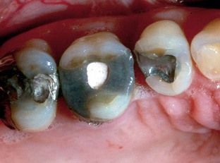
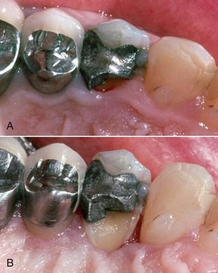
Avoiding Marginal Problems With Restorations

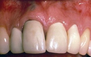
Excavation of caries below the free gingival margin or a fracture requires subgingival margin placement, or impressions and restorations will be faulty (Fig. 17-17). Exposure of all margins will ensure the chance for an excellent restorative result.

image

FIGURE 17-17 Failure to include entire tooth circumference in crown preparation, owing to lack of visibility of mesial aspect of tooth. Marginal leakage is also a result. Crown lengthening might have been the means to avoid this outcome.

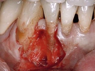
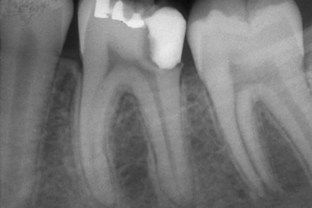
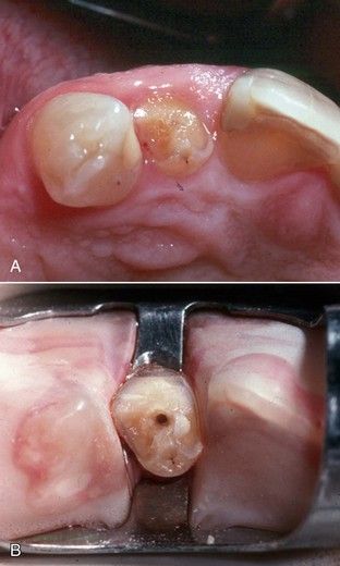
Managing Deep Fractures

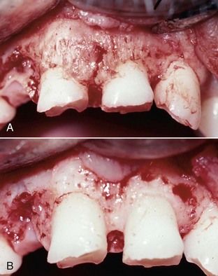
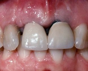
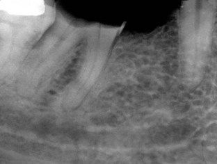
Many deep subgingival fractures cannot be restored adequately, and over time the tooth may develop periodontal complications. Crown lengthening addresses such complications prior to restoration, resulting in a routine procedure with an excellent, uncomplicated prognosis (Fig. 17-18).

image

FIGURE 17-18 A, Fractured palatal cusp on a maxillary first premolar. This would be a restorative challenge and a long-term periodontal liability. B, After crown lengthening, the periodontium is in a normal relationship, and the restoration will be routine.

Location of Obscure Root Canal Orifices

A specific endodontic indication is the case in which a coronal fracture has resulted in hyperplasia of the marginal gingiva, obscuring the location of a canal orifice. Often, if these fractures were present as mere cracks for any length of time, narrowing and calcification of adjacent root canal orifices occurred secondary to long-term bacterial ingress and pulp tissue irritation. After crown lengthening, orientation is greatly improved, enhancing access preparation and canal orifice location (Fig. 17-19).

image

FIGURE 17-19 A, A fractured maxillary lateral incisor with a calcified canal and hyperplastic gingiva covering part of the remaining tooth surface. B, Locating the canal is much easier after crown lengthening.

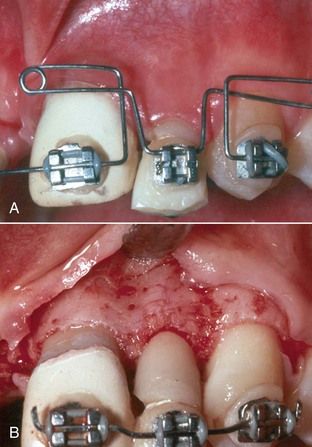
Following Orthodontic Extrusion of Teeth or Roots

The techniques for orthodontic extrusion are discussed in previous editions of this text and in the orthodontic literature. The periodontal consequence of extrusion is coronal movement of the attachment.5 Crown lengthening will usually be necessary to reposition the gingival margin apically for both esthetics and restorability (Fig. 17-20).

image

FIGURE 17-20 A, The attachment follows the tooth in orthodontic extrusion. B, Crown lengthening is indicated to reposition the gingival attachment apically for both restorative and esthetic purposes.

Potential Contraindications for Crown Lengthening

There are both general and local considerations in which crown lengthening may not be considered in the treatment planning for tooth retention.

Medical Status

Generally, the medical status and current medications prescribed for the patient are reviewed for the same conditions that would contraindicate any surgical procedure. The reader is referred to other texts on oral surgery for a discussion of these issues.

Esthetic Zone

Loss of a clinical crown through fracture is a common indication for crown lengthening. In the esthetic zone, however, apical repositioning of the gingival margin may be unacceptable esthetically,33 particularly in a patient with a prominent “gingival smile.” Even though the restorative results may be technically excellent, the crown may be unacceptably long (Fig. 17-21).

image

FIGURE 17-21 Crown lengthening alone on fractured teeth leads to unesthetically long crowns. Orthodontic extrusion should be considered first.

Minimal Vertical Dimension of the Root Trunk

On molar teeth, the root trunk is defined as the distance from the furcation to the level of crestal bone. If the trunk distance is less than 4 mm, crown lengthening cannot establish the 4 mm of tooth exposure without extending past and into the furcation11 (Fig. 17-22).

image

FIGURE 17-22 A severely carious molar with less than 4 mm of root trunk. Crown lengthening cannot be accomplished without exposure of the furcation.

Surgical Techniques for Crown Lengthening

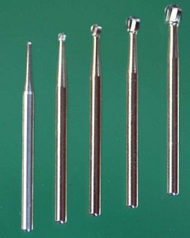
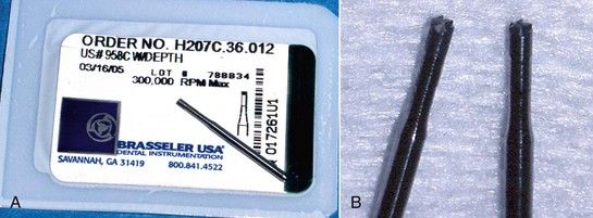
Surgical crown lengthening does not require an extensive list of instruments or supplies. Generally the procedure will include a standard oral surgery setup to incise, elevate, reflect, reposition, and suture tissue. In addition, a set of periodontal curettes, a small chisel, and a series of surgical-length round burs will be sufficient to complete most procedures. If the clinician is already equipped to perform periapical surgery, few additions to the instrument list will be necessary. A suggested instrument list is provided in Table 17-1; all instruments listed have functional substitutes.

TABLE 17-1 Suggested Instruments for Crown Lengthening Procedures

Instrument Suggested Choice
Scalpel Bard-Parker No. 15
Periosteal elevator No. 7 Spatula or Woodson No. 1*
Periosteal retractor Seldin No. 23 or Minnesota*
Periodontal curettes Gracey 1-2, 11-12, 13-15, and 15-16*
Surgical-length burs Round (USA) No. 2, 4, 6, 8; ISO 1.0, 1.4 1.8, and 2.3 mm (Fig. 17-23)
End-cutting bur Brasseler (USA) 958C WDEPTH (Fig. 17-24)
Chisel Wedelstadt 1-2
Needle holder Mayo-Hegar 140 mm, preferably with tungsten carbide
Suture 4-0 Resorbable

* Hu-Friedy Co., Chicago, Illinois, USA.

Brassler USA, Savannah, GA, USA.

CK Dental Industries, Orange, CA, USA.

image

FIGURE 17-23 Surgical-length round burs.

image

FIGURE 17-24 A, Brasseler end-cutting bur. B, Enlarged photo showing smooth sides and cutting flutes on ends only.

Surgical Procedures

The objective of the surgical procedure is to place a sound dentinal margin at least 2 mm above the free gingival margin. Since the average su/>

Only gold members can continue reading. Log In or Register to continue

Stay updated, free dental videos. Join our Telegram channel

Jan 2, 2015 | Posted by in Endodontics | Comments Off on 17: Problem Solving in the Management of Teeth With Subgingival Carious and Fractured Margins Including Radicular Defects

VIDEdental - Online dental courses

Get VIDEdental app for watching clinical videos