Overview of the Dentitions
Learning Objectives
• Define and pronounce the key terms of this chapter when discussing the teeth or portions of a tooth.
• Describe the two dentitions and their relationship to each other.
• Integrate the knowledge of the dentitions into the dental treatment of patients.
New Key Terms
Angle: line, point (kusp)
Anatomical crown, root (kusp)
Clinical crown, root (kusp)
Contact area (kusp)
Cusp (kusp)
D-A-Q-T System (den-tish-in)
Dental anatomy (den-tish-in)
Dentition period (den-tish-in)
Embrasures (em-bray-zhers)
Height of contour (in-ter-prok-si-mal)
International Standards Organization Designation System (in-ter-prok-si-mal)
Interproximal space (in-ter-prok-si-mal)
Occlusion (ah-kloo-zhun)
Palmer Notation Method (kwod-rints)
Quadrants (kwod-rints)
Ridges (sex-tants)
Root axis line, concavities (sex-tants)
Sextants (sex-tants)
Surfaces: distal (dis-tl),
incisal (in-sigh-zl),
occlusal (ah-kloo-zl),
masticatory (mass-ti-ka-tor-ee),
mesial (me-ze-il),
palatal (pal-ah-tal),
proximal (prok-si-mal)
Thirds
Universal Tooth Designation System
The Dentitions
The term dentition is used to describe the natural teeth in the jaws. The dentitions are initially discussed in this beginning of Unit IV. As described in Chapter 6 in relationship to tooth development, a person has two dentitions during a lifetime: primary dentition and permanent dentition.
The first dentition present is the primary dentition (Figure 15-1). Child patients and their supervising adults consider the primary teeth to be the baby teeth. An older term for the primary dentition is the deciduous dentition. This term is derived from the concept that the primary dentition is exfoliated, or shed (just as deciduous trees shed their leaves), and replaced entirely by the permanent dentition. Thus, the permanent dentition is the second dentition to develop (Figure 15-2). The permanent dentition is also sometimes considered the secondary dentition, and the permanent teeth are called the adult teeth. By recent convention (or convenience), clinicians seem to prefer to mix and match terms when referring to the two dentitions, as in primary dentition and permanent dentition.
The permanent dentition is also sometimes considered the succedaneous dentition because most of these permanent teeth succeed primary predecessors. However, dental professionals must remember that the molars of the permanent dentition are nonsuccedaneous because they are without any primary predecessors; only the anteriors and premolars of the permanent dentition are succedaneous. Development of the primary dentition, eruption and shedding of the primary teeth, and development of the permanent dentition are discussed further in Chapter 6.
Tooth Types
Teeth comprise around 20% of the surface area of the oral cavity, maxillary more so than mandibular teeth. Tooth types of both arches within the primary dentition include 8 incisors, 4 canines, and 8 molars, for a total of 20 teeth (see Figures 15-1 and 2-4). The anatomy of the primary dentition is discussed further in Chapter 18.
Tooth types of both arches within the permanent dentition include 8 incisors, 4 canines, 8 premolars, and 12 molars, for a total of 32 teeth (see Figure 15-2). Note that only the permanent dentition has premolars; in contrast, the primary dentition does not have premolars. The anatomy of the permanent dentition is discussed further in Chapter 16 (anterior teeth) and Chapter 17 (posterior teeth).
Each tooth type has a specific form, no matter which dentition it is in. This tooth form is related to the function during mastication for the tooth, as well as to its role in speech and esthetics. The form and function of each tooth type are similar for both the primary and permanent dentitions.
The incisors function as instruments for biting and cutting food during mastication, because of their triangular proximal form. The canines, because of their tapered shape and their prominent cusp, function to pierce or tear food during mastication.
The premolars, which are found only in the permanent dentition, function to assist the molars in grinding food during mastication because of their broad occlusal surface and their prominent cusps. The premolars also assist the canines in piercing and tearing food with their cusps. Finally, as the teeth with the largest and strongest crowns, the molars function in grinding food during mastication, assisted by the premolars. It is the wide occlusal surfaces of the molars, with their prominent cusps, that help with mastication.
 Clinical Considerations for Tooth Type
Variation of teeth within a particular tooth type is a given and is always of interest to clinicians.
The individual tooth form and its related function can be lost as a result of attrition, caries, and trauma. Functional tooth form that has been lost can be approximated by restorative treatment, in many cases, using crowns, bridges, partial or complete dentures, or implants (see Figure 14-23). If not restored, this change in form over time can affect mastication, especially in older patients, who may change to a softer diet that may be poor nutritionally.
Because the specific shape of a tooth varies in each person and possibly within a dentition, a mold of the restored crown is made when integrating artificial crowns (or caps) within an individual dentition. The goal is to match as closely as possible the shape of the patient’s teeth on the opposite side so the arch appears symmetrical and the crowns fit the space provided. This mold is selected by comparing it with a mold guide of white plastic model crowns provided by various manufacturers.
Tooth Designation
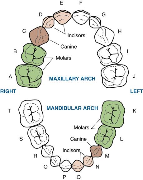
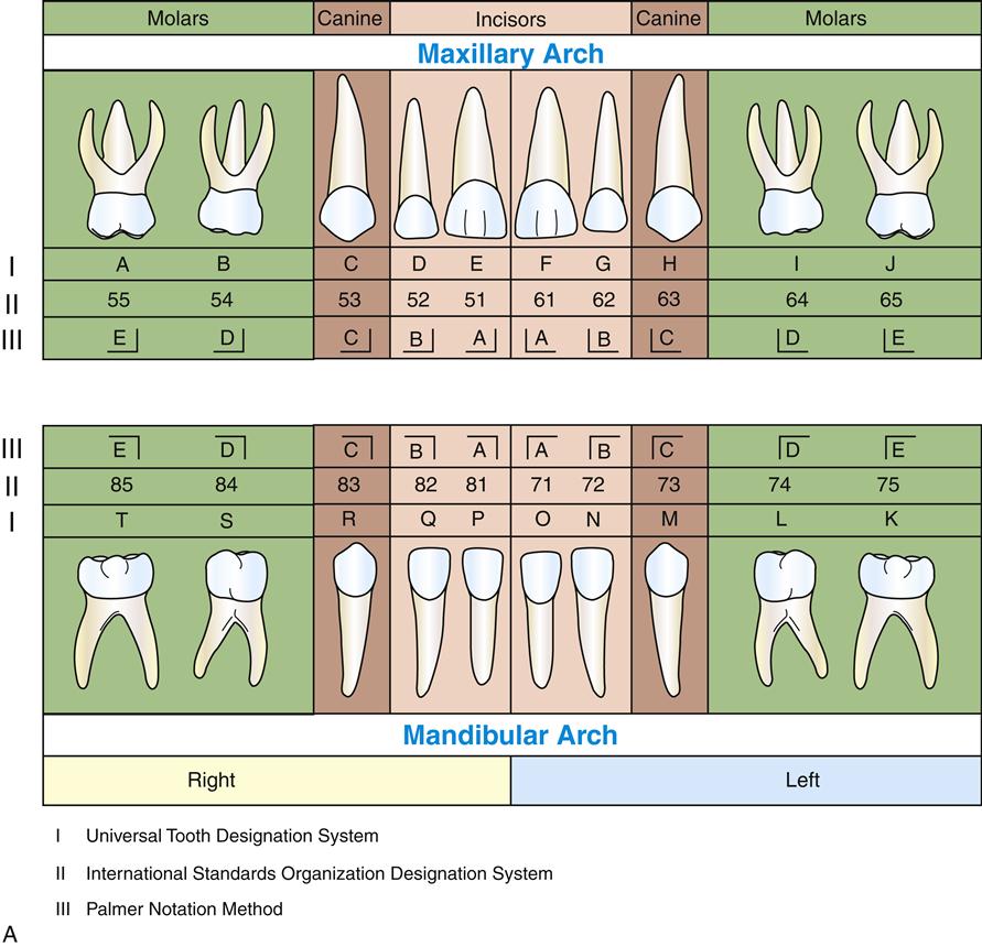
The primary teeth and the permanent teeth are both designated by the Universal Tooth Designation System (Figure 15-3). This system is the most widely used in United States for the designation of both dentitions because it is adaptable to electronic data transfer; this textbook uses the Universal System. With the Universal Tooth Designation System, the primary teeth are designated in a consecutive arrangement by using capital letters, A through T, starting with the maxillary right second molar, moving clockwise, and ending with the mandibular right second molar (see Figure 15-1).
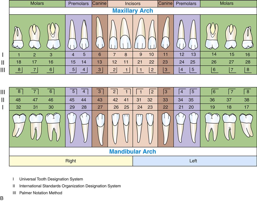
B: Universal Tooth Designation System, International Standards Organization Designation System, and Palmer Notation Method for the permanent teeth.
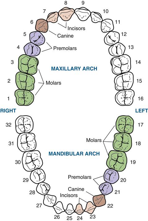
The permanent teeth are designated by the Universal Tooth Designation System in consecutive arrangement as the patient is observed from in front by using the digits 1 through 32, starting with the maxillary right third molar, moving clockwise, and ending with the mandibular right third molar (see Figure 15-2). The clockwise convention also is also used when charting restorations or periodontal conditions in the oral cavity for a patient.
However, the need for a system that can be used internationally, as well as by electronic data transfer, is recognized; thus the acceptance of the International Standards Organization Designation System (ISO System) by the World Health Organization (see Figure 15-3). With this system, the teeth are designated by using a two-digit code. The first digit of the code indicates the quadrant (see later discussion under general dental terms), and the second indicates the tooth in this quadrant. This is based on the system of the Fédération Dentaire Internationale (FDI).
With the ISO System, the digits 1 through 4 are used for quadrants in a clockwise manner in the permanent dentition, and digits 5 through 8 are used in a clockwise manner for those quadrants of the primary dentition. For the second digit, which indicates the tooth, the digits 1 through 8 are used for the permanent teeth, with this designation using the median line in a distal direction for numbering. The digits 1 through 5 are then used for the primary dentition, with this designation also using the median line in a distal direction for numbering.
Another system that is commonly used in orthodontics is the Palmer Notation Method also known as the Military Tooth Numbering System (see Figure 15-3). It is helpful with this dental specialty because it allows immediate discussion of the teeth that require prompt treatment, and it can produce a very graphical image, akin to a map of the dentition. In this system, the teeth are designated with a right-angle symbol indicating the quadrants with the tooth number inside, similar in numbering to the ISO System that superseded it.
Dentition Periods
Although there are only two dentitions, there are three dentition periods throughout a person’s lifetime, because the time period of the two dentitions overlaps (Table 15-1). Each patient should be assigned a dentition period to allow the most effective dental treatment for that period. This specificity is especially important with the consideration of orthodontic therapy, because growth during certain dentition periods is maximized to allow expansion of the jaws and movement of the teeth.
TABLE 15-1
Dentition Periods and Clinical Considerations
| PRIMARY DENTITION PERIOD | MIXED DENTITION PERIOD | PERMANENT DENTITION PERIOD | |
| Approximate time span | ~6 months to ~6 years | ~6 years to 12 years | After ~12 years |
| Teeth marking start of period | Eruption of primary mandibular central incisor | Eruption of permanent mandibular first molar | Shedding of the last primary tooth |
| Dentition present | Primary | Primary and permanent | U/> |
Stay updated, free dental videos. Join our Telegram channel
VIDEdental - Online dental courses


