15: Metabolic and Genetic Diseases

Metabolic and Genetic Diseases

Metabolic Conditions

Paget’s Disease

Paget’s disease, or osteitis deformans, is a chronic, slowly progressive metabolic disorder of bone of undetermined cause (Box 15-1). Etiologic theories include infection by paramyxovirus and mutations in several genes that are involved in osteoclastogenesis. Altered osteoclast development and function result in abnormal bone remodeling. Osteoblast function may also be abnormal in Paget’s disease. This condition generally progresses through several stages that include an initial resorptive phase, followed by a vascular phase, and eventually by a sclerosing phase.

Clinical Features

Paget’s disease is a hyperactive bone turnover state that typically occurs in patients older than 50 years. It is relatively common and has been reported to occur in 3% to 4% of the middle-aged population and in as many as 10% to 15% of the elderly. In approximately 14% of cases, a positive family history can be elicited. Paget’s disease has a 3 : 2 male predilection, and it seems to occur more often in patients of Northern European descent.

The most common sites of involvement include the pelvis, skull, tibia, vertebrae, humerus, and sternum. The jaws are affected in approximately 20% of patients, and the maxilla is involved twice as often as the mandible (Figure 15-1). At initial presentation, symptoms often relate to deformity or pain in the affected bone(s). Bone pain is described as deep and aching. A perception of elevated skin temperature over the affected bone is often noted because of the hypervascularity of the underlying bone. Neurologic complaints—including headache, auditory or visual disturbances, facial paralysis, vertigo, and weakness—may be related, in large part, to narrowing of the skull foramina, resulting in compression of vascular and neural elements. Approximately 10% to 20% of patients are asymptomatic and are incidentally diagnosed after radiographic or laboratory studies are performed for unrelated problems.

Classically, dental patients who wear complete dentures may complain of newly acquired poor prosthetic adaptation and function as the maxilla symmetrically enlarges. The alveolar ridge ultimately widens, with relative flattening of the palatal vault. When teeth are present, increased spacing, as well as loosening, is noted. In severe cases, continued enlargement of the maxilla or mandible can make closure of the lips difficult or impossible.

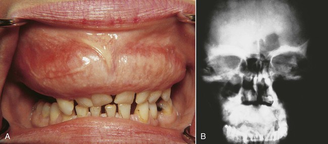
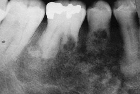
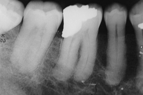
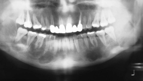
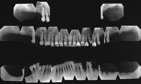
Classic radiographic findings in the late stage of Paget’s disease are due to bony sclerosis providing a patchy radiopaque pattern described as resembling cotton or wool. In the jaws, this pattern of bone change may be associated with hypercementosis or resorption of tooth roots, loss of lamina dura, and obliteration of the periodontal ligament space (Figures 15-2 and 15-3).

Histopathology

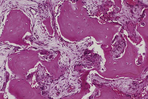
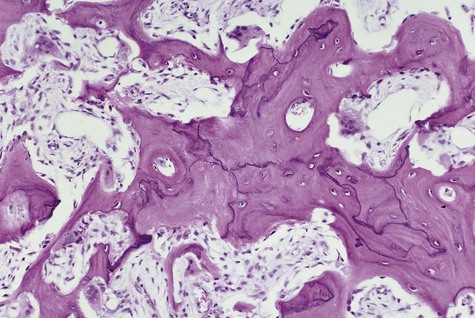
In the initial resorptive phase, random overactive osteoclastic bone resorption is evident. Resorbed bone is replaced by vascularized connective tissue in company with prominent osteolysis and osteogenesis. Bone eventually develops a dense mosaic pattern as a result of reversal lines in increasingly sclerotic bone, as osteoclasts give way to osteoblasts (Figures 15-4 and 15-5).

The laboratory can provide important information about the diagnosis of Paget’s disease. Serum calcium and serum phosphate levels are normal in the presence of markedly elevated alkaline phosphatase levels. The intense osteoblastic activity in this metabolically active bone is believed to be responsible for the elevated alkaline phosphatase levels. The amount of bone resorption may be correlated with increases in urinary calcium and hydroxyproline levels.

Treatment

The primary indicator for therapeutic intervention is patient discomfort. Elevation of alkaline phosphatase levels to twice normal levels is also an indication for treatment. Therapy has been directed at controlling osteoclast formation and function. The use of calcitonin and bisphosphonate has been effective. Both suppress bone resorption and deposition, as reflected in a reduction in the biochemical indices, including alkaline phosphatase and urinary hydroxyproline levels. A 50% reduction in either index constitutes a good therapeutic response (see Chapter 13 for complications of bisphosphonate therapy).

Paget’s disease is a slowly progressive disorder, but it is seldom fatal. Relief of symptoms, particularly bone pain, with oral or intravenous bisphosphonates is beneficial. Complications include skeletal deformity, weakened bones, neurologic deficits, and pathologic fracture. Heart failure may also be an important complication of Paget’s disease as a consequence of the hypervascular bone. In the early vascular phase, bleeding following any type of bone surgery (e.g., tooth extraction) can be problematic. In a small percentage of cases, malignant transformation into osteosarcoma may occur. Depending on the series reported, this has ranged from 1% to 15%.

Hyperparathyroidism

Hyperparathyroidism may be one of three types: primary, secondary, or hereditary (Box 15-2). Rarely, hyperparathyroidism may be associated with a Noonan-type syndrome, a complex, autosomal-dominant inherited trait comprising short stature, unusual facies, mental retardation, and cardiac defects.

Primary hyperparathyroidism is characterized by hypersecretion of parathyroid hormone from one or more hyperplastic parathyroid glands (3%), a parathyroid adenoma (90%), or, less commonly, an adenocarcinoma (3%). Characteristic abnormal laboratory findings include elevated parathormone levels and elevated calcium and alkaline phosphatase levels resulting from parathormone stimulation of osteoclast-mediated bone resorption, from decreasing calcium excretion in the kidneys, and from increased intestinal resorption.

Secondary hyperparathyroidism occurs as a compensatory response to hypocalcemia, as may be found in renal failure, in patients undergoing renal dialysis, and in those with intestinal malabsorption syndromes. In these patients, vitamin D3, which is activated in the kidney, is reduced. Vitamin D3 is required for calcium absorption and metabolism. The hereditary form has been shown to be an autosomal-dominant condition mapped to chromosome 1q21-q31, the location of the HRPT2 endocrine tumor gene.

Clinical Features

The disease spectrum of primary hyperparathyroidism ranges from asymptomatic cases (diagnosed by routine serum calcium determinations) to severe cases manifesting as lethargy and occasionally coma. The incidence increases with age and is greater in postmenopausal women. Early symptoms include fatigue, weakness, nausea, anorexia, arrhythmias, polyuria, thirst, depression, and constipation. Bone pain and headaches are often reported.

Several clinical features are associated with primary form of this disease, classically described as “stones, bones, groans, and moans.” Lesions of the kidneys, skeletal system, gastrointestinal tract, and nervous system are responsible for this syndrome complex. The renal component includes the presence of renal calculi or, more rarely, nephrocalcinosis associated with hypercalcemia.

Gastrointestinal manifestations include peptic ulcer resulting from the increase in gastric acid, pepsin, and serum gastrin levels. Rarely, pancreatitis may develop as a result of obstruction of the smaller pancreatic ducts by calcium deposits.

Neurologic manifestations may become evident when serum calcium levels are very high, exceeding 16 to 17 mg/dL. In such instances, coma or parathyroid crisis may occur. Loss of memory and depression are common, and, rarely, true psychosis may appear. Some of the neurologic findings may be attributed to calcium deposits in the brain.

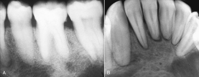
Severe osseous changes (called, in the past, osteitis fibrosa cystica) are the result of significant bone demineralization, with fibrous replacement producing radiographic changes that appear cystlike. In the jaws, these lesions microscopically resemble central giant cell granuloma. Less obvious radiographic changes may include an osteoporotic appearance of the mandible and maxilla, reflecting more generalized resorption (Figure 15-6). Loosening of the teeth may occur, as well as corresponding obfuscation of trabecular detail and overall cortical thinning. Partial loss of the lamina dura is seen in a minority of patients with hyperparathyroidism (Figures 15-7 and 15-8). Pulpal obliteration, with complete calcification of the pulp chamber and canals, has been reported in association with secondary hyperparathyroidism.

Hyperthyroidism

Hyperfunction of the thyroid gland, or hyperthyroidism, encompasses several conditions. It is characterized by excessive amounts of the thyroid hormones triiodothyronine (T3) and thyroxine (T4) or by increased levels of thyroid-stimulating hormone (TSH) and associated hypermetabolism. In adults, hyperthyroidism occurs with an incidence of 3 cases per 10,000 per year, with a distinct female preponderance of approximately 5 : 1.

The most common disorder leading to clinical hyperthyroidism is Graves’ disease, which accounts for 70% to 85% of all cases. The cause is believed to be auto-immune. It appears to be related to the production of abnormal thyroid stimulator (long-acting thyroid stimulator [LATS]), which differs chemically and functionally from TSH. LATS is able to bind the thyroid-TSH receptors in preference to TSH and to remain bound for prolonged periods. The LATS substance is immunoglobulin (Ig)G produced by B cells, which is capable of inducing thyroid hyperplasia and increasing iodine uptake by the thyroid, free of any pituitary gland influence. Thyrotoxicosis may also result from excess stimulation of the thyroid gland via the hypothalamic-pituitary axis, or by secretion of thyroid hormone from ectopic, endogenous, or exogenous sources.

Heat intolerance, hyperhidrosis, and palmar erythema are common findings. Fine motor tremor and muscle weakness, palpitations, diarrhea, anxiety, weight loss, and menstrual dysfunction are also commonly encountered. Patients may complain of an altered complexion and thinning, brittle hair. Ocular changes include upper lid retraction and so-called lid lag on normal blinking. The bright-eyed stare that often results from upper lid retraction may be further accentuated by exophthalmos. Pretibial myxedema and acropachy may be found in patients with Graves’ disease.

Cardiac manifestations are among the earliest and most consistent features of this disease. Increased metabolic activity places greater demand on the cardiovascular system; accordingly, increases in stroke volume, pulse rate, and cardiac output are usually observed.

Although the oral manifestations of this condition are not specific, they are consistent (Box 15-3). In children, premature or accelerated exfoliation of deciduous teeth and the concomitant rapid eruption of permanent teeth are often noted. In adults, osteoporosis of the mandible and maxilla may be found. On occasion, patients may complain of a burning tongue, as well as other, nonspecific symptoms. Of interest is a reported threefold increase in the incidence of dental erosion in these patients in comparison with euthyroid control subjects.

Medical treatment consists of thyroid-suppressive drug therapy or radioactive iodine administration, which essentially inactivates the hyperfunctional thyroid tissue. Thyroid-suppressive drugs include thiocarbamides such as propylthiouracil and methimazole. These drugs inhibit iodine oxidation and iodination of tyrosyl residues—two steps in the synthesis of thyroid hormones. Surgical treatment remains an option, although the potential for inadvertent parathyroid gland removal and subsequent hypoparathyroidism is a risk.

Of clinical importance is the need to reduce stress to minimize the risk of precipitating a thyroid crisis in patients with poorly controlled disease. The use of certain drugs such as epinephrine and atropine is contraindicated, because they may precipitate a thyroid storm, which is a life-threatening state of thyroid hormone–induced hypermetabolism.

Hypothyroidism

Hypothyroidism is a systemic condition that is caused by reduced production of thyroid hormone. This results from a number of factors including congenital defect, iodine deficiency goiter, autoimmune (Hashimoto’s) thyroiditis, diseases of the pituitary and hypothalamus, and idiopathic causes. The common result of these etiologic factors is cretinism when the condition occurs in children, and myxedema when it occurs in adults.

The key clinical features are listed in Box 15-4. Diagnosis is based on the history, the physical examination, and determination of serum levels of TSH, and tetraiodothyronine (T4). In typical hypothyroid patients with primary disease, T4 levels are low (sometimes normal) and TSH levels are high (compensatory pituitary reaction). In secondary disease in which the pituitary gland is malfunctioning, both T4 and TSH levels are low. Treatment is based on gradual replacement with synthetic and natural thyroid hormone preparations.

Hypophosphatasia

Hypophosphatasia represents a deficiency of alkaline phosphatase. This hereditary disorder is transmitted in an autosomal-recessive manner. Of dental significance is that this unusual genetic metabolic disease is one of the main causes of premature loss of the primary dentition. (Other conditions in which premature tooth exfoliation may be seen include cyclic neutropenia, idiopathic histiocytosis, juvenile periodontitis, acrodynia, rickets, and Papillon-Lefèvre syndrome.) Although the primary dentition is nearly exclusively involved, adolescent and adult patients with this condition may also experience dental abnormalities, including reduced marginal alveolar bone, abnormal root cementum, focal areas of dentin resorption, altered mineralization of coronal dentin, and large coronal pulp chambers of the molar dentition.

The chief clinical features of hypophosphatasia include enlarged pulp chambers of the primary teeth, alveolar bone loss with a predisposition for the anterior portion of the mandible and maxilla, and hypoplasia or aplasia of cementum over the root surface. Root development may be deficient, especially toward the apex. The crowns of the involved teeth demonstrate rickets-type changes, which are characterized chiefly by hypoplastic enamel defects. Enamel hypoplasia, increased pulp spaces, and premature tooth exfoliation are present in the permanent and primary dentitions. Dental abnormalities are the result of inadequate formation of both dentin and cementum.

Long bones show inadequate levels of mineralization with abnormally wide osteoid seams. Serum chemistry studies indicate a reduction in alkaline phosphatase levels, with concomitant urinary findings of detectable phosphoethanolamine. Tissue levels of alkaline phosphatase likewise are decreased in this disorder.

Four clinical types of hypophosphatasia have been recognized:

1. A congenital type has a 75% rate of neonatal mortality.

2. An early infantile type appears within the first 6 months of life, with a mortality rate of 50%. Renal calcinosis, as well as risks of cranial synostosis, delayed motor development, and premature loss of teeth, may accompany this disease.

3. A late infantile or childhood type begins between 6 and 24 months of age. Skeletal findings tend to be less pronounced, but abnormalities of long bone structures, including irregular ossifications at the metaphysis, may be observed, along with rickets-type changes at the costochondral junctions. Of importance in this form of the disease is premature loss of the anterior primary teeth, often the first sign of the illness.

4. The adult type, although distinctly uncommon, is characterized by bone pain, pathologic fractures, and a childhood history of rickets.

No successful treatment is known, apart from controlling the hypercalcemia that results from the hypophosphatasia. Large doses of vitamin D occasionally have produced partial improvement, although hypercalcemia and soft tissue calcinosis may result from such an approach. Genetic counseling of the family, as well as early diagnosis, is of great value.

Infantile Cortical Hyperostosis

Infantile cortical hyperostosis, or Caffey’s disease, is a self-limited, short-lived proliferative bone disease of undetermined origin. It is characterized by cortical thickening of various bones, most commonly the mandible (80% of cases), and less commonly the clavicles, long bones, maxilla, ribs, and scapulae. Pain, fever, and hyperirritability may precede or develop concurrently with the swelling. From 75% to 90% of cases demonstrate mandibular involvement, typically over the angle and ascending ramus symmetrically. Sporadic cases of infantile cortical hyperostosis almost always show mandibular involvement, and familial cases demonstrate such involvement approximately 60% of the time.

In addition to the osseous changes, swelling of overlying soft tissues usually occurs. There are no gender, racial, or geographic predilections. The characteristic age of onset is usually by the seventh month of life; the average age of onset is 9 weeks.

Radiographically, an expansile hyperostotic process is visible over the cortical surface, with rounding or blunting of the mandibular coronoid process. Initially, the hyperostotic element is separated from the underlying bone by a thin radiolucent line.

Diagnosis may be facilitated by the use of technetium (99mTc) scans, which are often positive, before routine radiographic detection is begun. Laboratory findings that are helpful in establishing the diagnosis include an elevated erythrocyte sedimentation rate, increased phosphatase levels, anemia, leukocytosis, and occasionally thrombocytopenia or thrombocytosis.

Infantile cortical hyperostosis is usually a self-limiting process, with treatment generally directed at supportive care. Systemic corticosteroids and nonsteroidal anti-inflammatory drugs have been used with some success. This disease has a tendency to follow an uneven, although predictable, course, with possible relapses and remissions. During such recurrences or relapses, nonsteroidal anti-inflammatory drugs have been recommended to control symptoms and halt progression of the disease, suggesting that prostaglandins may have a role in the origin. The resolution phase ranges from 6 weeks to 23 months, with an average duration of 9 months. Radiographic and histologic resolution may take up to several years, with a generally excellent prognosis, despite the possibility of recurrences and occasional residual effects, such as severe malocclusion and mandibular asymmetry.

Phantom Bone Disease

Phantom bone disease, also known as massive osteolysis, Gorham’s disease, or vanishing bone disease, is an unusual process characterized by post-traumatic or spontaneous slow, progressive, localized destruction of bone. It is a non-neoplastic condition characterized by a proliferative vascular and connective tissue response. Fibrovascular tissue may completely replace the involved bone, but the mechanism of bone destruction and resorption is unknown. This is a rare entity of unknown cause, with fewer than 150 cases reported since its initial description in 1838. The process has been described in virtually every bone in the body, with 15 cases reported in the maxillofacial region.

No ethnic or gender predilection has been noted. There appears to be no genetic basis for transmission. Various studies, including metabolic, endocrine, and neurologic tests, have not been helpful in determining the cause of phantom bone disease.

In most patients, the disease develops before the fourth decade of life, although it has been described in patients ranging from 18 months to 72 years of age. Onset of the disease is insidious; pain usually is not a feature unless concomitant pathologic fracture of the involved bone occurs. Progressive atrophy of the affected bone, resulting in significant deformity, constitutes a useful diagnostic sign of massive osteolysis. Although most cases involve a single bone, the disease may be polyostotic, usually affecting contiguous bones. This disease is progressive but variable—over time, the bone may completely disappear, or it may spontaneously stabilize. Significant regeneration has not been reported.

The earliest radiographic sign of the disease has been reported to be one or more intermedullary subcortical radiolucencies of variable size, usually with indistinct margins and thin radiopaque borders. In time, these foci enlarge and coalesce, eventually involving the cortex. A characteristic tapering ultimately occurs when long bones are affected.

Laboratory studies fail to show biochemical abnormalities. Microscopically, replacement of bone by connective tissue with many dilated capillaries and anastomosing vascular channels is noted. As the disease progresses, dissolution of both medullary and cortical bone is seen. A fibrotic band, thought to represent residual periosteum, persists.

No effective treatment for phantom bone disease is known, although moderate doses of radiation therapy (40 to 45 Gy in 2-Gy fractions) have resulted in good outcomes with few long-term complications. Limited success has been attained with bone grafts and implants.

Acromegaly

Acromegaly is a rare condition with a prevalence of approximately 50 to 70 cases per million population and an incidence of 3 cases per million per year. This disease is characterized by bony and soft tissue overgrowth and metabolic disturbances. These changes occur as a result of chronic hypersecretion of growth hormone subsequent to closure of the epiphyseal plates. If hypersecretion occurs before epiphyseal closure, gigantism results.

Clinical Features

Acromegaly presents most often in the fourth decade, with an even gender distribution and no racial or geographic predominance. This disorder is of insidious onset, and diagnosis is often delayed for many years. Younger patients have more aggressive tumors and develop clinically recognizable acromegaly more rapidly.

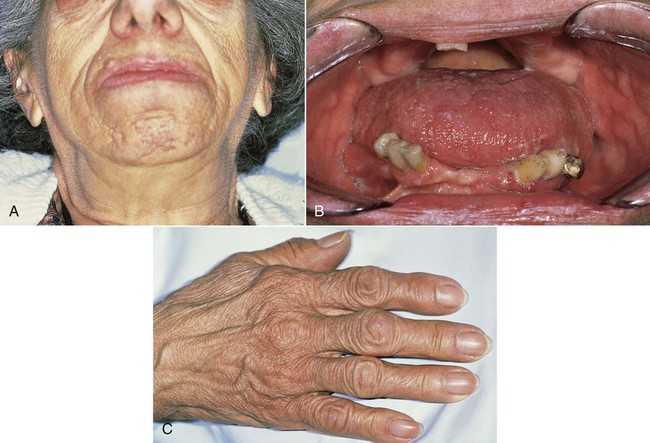
Clinical signs and symptoms result from local effects of the expanding pituitary mass and the effects of excess growth hormone secretion (Figure 15-9). Affected individuals present with hyperhidrosis; coarse body hair; muscle weakness; paresthesia, especially carpal tunnel syndrome; dysmenorrhea; and decreased libido or impotence. Sleep apnea, hypertension, and heart disease are also encountered. Skin tag formation is common and may be a marker for colonic polyps. In the facial bones and the jawbones, new periosteal bone formation may be seen, as well as cartilaginous hyperplasia and ossification. Resultant orofacial changes include frontal bossing, nasal bone hypertrophy, and relative mandibular prognathism or prominence. Enlargement of the paranasal sinuses, as well as secondary laryngeal hypertrophy, produces a rather deep, resonant voice, which is typical of acromegaly. Overall coarsening of the facial features is noted, resulting from connective tissue hyperplasia.

Oral manifestations include enlargement of the mandible and maxilla, with secondary separation of teeth resulting from alveolar overgrowth. Condylar hyperplasia with concomitant bone formation at the anterior portion of the mandible and a distinct increase in the gonial angle produces a rather typical dental malocclusion and prognathism. A complete posterior cross-bite is a common finding in such a circumstance. Thickened oral mucosa, increased salivary gland tissue, macroglossia, and prominent lips are also noted in most instances. It has been reported that with concomitant changes in mandibular structure, marked alterations in the diameter of the inferior alveolar canal, myofascial pain dysfunction syndrome, and speech abnormalities may result. The demonstration of growth hormone levels that are nonsuppressible by glucose loading is diagnostic. Computed tomography or magnetic resonance imaging of the sella turcica may help confirm the diagnosis of acromegaly-associated tumor. Radioimmunoassay studies of somatomedin C may be used as a routine screening test.

Genetic Abnormalities

Cherubism

Cherubism is a benign hereditary condition that is transmitted as an autosomal-dominant trait. Penetrance is 100% in males and 50% to 75% in females, with a 2 : 1 male predominance. Sporadic cases have also been seen. Mutations in the SH3BP2 gene have been reported in patients with this condition. Cherubism has been classified by some investigators as an autoinflammatory disease (i.e., a genetically determined chronic noninfectious inflammatory disorder).

Clinical Features

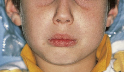
Cherubism affects the maxilla and/or mandible and usually is found in children by 5 years of age (Box 15-5). The term cherubism has been used to describe patients with cherubic facies of marked fullness of the jaws and cheeks and upwardly gazing eyes. The mandibular angle, ascending ramus, retromolar region, and posterior maxilla are most often affected. The coronoid process can also be involved, but the condyles are always spared. A vast majority of cases occur only in the mandible. The bony expansion is most often bilateral, although unilateral involvement has been reported. The specific gene maps to chromosome 4p16.3, which encodes the SH3-binding protein, SH3BP2.

Patients typically have painless symmetric enlargement of the posterior region of the mandible, with expansion of the alveolar process and ascending ramus. The clinical appearance may vary from barely discernible posterior swelling of a single jaw to marked anterior and posterior expansion of both jaws, resulting in masticatory, speech, and swallowing difficulties (Figures 15-10 and 15-11). Intraorally, a hard, nontender swelling can be palpated in the affected area.

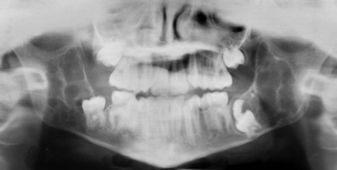
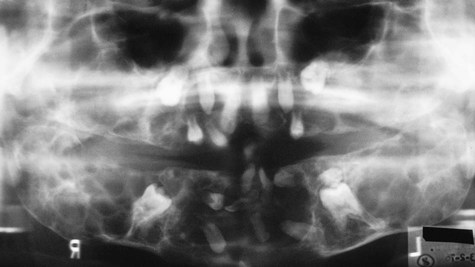
With maxillary disease, involvement of the orbital floor and the anterior wall of the antrum occurs. Superiorly directed pressure on the orbit results in increasing prominence of the sclera and the appearance of upturned eyes. The palatal vault may be reduced or obliterated. Maxillary involvement usually results in the greatest deformity. All four quadrants of the jaws may be simultaneously involved with this painless process of bony expansion (Figure 15-12). Premature exfoliation of the primary dentition may occur as early as 3 years of age. Displacement of developing tooth follicles results in poor development of selective permanent teeth and ectopic eruption or impaction. Permanent teeth may be missing or malformed, with the mandibular second and third molars most often affected. Significant malocclusions can be anticipated even with unifocal involvement.
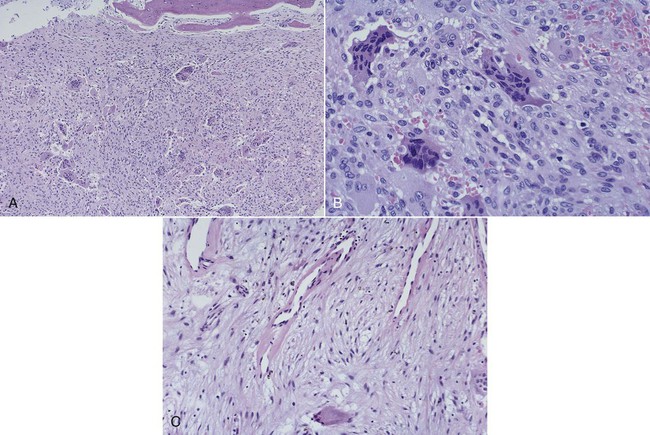
Submandibular and upper cervical lymphadenopathy are common, although reactive regional lymphadenopathy, particularly of submandibular lymph nodes, usually subsides after 5 years of age. Intelligence is unaffected. Serum calcium and phosphorous levels are within normal limits, but alkaline phosphatase levels may be elevated.

Radiographic surveys may provide the only signs of disease. Radiographic lesions characteristically appear as numerous well-defined multilocular radiolucencies of the jaws. The borders are distinct and are divided by bony trabeculae. Seen in the mandible are expansion and thinning of the cortical plate with occasional perforation; displacement of the inferior alveolar canal may be noted. An occlusal radiograph of the maxilla may give a soap bubble–like picture, with maxillary antrum obliteration. Unerupted teeth are often displaced and appear to be floating in the cystlike spaces.

Histopathology

Histologically, the lesions are composed of a vascularized fibrous stroma containing multinucleated giant cells, resembling central giant cell granuloma (Figure 15-13). Mature lesions exhibit a large amount of fibrous tissue and fewer giant cells. A distinctive feature that is often present is eosinophilic perivascular cuffing of collagen surrounding small capillaries throughout the lesion.

Osteopetrosis

Osteopetrosis, also known as Albers-Schönberg disease, is a rare hereditary bone condition characterized by a generalized symmetric increase in skeletal density due to defective bone resorption. Mutations in genes associated with osteoclastogenesis have been identified in patients with this disease. It can be divided into three clinical groups: (1) the infantile-malignant form is autosomal recessive in nature and is fatal within the first 2 to 3 years of life in the absence of treatment; (2) an intermediate autosomal-recessive type is nonfatal but clinically aggressive, with onset usually within the first decade; and (3) an autosomal-dominant form is the least severe, with full life expectancy but with considerable morbidity resulting from orthopedic alterations.

The characteristic feature of osteopetrosis />

Stay updated, free dental videos. Join our Telegram channel

Jan 12, 2015 | Posted by in Oral and Maxillofacial Pathology | Comments Off on 15: Metabolic and Genetic Diseases

VIDEdental - Online dental courses

Get VIDEdental app for watching clinical videos