15: Medical Conditions

Medical Conditions

Safe management of the oral and maxillofacial surgery patient requires an understanding of medial comorbidities that may complicate the perioperative or long-term outcome of procedures. Although it is not possible to describe the large array of medical conditions that may have surgical implications in oral and maxillofacial surgery, in this chapter we have elected 12 essential medical conditions that are encountered in the routine practice of any surgical subspecialty.

As the scope and depth of medicine and surgery expand, maintaining a command of all new developments becomes even more challenging. When necessary, consultation with practitioners in other medical or surgical subspecialties (e.g., cardiology, infectious diseases, ear/nose/throat) should be requested to obtain adequate information for safe surgical treatment.

Congestive Heart Failure

PMHX/PDHX/Medications/Allergies/SH/FH

The patient has a history of myocardial infarction 6 years earlier, with subsequent coronary artery bypass graft surgery without complications (documented previous coronary artery disease [CAD]). He also has a previous diagnosis of essential (idiopathic) hypertension, which is controlled by a single-drug regimen using hydrochlorothiazide (thiazide diuretic). He admits to poor compliance with his medications (risk factor for congestive heart failure [CHF] exacerbation) and had been previously diagnosed CHF, with a documented ejection fraction of 35% (the ejection fraction is the percentage of blood ejected forward into the systemic circulation by the left ventricle with each contraction; normal is greater than 55%). He is taking lovastatin (an HMG-CoA reductase inhibitor [cholesterol-lowering medication]) for hypercholesterolemia (risk factor for CAD). The patient has a 30 pack-year history of smoking (risk factor for CAD); he quit smoking after his myocardial infarction.

CAD involves varying degrees of impaired blood supply (oxygen) to the myocardium (causing ischemic heart disease), which puts the heart at risk for ischemic events (angina, myocardial infarction), with potential functional impairment of the myocardium and subsequent systolic dysfunction. This systolic dysfunction (heart failure) leads to the backup of blood (congestion); hence the term congestive heart failure. Risk factors for CHF include CAD (causes 50% to 75% of cases), uncontrolled systemic hypertension, valvular heart disease, cardiomyopathy (dilated or hypertrophic), stress (takotsubo), drugs ( alcohol, cocaine, chemotherapy), and infections (viral myocarditis).

Examination

Advanced trauma life support (ATLS) primary survey. Negative except for moderate respiratory distress. The patient was immediately placed on supplemental oxygen via a nasal cannula and showed improvement of his work of breathing.

General. The patient is awake, alert, and oriented to person, time, and place. He shows an increase in respiratory effort, evidenced by use of accessory muscles of respiration.

Vital signs. Blood pressure is 110/75 mm Hg, heart rate 120 bpm (tachycardia), respirations 28 per minute (tachypnea), and temperature 37.1°C.

Maxillofacial. Findings consistent with a mildly displaced Le Fort I fracture (see the section Le Fort I Fracture in Chapter 8).

Cardiovascular. Fast but regular rhythm with normal S1 sound (closure of mitral and tricuspid valves) and S2 sound (closure of aortic and pulmonic valves). An S3 sound is auscultated at the left sternal border at the fifth intercostal space (an S3 sound is heard in early diastole and is secondary to rapid ventricular filling in a dilated cardiac chamber). The point of maximum impulse (generated by the left ventricle as it touches the inner chest wall during systole) is laterally displaced with a parasternal heave (elevation of the chest wall to the left of the sternum). The jugulovenous pressure is elevated at 15 cm (normal is less than 9 cm) with a positive hepatojugular reflex (distention of the jugular veins on application of pressure in the right upper abdominal quadrant). Hepatojugular reflex and elevated jugulovenous pressure are signs of venous congestion observed in association with heart failure and volume overload.

Pulmonary. Use of the accessory muscles of respiration (sternocleidomastoid, scalenes, pectoralis major and minor, and serratus anterior muscles). Dyspnea is exacerbated when patient assumes the supine position (orthopnea). Bilateral basilar rales (fluid in the alveolar spaces) with dullness to percussion (due to pleural effusions) in the lung bases (fluid accumulation in the lungs is secondary to left-sided heart failure).

Abdominal. Nontender and nondistended, with hepatomegaly. Liver was percussed at 10 cm below the costal margin (hepatic congestion due to right-sided heart failure).

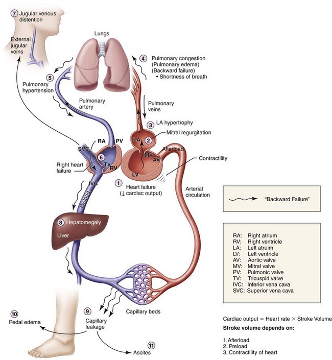
Extremity. Lower extremities show 3+ pitting edema at the ankles (significant fluid in the extravascular compartments due to venous congestion, causing capillary leakage; this is usually first noted in the lower extremities due to the added effect of gravity; Figure 15-1).

Imaging

A chest radiograph is the minimum imaging modality for the evaluation of CHF exacerbation. This is valuable for the evaluation of pulmonary edema and infiltration and for the approximation of the heart size. Echocardiography (transthoracic or transesophageal) is also useful for the evaluation of ventricular and valvular function and determination of the ejection fraction. The earliest finding of left-sided heart failure on the chest radiograph is cephalization of the pulmonary vessels. Normally, the vessels in the lung bases are larger and more numerous than those in the lung apices. This is secondary to the effects of gravity and the anatomically larger volume of the lungs at the base. With the progression of heart failure, the increased pressure is transmitted “backward” to the pulmonary veins and capillaries (hence the term “backward failure”). The lung bases are affected first; therefore, blood is preferentially “shunted” to the upper, or more cephalad, lobes, giving the radiographic appearance of cephalization. If the pressure in the vessels continues to rise, the fluid in the interstitium becomes radiographically evident as interstitial edema, bronchial wall thickening, and interlobular septa. The most noticeable are the Kerley B lines. These are short, thin, perpendicular lines extending to the pleura at the lung bases on a chest radiograph. The following imaging findings were noted for the current patient.

Chest radiograph. Bilateral blunting of the costophrenic angles with pronounced infiltrates in the lower lobes (consistent with bilateral pleural effusions and pulmonary edema).

Cephalization of the pulmonary vessels bilaterally. Increased cardiac silhouette (an increased cardiac silhouette, spanning more than one third of the thoracic cavity on an anteroposterior film, is indicative of an enlarged heart or dilated cardiomyopathy).

Transthoracic echocardiography. Dilated left ventricle consistent with dilated cardiomyopathy with decreased wall motion (systolic dysfunction) and mild mitral regurgitation. The pulmonic, aortic, and tricuspid valves were without stenosis or regurgitation. The ejection fraction was estimated at 25% (compromised ventricular function). No pericardial fluid and normal wall thickness were seen in all four chambers. Moderate elevation of the pulmonary artery pressure was noted.

CT (maxillofacial). Mildly displaced fracture at the Le Fort I level. (A CT scan of the chest can also be used to further evaluate the pulmonary parenchyma and cardiac structures.)

Labs

Brain natriuretic peptide level was 2,000 pg/ml (normal is less than 100 pg/ml).

With CHF, increased pressure and workload on the heart trigger the myocardial cells to secrete natriuretic peptides. Atrial myocytes secrete increased amounts of atrial natriuretic peptide, and the ventricular myocytes secrete both atrial and brain natriuretic peptides in response to the high atrial and ventricular filling pressures. Both of these peptides work as natriuretic, diuretic, and vasodilator agents and help reduce both preload and afterload. The plasma concentrations of both hormones are increased in patients with asymptomatic and symptomatic CHF.

Electrocardiogram Findings

The electrocardiographic findings for the current patient were as follows:

• Rate. Tachycardic at 120 bpm

• Rhythm. Regular; each P wave followed by a QRS complex; each QRS complex preceded by a P wave; QRS complexes occurring at regular intervals

• Axis. Positive deflection in lead I; negative deflection in lead aVF (indicative of left-axis deviation secondary to left ventricular hypertrophy)

• Intervals. PR interval less than 0.20 second, or 5 small boxes on electrocardiograph paper (more than 5 small boxes is consistent with first-degree atrioventricular node block); QRS complex less than 0.12 second, or 3 small boxes (more than 3 small boxes indicates widened QRS complex); QT interval less than half the distance from QRS complex to QRS complex (normal)

• Infarctions. Q waves in leads V1 through V5 (hallmark of old anteroseptal myocardial infarction); no flipped T waves, and no ST-segment elevation or depression (signs of acute ischemic events)

• Other. Loss of precordial R wave progression in leads V1 through V6 (suggestive of old anteroseptal MI and loss of anterior electrical forces)

Treatment

The cardiology service was consulted, and the patient was treated with fluid and salt restriction, intravenous Lasix (loop diuretic), oral lisinopril (angiotensin-converting enzyme [ACE] inhibitor), and oral carvedilol (β-blocker). Inotropic support was not required due to preserved perfusion and absence of cardiogenic shock. The patient’s cardiovascular symptoms and findings gradually improved within 36 hours (decreased shortness of breath, orthopnea, and paroxysmal nocturnal dyspnea; resolution of peripheral edema and pleural effusions; decrease in cardiac biomarker brain natriuretic peptide [BNP] level). He remained chest pain free and hemodynamically and electrically stable (no arrhythmias). After a careful perioperative cardiac risk assessment, the patient was taken to the operating room for fixation of the Le Fort fracture under general anesthesia.

Discussion

Heart failure can result from any structural or functional cardiac disorder that impairs the ability of the heart to pump blood. It is characterized by several symptoms, such as dyspnea, fatigue, edema, and weight gain. There are several etiologies for heart failure, including myocarditis/endocarditis (viral/bacterial infections), ischemic heart disease, infiltrative disease (amyloidosis, sarcoidosis), peripartum cardiomyopathy, hypertension, human immunodeficiency virus (HIV) infection, connective tissue disorders, substance abuse, certain chemotherapy drugs, and idiopathic origin.

Fluid retention in heart failure is initiated by the fall in cardiac output, leading to edema and decreased effective arterial volume. The reduction of cardiac output sets into motion a cascade of hemodynamic and neurohormonal derangements that provoke activation of the renin-angiotensin-aldosterone and sympathetic nervous systems. Although initially beneficial in the early stages of heart failure, these compensatory mechanisms eventually lead to a vicious cycle of worsening heart failure, fluid retention, and volume overload.

The classification systems used for the management of CHF are based on the severity of the condition, systolic versus diastolic dysfunction, and left-sided versus right-sided failure. Heart failure is classified by severity of symptoms using the New York Heart Association (NYHA) classification (most commonly used). This categorizes patients into one of four functional classes, depending on the degree of effort needed to elicit symptoms:

The American College of Cardiology/American Heart Association classification and guidelines for the treatment of heart failure can be found in Table 15-1.

Table 15-1

Classification and Guidelines for the Treatment of Heart Failure

Stage Characteristics Treatment
A High risk for heart failure without structural heart disease or symptoms ACEI or ARB
B Structural heart disease without signs or symptoms ACEI or ARB + β-blocker (devices such as defibrillators in appropriate patients)
C Structural heart disease with prior/current symptoms of heart failure. Routine use of diuretics, ACEI, or β-blockers, and aldosterone antagonist, ARB, digitalis, or hydralazine/nitrates, and devices such as biventricular pacing or implantable defibrillator in selected patients
D Advanced heart disease Medications from stages A through C, with optional use of palliative care, heart transplantation, chronic inotropes, permanent mechanical support, or experimental surgery or drugs

ACEI, Angiotensin-converting enzyme inhibitors; ARB, angiotensin receptor blocker.

Data from Hunt SA, Abraham WT, Chin MH, et al: ACC/AHA 2005 guideline update for the diagnosis and management of chronic heart failure in the adult: a report of the American College of Cardiology/American Heart Association Task Force on Practice Guidelines (Writing Committee to Update the 2001 Guidelines for the Evaluation and Management of Heart Failure). American College of Cardiology Web Site. Available at: http://www.acc.org/clinical/guidelines/failure//index.pdf.

When using the right-sided or left-sided designation as the etiology of heart failure, it is the symptomatology and physical and echocardiographic findings that determine the anatomic side of the failing heart. With right-sided heart failure, there is elevation of the jugulovenous pressure, peripheral edema, and hepatomegaly. In left-sided heart failure, fluid backs up in the lungs, causing dyspnea, cough, pleural effusions, and rales. Sustained left-sided heart failure eventually causes right-sided heart failure. After all, the most common cause of right-sided heart failure is left-sided failure.

The systolic versus diastolic designation of heart failure involves determining whether failure is the result of impaired contraction or inefficient relaxation. Systolic dysfunction is commonly defined as heart failure secondary to impaired ejection fraction, whereas diastolic dysfunction is characterized by heart failure in the setting of preserved ejection fraction. Causes of diastolic dysfunction include myocardial hypertrophy and infiltrative cardiomyopathy (cardiac amyloidosis).

The pharmacotherapy of heart failure (HF) is aimed at improving cardiac function (contractility and stroke volume) and reducing the workload of the heart (preload and afterload reduction). Most patients with HF should be routinely managed with a combination of three types of drugs: a diuretic, an ACE inhibitor (ACEI) or an angiotensin receptor blocker (ARB), and a β-blocker. The value of these drugs has been established by the results of numerous large-scale clinical trials. Patients with evidence of fluid retention should take a diuretic until a euvolemic state is achieved, and diuretic therapy should be continued to prevent the recurrence of fluid retention. Diuretics interfere with the sodium retention of HF by inhibiting the reabsorption of sodium or chloride at specific sites in the renal tubules.

Treatment with an ACEI or an ARB should be initiated and maintained in patients who can tolerate them. ACEIs are the best-studied class of agents in HF, with multiple mechanisms of benefit for both HF and coronary artery disease. They have been shown to favorably influence the long-term prognosis of HF.

β-blockers act principally to inhibit the adverse effects of the sympathetic nervous system in patients with HF. Sympathetic activation can increase ventricular volumes and pressure by causing peripheral vasoconstriction and by impairing sodium excretion by the kidneys. Long-term treatment with β-blockers can lessen the symptoms of HF, improve the clinical status of patients, and enhance the patient’s overall sense of well-being. In addition, as do ACEIs, β-blockers can reduce the risk of death and the combined risk of death and hospitalization.

Therapy with digoxin as a fourth agent may be initiated at any time to reduce symptoms, prevent hospitalization, control rhythm, and enhance exercise tolerance. Digoxin is a digitalis glycoside that inhibits sodium-potassium (Na+-K+) adenosine triphosphatase. Inhibition of this enzyme in cardiac cells and vagal afferent fibers results in an increase in the contractile state of the myocardium and reduction in sympathetic outflow from the central nervous system, respectively.

Other treatments that can be initiated in heart failure patients include use of an aldosterone antagonist and use of a combination of hydralazine and isosorbide dinitrate. Spironolactone is the most widely used aldactone antagonist. It has been shown in long-term trials to reduce death and heart failure hospitalizations and to improve the functional class for patients with NYHA class III or class IV symptoms.

The combination of hydralazine and isosorbide dinitrate reduces mortality in patients with heart failure who remain symptomatic despite optimal medical therapy, particularly in the African American cohort of patients. Hydralazine and isosorbide are arterial and venodilators that act by lowering systemic vascular resistance (afterload). Other advanced therapies for heart failure include intraaortic balloon counterpulsation, cardiac resynchronization, and left ventricular assist devices.

Bibliography

Dickstein, K, Cohen-Solal, A, Filippatos, G, et al. ESC guidelines for the diagnosis and treatment of acute and chronic heart failure 2008: the Task Force for the Diagnosis and Treatment of Acute and Chronic Heart Failure 2008 of the European Society of Cardiology. Developed in collaboration with the Heart Failure Association of the ESC (HFA) and endorsed by the European Society of Intensive Care Medicine (ESICM). Eur J Heart Fail. 2008; 10(10):933–989.

Grady, KL, Dracup, K, Kennedy, G, et al. Team management of patients with heart failure: a statement for healthcare professionals from the Cardiovascular Nursing Council of the American Heart Association. Circulation. 2000; 102:2443.

Hunt, SA, Abraham, WT, Chin, MH, et al. Focused update incorporated into the ACC/AHA 2005 guidelines for the diagnosis and management of heart failure in adults: a report of the American College of Cardiology Foundation/American Heart Association Task Force on Practice Guidelines developed in collaboration with the International Society for Heart and Lung Transplantation. J Am Coll Cardiol. 2009; 53(15):e1–e90.

Knudsen, CW, Omland, T, Clopton, P, et al. Diagnostic value of B-type natriuretic peptide and chest radiographic findings in patients with acute dyspnea. Am J Med. 2004; 116:363.

Young, JB, Gheorghiade, M, Uretsky, BF, et al. Superiority of “triple” drug therapy in heart failure: insights from the PROVED and RADIANCE trials—prospective randomized study of ventricular function and efficacy of digoxin; randomized assessment of digoxin and inhibitors of angiotensin-converting enzyme. J Am Coll Cardiol. 1998; 32(3):686–692.

Acquired Immunodeficiency Syndrome

PMHX/PDHX/Medications/Allergies/SH/FH

The patient tested positive for HIV 6 years ago. He believes he acquired the virus through unprotected sexual contact. On his most recent hospital admission, his CD4 count was 108 cells/µl; therefore, he was diagnosed with acquired immunodeficiency syndrome (AIDS). (An absolute CD4 count below 200 cells/µl is an AIDS-defining feature in adults; in pediatric patients, the CD4 cell percentage is more accurate.) The patient’s internist indicated that the patient’s recent episode of pneumonia was due to the recent decline in his CD4 count.

There are no particular preoperative criteria for maxillofacial surgery in patients with HIV infection or AIDS. However, each patient should be assessed for risks and benefits of surgery. Some of the common concerns in a surgical patient are the hemoglobin level and platelet count. Despite popular belief, neither the lymphocyte count nor the viral load alters maxillofacial surgical intervention. Nonetheless, a patient with a rapidly declining lymphocyte count or a rise in viral load should be reassessed before any surgical intervention.

Examination

General. The patient is a cooperative man with evidence of muscle wasting (cachexia) who appears anxious (generalized muscle wasting is seen with advancing AIDS).

Vital signs. Blood pressure is 121/79 mm Hg (hypotension), heart rate 90 bpm, respirations 14 per minute, temperature 37.8°C (with advanced AIDS, it is not uncommon to have a normal temperature despite the presence of an acute infection; this is due to the failure and/or deficiency of available white blood cells to mount an appropriate inflammatory response, which results in fever), and SaO2 92% on room air (a recent history of pneumonia can result in a decrease in baseline oxygen saturation on room air).

Maxillofacial. There is no evidence of hair loss or patchy ulceration on the scalp. (Scalp ulceration may be seen in patients with disseminated fungal diseases, such as cryptococcal infection. These ulcerations may also present in the oral cavity or on the peripheral extremities. A biopsy can be used to confirm the diagnosis. This finding can be used as a sign to evaluate other organs for fungal invasion.) Bilateral temporal wasting and prominent zygomatic arches are noted. Examination of the neck reveals a soft dorsal hump. (Lipodystrophy may result either directly from HIV or as a side effect of antiretroviral therapy, particularly protease inhibitors. Other physical signs to investigate are seborrheic dermatitis [dandruff], vesicular rash with central umbilication in the forehead [molluscum contagiosum], and enlargement of the parotid glands [differential diagnoses consist of lymphoepithelial cyst, lipodystrophy, and lymphoma].)

Intraoral. Oral hygiene is poor, and the breath is fetid. A generalized inflammation of the maxillary and mandibular gingivae, exposed buccal bone, and mobility of the teeth are noted. Gentle palpation of the gingival tissue results in bleeding and pain. There is no facial or intraoral fluctuance and no obvious swelling. (In addition to HIV gingivitis and HIV periodontitis, the oral examination should concentrate on the presence of warts [human papillomavirus]; oral neoplastic growths, such as Kaposi sarcoma [human herpes virus 8]; hairy leukoplakia [Epstein-Barr virus]; candidiasis; and other fungal infections. Aphthous ulcers, lymphoma, herpes, and cytomegalovirus ulcerations also may be seen in patients with AIDS.)

Chest. Chest is clear on auscultation (if the patient presents with crackles, a chest radiograph is indicated).

Cardiovascular. Regular rate and rhythm, S1 and S2, and no gallops, rubs, or murmurs (HIV may affect the heart, resulting in dilated cardiomyopathy, but this is not common).

Labs

During the perioperative evaluation of a patient with HIV infection, a complete blood count is valuable but should be used with caution. A rise in the white blood cell count may not be seen in response to physiologic or inflammatory demands, as would be seen in immune-competent individuals. The lymphocyte subset can be used to assess the susceptibility to opportunistic infections. The neutrophil count may be elevated with bacterial infections. The hemoglobin and hematocrit levels may be used to assess volume status (hemoglobin would be falsely elevated). Hemoglobin and platelet levels may be depleted, and patients may require packed red blood cell or platelet transfusions before major surgery.

Additional tests may be helpful, depending on the extent of the surgical procedures or the presence of concurrent comorbidities associated with immune suppression. Arterial blood gas analysis is helpful for assessing the pulmonary status of a patient with active pneumonia. A basic metabolic panel may be used to evaluate intravascular fluid status in dehydrated and volume-depleted patients. A blood urea nitrogen to creatinine ratio greater than 20 is suggestive of volume depletion. The coagulation factors are rarely depleted in patients with HIV, but HIV or other infections may predispose a patient to disseminated intravascular coagulation (DIC). Coagulation studies should be obtained as needed. Evaluation of hepatic function is also important. Although HIV may affect liver function directly, of more concern is a concurrent infection with hepatitis B or C virus (note that the route of transmission is similar for HIV and the hepatitis B and C viruses).

Treatment

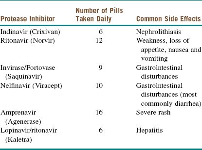
There is some controversy regarding the optimal time to initiate treatment for patients who are seropositive for HIV. Most infectious disease specialists start pharmacotherapy when the CD4 count drops below 50 cells/µl. Regardless of the CD4 count, treatment should be initiated as soon as possible in patients with HIV nephropathy, pregnant patients, and those coinfected with hepatitis B. Current recommendations consist of a combination of a nucleoside reverse transcriptase inhibitor (NRTI; Table 15-2), a non–nucleoside reverse transcriptase inhibitor (NNRTI; Table 15-3), protease inhibitors (Table 15-4), integrase inhibitors, fusion inhibitors, and CCR5 antagonists. A combination of these medications is commonly referred to as highly active antiretroviral therapy (HAART).

Table 15-2

Nucleoside Reverse Transcriptase Inhibitors

Nucleotide Reverse Transcriptase Inhibitor Abbreviation Side Effects
Retrovir AZT Anemia, neutropenia
Videx DDI Pancreatitis, peripheral neuropathy (PN)
Hivid DDC Pancreatitis, PN
Zerit D4T Pancreatitis, PN
Epivir 3TC Also used for hepatitis B virus (HBV) infection
Ziagen ABC Rash, death

Table 15-3

Nonnucleoside Reverse Transcriptase Inhibitors

Nonnucleoside Reverse Transcriptase Inhibitor Side Effects
Nevirapine (Viramune) Hepatotoxicity, hepatic necrosis during the first 4 weeks
Delavirdine (Rescriptor) Rash, headache
Efavirenz (Sustiva) Teratogenic; Stevens-Johnson rash, hallucinations, nightmares

Table 15-4

Protease Inhibitors

image

When a patient presents with a CD4 count below 200 cells/µl, they are started on trimethoprim-sulfamethoxazole for Pneumocystis jirovecii (formerly called Pneumocystis carinii) prophylaxis. At a CD4 count below 100 cells/µl, the patient is increasingly susceptible to toxoplasmosis infections. Because this is treated with the same medication, no additional prophylactic is needed. When the CD4 count drops below 50 cells/µl, there is a high risk of Mycobacterium avium complex infection. This is empirically treated with clarithromycin or azithromycin (macrolide antibiotics). Viral infections, such as herpes simplex virus, are treated with famciclovir or acyclovir (there is prophylactic treatment; patients are treated only in the face of infection). Some infections, such as Candida species, may present at a CD4 count below 500 cells/µl, but prophylactic treatment of fungal infections is not recommended.

The initial treatment of acute necrotizing ulcerative periodontitis consists of fluid resuscitation as needed, analgesics, and antibiotic therapy. With generalized severe periodontal disease, full mouth extraction is both definitive and curative. Patients with HIV infection are treated aggressively to prevent any odontogenic sources of infection, which may cause severe complications in a patient with a declining immune system.

The current patient underwent outpatient full mouth extraction under intravenous sedation. After surgical treatment, the alveolar ridges healed without complications.

Immune suppression can be due to defects in various aspects of the immune system. Concerns and cautions are not the same when dealing with different defects of the immune system. For example, a patient who is neutropenic is more susceptible to bacterial infection, whereas T-lymphocyte deficiency increases susceptibility to fungal, viral, and parasitic infections.

In oral surgical procedures, there are more complications associated with neutropenia (sepsis, oral ulceration, periodontal disease) than with lymphopenia. For the neutropenic patient, preoperative antibiotics are used to prevent sepsis. Postoperative antibiotics may also be used.

Complications

Patients affected by HIV infection may present with thrombocytopenia. This can be due to idiopathic thrombocytopenic purpura or thrombotic thrombocytopenic purpura. Idiopathic thrombocytopenic purpura is an autoimmune disorder, resulting from antibodies to glycoprotein platelet 2β3α-receptors. This may present as an acute condition (mostly in children) or a chronic condition (mostly in adult women). Treatment consists of prednisone, intravenous immunoglobulin, splenectomy, azathioprine, or vincristine. Thrombotic thrombocytopenic purpura presents as a combination of five symptoms: renal failure, central nervous system abnormalities, fever, thrombocytopenia, and anemia. The exact etiology is not well understood. A similar syndrome, hemolytic uremic syndrome, is caused by Escherichia coli O157:H7. In the presence of neurologic symptoms, the diagnosis of thrombotic thrombocytopenic purpura is made; however, when renal failure is the prominent feature, it is usually due to hemolytic uremic syndrome. The treatment for thrombotic thrombocytopenic purpura is plasmapheresis.

There also are other causes of both idiopathic thrombocytopenic purpura and thrombotic thrombocytopenic purpura. Idiopathic thrombocytopenic purpura may be caused by any viral infection, leukemia, lupus erythematosus, cirrhosis, antiphospholipid syndrome, and medications (quinine, heparin). Thrombotic thrombocytopenic purpura can be caused by cancer, bone marrow transplantation, pregnancy, and medication (ticlopidine, clopidogrel, cyclosporine, mitomycin, tacrolimus/FK-506, interferon-α).

The risk of postoperative infection in a patient with HIV infection or AIDS who undergoes maxillofacial surgery is controversial at best. Although earlier studies showed an increased risk of infection in those with HIV, this has not been confirmed by more recent studies. Most studies were conducted before the advent of HAART. There is a need for prospective evaluation for the risk of infections after various maxillofacial surgeries. Currently there are no recommendations regarding the presurgical or postsurgical care of a patient with HIV infection compared with that of healthy control patients.

Discussion

Immune deficiency may be an inherited, acquired, or iatrogenic disorder. Inherited defects may result from quantitative or qualitative defects of the cells or cellular pathways involved in immunity (neutrophil, macrophages, complement, lymphocytes). Immune suppression is also seen with organ transplantation for prevention of host-versus-graft or graft-versus-host disease and in chemotherapy. Acquired immune deficiency is seen with conditions such as diabetes, leukemia, and AIDS.

AIDS was recognized in 1981 after multiple homosexual male patients were diagnosed with Pneumocystis pneumonia and Kaposi sarcoma (more recently found to be also associated with human herpes virus 8). Before 1981, Pneumocystis pneumonia was commonly seen in cancer patients, and Kaposi sarcoma was endemic to Africa and the Mediterranean region. In 1984, HIV, a retrovirus belonging to the Lenti virus family, was discovered concurrently by French and American scientists. The virus can be transmitted via exposure of body fluids through sexual contact, sharing of needles or paraphernalia, blood transfusions (horizontal transmission), or from mother to fetus (vertical transmission). Once in the bloodstream, HIV targets the lymphocytes and macrophages, which are the only cells with CD4+ receptors. T cells with a CXCR4 chemokine coreceptor are called T tropic, and macrophages with CCR5 are called M tropic. The virus is unable to infect these cells in the absence of these coreceptors. Interaction of the CD4 receptor with the viral glycoprotein 120 changes its stereochemistry, exposing glycoprotein 41, which binds to heparin sulfate in the membrane of the host cells, fusing the viral envelope with the cell membrane. Once genetic material enters the cell, a complementary DNA or coda is made from the original RNA by the enzyme reverse transcriptase. This complementary DNA joins the host DNA by using the enzyme integrase, forcing the cell to make necessary proteins to replicate the virus. Finally, the packaged virus leaves the host cell, using the cell membrane as a viral envelope, and subsequently infecting other cells. CCR5 inhibitors, fusion inhibitors, integrase inhibitor, NRTIs and NNRTIs are respectively used to inhibit every step in this process.

After inoculation, a patient typically seroconverts in approximately 3 weeks, although the time period can range from 9 days to as long as 6 months. Routine laboratory testing for HIV before this date results in a negative test. The patient may be asymptomatic or may develop flulike symptoms. The viral load rapidly rises and then falls during this period, and the CD4 count rapidly drops before returning to nearly its original level. During the following years, if the infection goes untreated, there is a steady decrease in the CD4 cell count, along with an increase in the viral load. In general, the viral load is a reflection of the speed of progression of AIDS, whereas the CD4 cell count reflects the current immune status and is used to evaluate susceptibility to opportunistic infections.

More than 1 million Americans (1,106,400) had been infected with HIV by 2006, according to the Centers for Disease Control and Prevention (CDC). This population is increasing at the rate of 40,000 new infections each year. Among oral and maxillofacial surgery patients, the prevalence of HIV is estimated to be as high as 4.8% in certain demographic areas. Although HAART has resulted in a significant drop in opportunistic infections, such as those presenting with oral manifestation, it has not eradicated these complications.

Oral manifestation of HIV may aid in both the diagnosis and prognosis of the disease. These lesions are seen in up to 30% to 80% of patients who have tested positive for HIV, and they may be infectious (bacterial, viral, fungal, and parasitic), neoplastic, or idiopathic. A study published by Diz Dios and colleagues found a reduction in oral lesions from 74.2% to 28.5% after the use of HAART. However, in that study there was a significant drop in patient follow-up due to death or relocation. A study by Aguirre and associates evaluated 72 patients with various oral diseases. A significant improvement was seen in the prevalence of pseudomembranous candidiasis (from 80% to 32%) with the use of HAART. Only a small change was seen in acute necrotizing periodontitis. Tappuni and Flemming evaluated 284 patients infected with HIV and concluded that oral lesions are seen significantly less often in patients receiving monotherapy compared with patients receiving no therapy. Furthermore, the use of HAART resulted in a statistically significant reduction in lesions compared with the use of monotherapy.

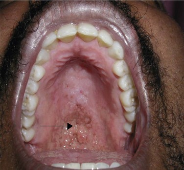
The differential diagnosis of oral lesions seen with HIV may be divided into bacterial, viral, fungal, neoplastic, and idiopathic categories. Bacterial infections may present as acute necrotizing gingivitis or periodontitis. Viral infections, such oral papillomas, are caused by the human papillomavirus and are commonly seen. Fungal infections, such as histoplasmosis or cryptococcosis (Figure 15-2), can cause oral ulcerations. An example of intraoral neoplastic disease is Kaposi sarcoma, caused by human herpes virus 8, which is most commonly seen in homosexual males. Idiopathic xerostomia is commonly seen, resulting in cervical caries.

The surgical management of patients with HIV infection or AIDS requires an understanding of the pathophysiology, medications, and associated disease processes.

Bibliography

Aguirre, JM, Echebrria, MA, Ocina, E. Reduction of HIV-associated oral lesion after highly active antiretroviral therapy. Oral Surg Oral Med Oral Pathol Oral Radiol Endod. 1999; 88:114–115.

Carey, JW, Dodson, TB. Hospital course of HIV-positive patients with odontogenic infections. Oral Surg Oral Pathol Oral Med Oral Radiol Endod. 2001; 91:23–27.

Centers for Disease Control and Prevention. HIV/AIDS surveillance report. Atlanta: the CDC; 2008.

Depoala, LG. Human immunodeficiency virus disease: natural history and management. Oral Surg Oral Med Oral Pathol Oral Radiol Endod. 2000; 90:266–270.

Diz Dios, P, Ocampo, A, Miralles, C, et al. Changing prevalence of human immunodeficiency virus–associated oral lesion. Oral Surg Oral Med Oral Pathol Oral Radiol Endod. 2000; 90:403–404.

Dodson, TB. HIV status and the risk of post-extraction complications. J Dent Res. 1997; 76:1644–1652.

Dodson, TB. Predictors of postextraction complications in HIV-positive patients. Oral Surg Oral Med Oral Pathol Oral Radiol Endod. 1997; 84:474–479.

Dodson, TB, Nguyen, T, Kaban, LB. Prevalence of HIV infection oral and maxillofacial surgery patients. Oral Surg Oral Med Oral Pathol Oral Radiol Endod. 1993; 76:272–275.

Dodson, TB, Perrott, DH, Gongloff, RK, et al. Human immunodeficiency virus serostatus and the risk of postextraction complications. Int J Oral Maxillofac Surg. 1994; 23:100–103.

Frame, PT. HIV disease in primary care. Prim Care. 2003; 30:205–237.

Mehrabi, M, Bagheri, S, Leonard, MK, Jr., et al. Mucocutaneous manifestation of cryptococcal infection: report of a case and review of the literature. J Oral Maxillofac Surg. 2005; 63:1543–1549.

Miller, EJ, Jr., Dodson, TB. The risk of serious odontogenic infection in HIV-positive patients: a pilot study. Oral Surg Oral Med Oral Pathol Oral Radiol Endod. 1998; 86:406–409.

Panel on Antiretroviral Guidelines for adult and Adolescents. Guidelines for the use of antiretroviral agents in HIV-1 infected adults and adolescents. http://aidsinfo. nih. gov/contentfiles/lvguidelines/adultandadolescentgl. pdf.

Patton, LL. Hematologic abnormalities among HIV-infected patients: associations of significance for dentistry. Oral Surg Oral Med Oral Pathol Oral Radiol Endod. 1999; 88:561–567.

Ray, N, Doms, RW. HIV-1 coreceptors and their inhibitors. Curr Top Microbiol Immunol. 2006; 303:97–120.

Schmidt, B, Kearns, G, Perrott, D, et al. Infection following treatment of mandibular fractures in human immunodeficiency virus seropositive patients. J Oral Maxillofac Surg. 1995; 53:1134–1139.

Shanti, RB, Aziz, SR. HIV-associated salivary gland disease. Oral Maxillofac Surg Clin North Am. 2009; 21:339–343.

Sleasman, JW, Goodenow, MM. HIV-1 infection. J Allergy Clin Immunol. 2003; 111:S582–S592.

Tappuni, AR, Flemming, JP. The effect of antiretroviral therapy on the prevalence of oral manifestations in HIV-infected patients: a UK study. Oral Surg Oral Med Oral Pathol Oral Radiol Endod. 2001; 92:623–628.

Chronic Kidney Disease

Examination

General. The patient is a mildly obese woman in mild distress secondary to pain.

Vital signs. Blood pressure is 162/98 mm Hg, heart rate 104 bpm, respirations 18 per minute, and temperature 38.8°C.

Neurologic. Patient is alert and oriented to place, time, and person.

Maxillofacial. There is fluctuant, tender, and erythematous right-sided facial swelling extending from the angle of the mandible to the right submandibular space. The floor of the mouth and the oropharyngeal airway are normal. The right mandibular third molar (tooth #32) is impacted, with swelling of the surrounding operculum. Right submandibular lymphadenopathy is noted.

Pulmonary. The chest is clear on auscultation bilaterally.

Cardiovascular. Regular rate and rhythm with no murmurs, rubs, or gallops.

Extremity. There is no swelling, edema, or muscle weakness.

Labs

Laboratory tests are ordered based on the severity and acuity of symptoms related to chronic kidney disease in conjunction with the patient’s nephrologist/internist. A baseline complete blood count, metabolic panels, liver function tests, and coagulation studies are usually obtained.

The following laboratory study results were obtained for the current patient:

The laboratory findings are characteristic of chronic renal disease. The hemoglobin and hematocrit are decreased secondary to the decreased production of erythropoietin by the kidneys. The elevated blood urea nitrogen and creatinine levels reflect the decreased glomerular filtration rate, which is also responsible for the elevated serum potassium. Proteinuria is a result of increased glomerular permeability. The decreased calcium is a result of decreased gastrointestinal absorption secondary to decreased renal production of active vitamin D.

Treatment

The management of a patient with chronic kidney disease is often complicated. Of particular concern is fluid status and electrolyte balance. Correction of metabolic and fluid abnormalities should be done in conjunction with the nephrologist before any surgical intervention. This may be accomplished by judicious hydration, careful electrolyte replacement, and medications. As chronic renal disease progresses to end-stage renal disease (ESRD), either peritoneal dialysis or hemodialysis becomes necessary. Hypertension is typically very difficult to adequately treat with medication alone and usually involves a salt restriction. In some cases, some degree of hypertension may need to be tolerated. Many medications, particularly antibiotics, need to be appropriately dosed for the reduced glomerular filtration rate or avoided altogether. All patients able to take oral nutrition should have a renal diet low in sodium, potassium, and protein. Patients requiring dialysis are best scheduled for surgery the day after their dialysis treatment (to optimize fluid and electrolyte balance) with a resumption of their usual dialysis the day after surgery. Because patients frequently are heparinized for dialysis, a minimum of 6 hours is prudent after cessation of heparin. In emergency cases in which surgery cannot wait, the dialysis treatment can be done without heparin. Patients with successful renal transplants may be considered to have adequate renal function but are commonly receiving immunosuppressive drugs, including corticosteroids, placing them at increased risk for infections and adrenal insufficiency in the perioperative period.

The initial management of the current febrile and anorexic patient included judicious fluid resuscitation. Normal saline (500 ml bolus) was given, followed by a maintenance rate of 50 ml/hr. Fluid resuscitation reduced the serum creatinine to 3.4 mg/dl, suggesting that some of the renal insufficiency was secondary to dehydration. In patients with end-stage renal disease who are dialysis dependent, fluid resuscitation may not be necessary. The patient’s elevated temperature was treated with acetaminophen (avoid nonsteroidal antiinflammatory drugs in chronic kidney disease, because they decrease renal blood flow). Although the degree of hyperkalemia was only mild, an electrocardiogram was performed to evaluate for loss of P waves, widened QRS complex, and peaked T waves (none of which were present). Kayexalate was given orally to lower the serum potassium level. The patient was begun on intravenous clindamycin. No dosing adjustment was needed, because the clearance of this drug is largely hepatic. Preoperative pain control was achieved primarily with a scheduled hydrocodone/acetaminophen combination, with morphine for breakthrough pain. Hydrocodone and morphine are metabolized hepatically via conjugation, but their metabolites are renally excreted. The half-lives, therefore, tend to increase in chronic kidney disease, and a reduction in the frequency of administration (every 8 hours) was needed to avoid toxicity and excessive sedation.

Hyperglycemia was initially treated with sliding scale insulin. On the second day after admission, incision and drainage of the right submandibular abscess and removal of the right mandibular third molar were performed under a general anesthetic. The postoperative course was uneventful. The patient was placed on a renal diet (low protein) with a caloric restriction as soon as she was able to eat, and at that time her usual insulin regimen was begun.

Complications

The development of uremic syndrome due to end-stage renal disease (often when the glomerular filtration rate is less than 10 ml/minute) is associated with a variety of symptoms (Table 15-5) and herald the urgent need for dialysis. This syndrome is due to the combined effects of the accumulation of various metabolites (not just urea). Other indications for dialysis include acidosis, electrolyte abnormalities, drug toxicity, and fluid overload. Thrombosis, hypotension, occlusion through blood pressure cuffs, and improper tucking of extremities may compromise the vascular access (arteriovenous fistulas, synthetic grafts, and central venous catheters) used to perform dialysis and should be avoided.

Table 15-5

Symptoms of Uremic Syndrome Due to End-Stage Renal Disease

System Symptoms
Central nervous Irritability, insomnia, lethargy, seizures, coma
Musculoskeletal Weakness, gout, pseudogout, renal osteodystrophy
Hematologic Anemia, coagulopathy
Pulmonary Noncardiogenic pulmonary edema, pneumonitis
Cerebrovascular Pericarditis, arrhythmias, cardiomyopathy, atherosclerosis
Gastrointestinal Nausea, vomiting, anorexia, gastrointestinal bleeding
Acid-base/volume Hyperkalemia, volume overload (other electrolyte disturbances)
Endocrine Hyperparathyroidism, hyperlipidemia, increased insulin resistance
Dermatologic Pruritus, skin discoloration (yellow)

The single most feared complication of chronic renal disease is the development of acute renal failure (ARF) on top of the underlying chronic renal insufficiency. A quick assessment of kidney function can be determined by measuring the urine output. Normal urine output is 0.5 ml/kg/hr in adults; 1 ml/kg/hr in children; and 2 ml/kg/hr in infants. Causes of acute renal failure can be divided into prerenal, renal, and postrenal causes. The most likely cause of prerenal failure is hypovolemia secondary to blood loss or dehydration (as in the current patient). Laboratory indices that suggest a prerenal source include a blood urea nitrogen to creatinine ratio greater than 20 and a fractional excretion of sodium of less than 1%. Furthermore, rapid improvement in the serum creatinine level with fluid resuscitation is highly suggestive. Renal causes of acute renal failure include acute tubular necrosis, acute interstitial nephritis, and acute glomerulonephritis. Acute tubular necrosis may occur secondary to either hypoperfusion or toxic agents, such as myoglobinuria (rhabdomyolysis), contrast agents, drugs (aminoglycosides, amphotericin), crystals (acyclovir, sulfonamides), and uric acid (tumor lysis syndrome). The hallmark laboratory feature of acute tubular necrosis is muddy brown casts in the urine. Acute interstitial nephritis may occur with many drugs and is a potential concern in any patient with chronic kidney disease. Drugs that can cause acute interstitial nephritis include cephalosporins, β-lactams, penicillins, sulfamethoxazole-trimethoprim (Bactrim), diuretics, and nonsteroidal antiinflammatory drugs. The presence of eosinophils in the urine is highly suggestive. The last potential cause of acute renal failure is postrenal obstruction. This is usually secondary to urethral obstruction from calculi or prostatic hypertrophy. It can also occur temporarily after removal of a regular urethral Foley catheter in an otherwise healthy individual. Narcotics are another cause for acute renal retention. A postrenal cause of acute renal failure that is distal to the ureteral orifices can be diagnosed with measurement of the postvoid residual. This can be measured by having the patient void naturally and then placing a temporary catheter in the bladder or by using bladder ultrasound to record the volume of remaining urine. A volume of less than 50 ml is considered normal.

Drug toxicity, another common complication of chronic kidney disease, can occur when there is a failure to adjust medication doses for the degree of renal insufficiency. Most drugs are metabolized in the liver and ultimately excreted by the kidney. Many metabolites of hepatically metabolized drugs are themselves metabolically active to some degree. The net result is an increase in the half-life of many drugs. In the presence of chronic kidney disease, it is possible to develop drug toxicity from failing to adjust either the drug dose or, more important, the frequency of drug administration. Drugs that are primarily renally cleared are also likely to accumulate if not dosed appropriately and need to be adjusted accordingly. Although many drugs are relatively nontoxic, failure to renally dose a drug can result in significant morbidity and mortality.

Discussion

There are many causes of chronic kidney disease. Diabetes mellitus is a common cause and, as in the current patient, may be complicated by uncontrolled hypertension and hypercholesterolemia. General management of the patient with chronic kidney disease includes a low-protein diet (less than 50 g/day), sodium restriction (less than 2 g/day), potassium restriction, fluid restriction, correction of hyperkalemia/hypokalemia, and either peritoneal or hemodialysis as required. The management of hyperkalemia requires an electrocardiogram (wide QRS, peaked T waves, loss of P waves), moderate intravenous hydration, Kayexalate and, in severe cases, intravenous calcium gluconate or chloride to stabilize the myocardium, combined with dextrose and insulin to lower the serum potassium. Hypokalemia is typically a result of excessive loop diuretic and requires judicious oral or parenteral replacement. Furthermore, perioperative care may complicated by impaired drug excretion, corticosteroids or immunosuppressive drugs, hypertension, anemia, and arrhythmias related to hyperkalemia. Bleeding may also complicate chronic kidney disease as a result of uremia. Bleeding time is typically elevated due to platelet dysfunction and von Willebrand factor abnormalities. Uremia is best controlled through dialysis, whereas von Willebrand factor levels may be increased with 1-deamino-8-D-arginine vasopressin (DDAVP), cryoprecipitate, or fresh frozen plasma.

Recent studies have shown that patients who experience ARF have a higher risk for chronic renal failure and ESRD. Although there is no consensus on a standardized risk stratification system, the adverse outcomes of ARF have significant morbidity and mortality if they are not recognized early and treated properly. Therefore, it is important to obtain an early nephrology consultation in patient management.

Liver Disease

Examination

General. Generalized muscle wasting (secondary to poor nutrition and protein catabolism) and lethargy (secondary to hepatic encephalopathy).

Vital signs. Blood pressure is 155/92 mm Hg (elevated blood pressure), heart rate 72 bpm, respirations 22 per minute (tachypnea), and temperature 36.2°C.

Neurologic. The patient is alert and orientated times three (person, place, and time) but intermittently confused, with asterixis (flapping of the hands with the arms and palms fully extended, a sign of hepatic encephalopathy).

Maxillofacial. Scleral icterus (due to hyperbilirubinemia), fetor hepaticus (due to elevated serum ammonia level), enlarged parotid glands (due to metabolic and nutritional derangements associated with chronic alcoholism), decreased sensation to light touch and direction of left V3, and left mandibular angle swelling and ecchymosis.

Chest and pulmonary. Bilateral crackles in the lung bases (fluid in the alveolar spaces), bilateral gynecomastia (enlarged breasts secondary to increased levels of estrogen), and hair loss over the chest.

Cardiovascular. Regular rate and rhythm, with no murmurs, gallops (S3 or S4), or rubs.

Abdominal. The abdomen is nontender and distended, with shifting dullness (due to ascites) and splenomegaly (due to portal hypertension secondary to liver cirrhosis). Nodular hard hepatomegaly and caput medusa (tortuous periumbilical veins secondary to portal hypertension) are also noted.

Extremity. Bilateral lower extremity 1+ pitting edema (secondary to hypoalbuminemia), Dupuytren contracture in the right index and middle finger (flexion deformity of the fingers secondary to flexor tendon fibrosis), and palmar erythema.

Skin. Multiple small petechiae, spider angiomas, and testicular atrophy (all secondary to decreased hepatic metabolism of estrogen) are present.

Labs

The laboratory test in the work-up of liver disease can be complex and crucial to the evaluation of the extent of liver injury and the degree of dysfunction with associated systemic involvement. A complete metabolic panel includes hepatic transaminases. Elevated hepatic enzymes reflect hepatocellular dysfunction. In the current patient, both aspartate aminotransferase and alanine aminotransferase levels are elevated (the aspartate aminotransferase–alanine aminotransferase ratio usually is greater than 2 : 1 with alcoholic hepatic damage). Ordered separately, elevated alkaline phosphatase and γ-glutamyl transpeptidase levels are also seen (reflecting biliary system abnormalities). Elevated blood urea nitrogen and creatinine levels can be seen, especially if there is associated hepatorenal syndrome (HRS). Hypokalemia and hypomagnesemia are also common with malnutrition and need to be corrected.

A complete blood count generally shows a macrocytic anemia (mean corpuscular volume is greater than 100/µm3) (secondary to vitamin B12 and folate deficiency) with thrombocytopenia (secondary to hypersplenism, increased sequestration, and decreased hepatic production of thrombopoietin). An elevated prothrombin time, partial thromboplastin time, and international normalized ratio are secondary to decreased synthesis of coagulation factors. The prothrombin time is often elevated first because of the shorter half-life of the vitamin K–dependent factor VII that is part of the extrinsic pathway measured best by the prothrombin time (even small decreases in factor VII result in increased prothrombin time) or the international normalized ratio (INR). High blood ammonia levels reflect the inability of the liver to convert ammonia to urea for excretion by the kidneys. Hypoalbuminemia is reflective of decreased albumin production in the liver. Finally, unconjugated hyperbilirubinemia (causing scleral icterus) is seen because of decreased bilirubin conjugation by the liver.

For the current patient, the following laboratory tests were obtained:

Treatment

Preoperative preparation of patients with severe liver disease is of paramount importance to prevent perioperative complications. Preoperative management includes administration of thiamine 100 mg (to prevent Wernicke encephalopathy, characterized by ophthalmoplegia, ataxia, and memory impairment), a nutritious diet, and multivitamins with folic acid and vitamin B12 supplementation (excess alcohol consumption is often associated with nutritional deficiencies). Any coagulopathy needs to be addressed preoperatively (see Complications section).

In the current patient, hypokalemia and hypomagnesemia were corrected with potassium chloride and magnesium sulfate infusions. Librium, a benzodiazepine, was given as a taper over 4 days to prevent life-threatening alcohol withdrawal (delirium tremens). Due to the risk of aspiration (increased in alcoholics), the patient was also continued on a proton pump inhibitor (decreases gastroesophageal reflux and the degree of chemical pneumonitis should aspiration occur). Due to the patient’s obvious respiratory distress as a result of the ascites, paracentesis (removal of peritoneal fluid) was performed; the removal of 4 L of fluid (with care taken to prevent hypotension) brought an immediate reduction in the work of breathing and the respiratory rate. The patient was started on furosemide and spironolactone to reduce the severity and frequency of recurring ascites. The hepatic encephalopathy was treated with administration of lactulose (to decrease ammonia production by enteric bacteria). The coagulopathy was treated with 6 units of fresh frozen plasma (to overcome deficiencies of multiple coagulation factors) and 4 units of platelets (to increase the platelet numbers to greater than 100,000 cells/µl). Subsequently, the patient underwent open reduction with internal fixation of the fracture without complications.

Complications

Complications for patients with liver disease are inherently dependent on the degree of functional impairment of the liver and concomitant preoperative systemic conditions.

Patients tend to be protein depleted, fluid overloaded, vitamin deficient, and coagulopathic, with electrolyte abnormalities, and often have an impaired ability to metabolize medications.

Adjunctive enteral feeding (nasogastric or orogastric tube) may be necessary in the perioperative period to meet caloric needs, especially in the setting of oral and maxillofacial surgery, when chewing may be difficult (e.g., intermaxillary fixation, swelling, pain). Parenteral nutrition may also be considered, but only in the setting of compromised gastrointestinal function (if the gut works, use it). Caloric requirements should be calculated with consideration to reducing the protein/amino acid content to prevent exacerbation of any encephalopathy. The latter is thought to relate to the blood ammonia level, which can be further reduced with the use of lactulose. Malnutrition and impaired protein synthesis impair wound healing, which can present as increased wound breakdown and delayed healing.

Coagulopathy may be the result of decreased platelets from splenic sequestration (hypersplenism occurs secondary to portal hypertension, which is secondary to liver cirrhosis). Platelet transfusion is the only treatment for thrombocytopenia. Spontaneous bleeding is seen with platelet counts less than 30,000/µl; for most minor procedures, a count greater than 50,000/µl is appropriate. Ideally, the patient should be transfused to a platelet count greater than 100,000/µl for major surgeries and procedures.

Coagulopathy may also be the result of decreased hepatic synthesis of clotting proteins, as is often the case with end-stage liver disease, or it may be the result of decreased absorption of fat-soluble vitamins (vitamins A, D, E, and K) from the gastrointestinal tract. The latter is more common with cholestatic liver disease (decreased bile salts reduce the absorption of fat and fat-soluble vitamins). In this situation, vitamin K can be administered, with an appropriate increase in the synthesis of vitamin K–dependent coagulation factors (factors II, VII, IX, and X). The end point of management is a substantial improvement in or normalization of the prothrombin time or INR. When decreased hepatic synthesis of coagulation proteins is the result of intrinsic liver disease (as in the current patient), transfusion with fresh frozen plasma is the treatment of choice. Care must be taken to avoid worsening of the total body fluid overload, which is typical of ascites and may precipitate pulmonary edema.

Liver failure may also be associated with hepatopulmonary syndrome, hepatorenal syndrome (with electrolyte disturbances), upper gastrointestinal bleeding, nonalcoholic steatohepatitis, and subacute bacterial peritonitis. Most drugs are metabolized by the liver and as such may need to be dosed appropriately or avoided altogether. Drugs that are renally excreted are preferable to those that require hepatic metabolism.

End-stage liver disease (ESLD) can be treated with liver transplantation, although most patients die of liver disease or are not eligible for transplantation. In 2002, the Mayo Clinic began to stratify ESLD liver transplant recipients with an objective calculator to determine the severity of liver dysfunction. The Model for End-Stage Liver Disease (MELD) score can be used to predict morbidity and mortality in patients needing nonliver surgery. The MELD score is readily calculated using the patient’s INR, bilirubin and creatinine. The formula is available at the website www.mayoclinic.org/meld/mayomodel5.html. Elective nonliver surgery is acceptable in a patient with a MELD score below 10; a score between 10 and 15 necessitates careful assessment of risks and benefit; and a score above 16 effectively eliminates elective procedures. Our patient, despite a MELD score of 16, was a candidate for open reduction and internal fixation of his fractured mandible, because this is not elective surgery. However, elevated MELD scores are associated with increased perioperative morbidity and mortality. Successful liver transplant recipients have a functionally normal liver but are immunosuppressed to prevent graft rejection. This may result in an increase in both opportunistic and perioperative infections.

Discussion

Liver disease can be the result of many insults. The most common causes are alcohol consumption and viral hepatitis. Hepatitis C is more common than hepatitis B, with an estimated 4 million cases in the United States. As many as 90% of these cases are chronic. Viral hepatitis also poses a risk for transmission to the surgeon and operating room staff from needle stick injury. Particular care should be taken to reduce this risk. The causes of liver dysfunction are many, but the consequences are often similar. Cirrhosis is the final common pathway of chronic inflammation and is irreversible. Alcoholic cirrhosis may coexist with alcoholic hepatitis. Liver dysfunction is associated with malnutrition, protein catabolism, poor wound healing, coagulopathy, portal hypertension, splenomegaly, ascites, portosystemic venous shunts (esophageal, periumbilical, retroperitoneal and hemorrhoidal shunts), encephalopathy, and impaired drug metabolism and clearance. A proper history and physical examination, in addition to appropriate laboratory tests, are critical in the perioperative period for the surgeon. All these factors combine to make management of the patient with liver disease a challenging and difficult task.

Von Willebrand Disease

Stay updated, free dental videos. Join our Telegram channel

Jan 12, 2015 | Posted by in Oral and Maxillofacial Surgery | Comments Off on 15: Medical Conditions

VIDEdental - Online dental courses

Get VIDEdental app for watching clinical videos