15: Dental Caries

Dental Caries, Pulp and Periapical Lesions

Definition and Etiology

Dental caries is a microbial disease of the calcified tissues of the teeth, characterized by demineralization of the inorganic portion and destruction of the organic substance of the tooth.

The World Health Organization (WHO) defines caries as a localized post-eruptive, pathological process of external origin involving softening of the hard tooth tissue and proceeding to the formation of a cavity.

Dental caries is derived from the Latin word caries which means decay or rotten.

Miller’s Chemicoparasitic Theory/Acidogenic Theory

Willoughby Miller in 1882 suggested that dental decay is a chemoparasitic process. He believed that caries was caused by a variety of microorganism and the acids in the oral cavity were synthesized by the action of microorganisms.

He recognized four important factors in his study of the carious process which are: role of microorganisms, role of carbohydrate substrate over the tooth surface, role of acids and the role of dental plaque.

According to Miller, the carious process occurs in two distinct steps: in the initial stages there is decalcification of enamel and destruction of dentin, and in the second stage there is dissolution of the softened residue of the enamel and dentin.

The acidic attack which causes destruction and dissolution of the residue is carried by the proteolytic action of the bacteria. This two-step process is supported by the presence of carbohydrates, microorganisms and dental plaque.

Role of carbohydrates

According to Miller’s observations, teeth decalcify when incubated in saliva and bread/sugar mixture and show no change when incubated with fat. His simple experiment demonstrated the cariogenic effects of carbohydrates.

However, the cariogenic potential of dietary carbohydrates varies on their physical form, chemical composition and frequency of intake. It is a well known fact that carbohydrates which have a slow clearance rate from the oral cavity are more cariogenic than those which have a higher clearance rate. The risk of caries incidence increases greatly if the dietary sugar is sticky in nature, and when there is increased frequency of ingestion of the sugars.

Polysaccharides are less easily fermented by plaque bacteria than monosaccharides and disaccharides. Glucose, sucrose and fructose which are highly fermentable have a greater role to play in the causation of dental caries. These sugars are readily broken down by the bacteria to produce acids that in turn cause the dissolution of the hydroxyapatite crystals of the enamel and dentin.

Sucrose is readily fermented by the cariogenic bacteria (mainly Streptococcus mutans) to produce acids, which can demineralize the tooth. S. mutans use sucrose to synthesize an extracellular insoluble polysaccharide with the help of the enzyme ‘dextran’, which helps in adhering the plaque firmly on to the tooth surface.

The pH of plaque falls to 4.5–5 in about 1–3 minutes of sugar intake. It is estimated that pH returns to neutral levels in approximately 30 minutes.

The physical nature of the diet intake also has a bearing on the incidence of carious lesions. Coarse fibrous foods aid in greater clearance rate from the oral cavity thereby minimizing the carious incidence. However, sticky refined food and sweetened soft drinks predispose to debris accumulation and the increased likelihood of carious lesions.

Proteolytic Theory

The initial work on the proteolytic theory was done by Heider and Bodecker in 1878 and Abbott in 1879. Studies showed that the organic portion of the tooth plays an important role in the development of dental caries.

It is believed that enamel lamellae and enamel rods which are composed of organic material form the pathways for the advancing microorganisms.

It has been established that enamel contains 0.56% of organic matter of which 0.18% is keratin and 0.17% is a soluble protein.

Gottlieb in 1944 suggested that the initial lesion of the carious process is due to the proteolytic enzymes attacking the lamellae, rod sheaths, tufts and walls of tubules.

He believed that the yellow pigmentation that is characteristic of caries was due to pigment produced by the proteolytic organisms. He also proposed that the Staphylococcus plays a vital role in initiating proteolytic activity.

Pincus in 1949 proposed that the proteolytic breakdown of the dental cuticle is the first step in the carious process. He proposed that the Nasmyth’s membrane and the enamel proteins are mucoproteins, which are acted upon by the sulfatase enzyme of the bacilli yielding sulfuric acid. The sulfuric acid thus produced combines with the calcium of the hydroxyapatite crystal and destroys the inorganic component of the enamel.

Proteolysis-Chelation Theory

Some of the minor flaws of the acidogenic and the proteolytic theories were addressed in the proteolysis-chelation theory. This theory was put forward by Schatz and coworkers in 1955.

Chelation is a process in which there is complexing of the metal ions to form complex substance through coordinated covalent bond which results in poorly dissociated and/or weakly ionized compound. Chelation is independent of the pH of the medium.

The proteolysis-chelation theory considers dental caries to be a bacterial destruction of organic components of enamel and the breakdown products of these organic components to have chelating properties and thereby dissolve the minerals in the enamel even at the neutral/alkaline pH. Mucopolysaccharides, lipids and citrates may also act as secondary chelators.

Schatz and coworkers observed that there is a simultaneous microbial degradation of organic component by proteolysis and the dissolution of inorganic part by the process of chelation.

Contributory Factors in Dental Caries

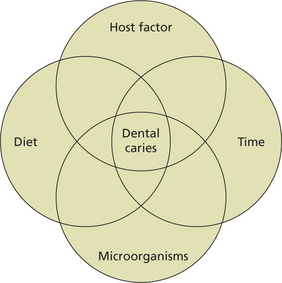
The four factors contributing to the caries process are (Figure 1):

Tooth Factor

Morphology and position in arch

Compared to the smooth surfaces of teeth, deep pits and fissures are more prone to carious attack because of food lodgment and bacterial stagnation. Owing to their, complex occlusal morphology consisting of numerous pits and fissures, the permanent mandibular first molars followed by the maxillary first molars and mandibular and maxillary second molars are more prone to carious attack.

Apart from the morphology of the tooth, the position of the tooth in the arch has a heavy bearing on the incidence of carious lesions.

Irregularities in the arch form, crowding and overlapping of the teeth also favor the development of caries as these regions provide an excellent environment for plaque accumulation.

Partially impacted third molars are more prone to caries and so are the buccally or lingually placed teeth.

Saliva

Composition

Hay et al (1982) and Lagerlof (1983) in their reports reiterated the fact that human salivary secretions are supersaturated on calcium and phosphate.

The concentrations of inorganic calcium and phosphorus show considerable variation within resting and stimulated saliva. Caries prone individuals have low calcium and phosphorus levels. Salivary proteins such as statherin, acidic proline-rich proteins (PRPs), cystatins, and histatins help in the maintenance of the homeostasis of the supersaturated state of saliva. According to Hay and Moreno (1989), statherin is present in stimulated saliva in concentrations sufficient to inhibit the precipitation of calcium and phosphate salts. Studies by Gibbons and Hay (1988) have shown that statherin may contribute to the early colonization of the tooth surfaces by certain bacteria, such as Actinomyces viscosus.

The acidic PRPs account for 25–30% of all proteins in saliva, and they have high affinity for hydroxyapatite in vitro (Hay and Moreno, 1989). The acidic PRPs bind free calcium, adsorb to hydroxyapatite surfaces, inhibit enamel crystal growth, and regulate hydroxyapatite crystal structure (Hay and Moreno, 1989).

The amount and quality of acidic PRPs and agglutinins are found to be different in caries-free and caries-active individuals as shown by the studies of Rosan et al (1982) and Stenudd (1999).

The role of cystatins in the caries process is still unclear. However, they may play a minor role in the regulation of calcium homeostasis in saliva. Phosphorylated and non-phosphorylated cystatins bind to hydroxyapatite.

Salivary flow rate, pH and buffer capacity

Saliva has the most important function of caries prevention by way of its flushing and neutralizing effects, commonly referred to as ‘salivary clearance’ or ‘oral clearance capacity’. As a thumb rule, the higher the flow rate, the faster the clearance and the higher the buffer capacity. Reduced salivary flow rate and the concomitant reduction of oral defense systems may cause severe caries and mucosal inflammation. Though, patients with impaired saliva flow rate often show high caries incidence (Papas et al, 1993; Spak et al, 1994) or caries susceptibility, it is still a mystery as to how much saliva is adequate enough.

The pH of saliva at which it ceases to be saturated with calcium and phosphorus is referred to as the ‘critical pH’. Normally, the critical pH is 5.5. Below this value, the inorganic content tends to demineralize. The normal pH of resting saliva is 6–7.

Buffering capacity

The buffer capacity of both unstimulated and stimulated saliva involves three major buffer systems: the bicarbonate (HCO−3), the phosphate, and the protein buffer systems. These systems have different pH ranges of maximal buffer capacity. The bicarbonate and phosphate systems have pH values of 6.1–6.3 and 6.8–7.0, respectively.

Since most of the salivary buffering capacity operative during food intake and mastication is due to the bicarbonate system, sufficient saliva flow provides the oral cavity with the neutralizing components.

The phosphate and protein buffer systems make a minor contribution to the total salivary buffer capacity, relative to the bicarbonate system. The phosphate system is, in principle, analogs to the bicarbonate system but without the important phase-buffering capacity, and it is relatively independent of the salivary secretion rate.

Lagerlof and Oliveby in 1994 showed that a low flow rate combined with a low or moderate buffer effect indicated poor salivary resistance against microbial attack.

It is a well-established fact that the buffer capacity of the saliva and the caries experience are inversely related.

The buffer effect of saliva is influenced by the hormonal and metabolic changes, as well as by altered general health. It is generally accepted that the buffer effect is greater in men than in women (Heintze et al, 1983). In women, the buffer effect decreases gradually, independent of flow rate, toward late pregnancy and promptly recovers after delivery. Introduction of either hormone replacement therapy in menopausal women (Laine and Leimola-Virtanen, 1996) or low-dose oral contraceptives (Laine et al, 1991) can slightly increase the buffer capacity.

Carbonic anhydrases (CAs) participate in the maintenance of pH homeostasis in various tissues and biological fluids of the human body by catalyzing the reversible hydration of carbon dioxide. Recent research suggests that salivary CA VI plays a role in protecting the teeth from caries (Kivela et al, 1999a, b). CA VI has been reported to bind to the enamel pellicle and retain its enzymatic activity on the tooth surface.

It is also believed that the urea and saline in saliva become hydrolyzed to produce ammonia and the later can cause rise in the salivary pH. This rise in pH can counter the attacks on the tooth surface during the progression of caries.

Antibacterial activity

The primary oral innate defense factors are peroxidase systems, lysozyme, lactoferrin, and histatins. In vitro studies have shown that these proteins are known to limit bacterial or fungal growth, interfere with bacterial glucose uptake or glucose metabolism and promote aggregation and, thus eliminate bacteria. Hanstrom et al (1983) and Tenovuo and Larjava (1984) reported that the salivary peroxidase and myeloperoxidase systems eliminate H2O2, which is highly toxic for mammalian cells.

The immunoglobulins, IgG, IgM, IgA, and secretory IgA (sIgA), form the basis of the specific salivary defense against oral microbial flora, including Streptococcus mutans. The most abundantly available immunoglobulin in saliva is dimeric slgA, which is produced by plasma cells located in the salivary glands. Two IgA subclasses are present in saliva; IgAl forms the major component of immunoglobulins, although the relative amount of IgA2 is higher in saliva than in other secretions (Tappuni and Challacombe, 1994).

In human beings, IgG, mainly of maternal origin, is the only detectable immunoglobulin in the saliva of neonates. Salivary IgA is absent at birth but is generally detectable by the age of 1 week.

The IgG concentration decreases to non-detectable levels after some months but appears again after tooth eruption (Brandtzaeg, 1989). Low concentrations of IgG can be detected in stimulated parotid saliva (Brandtzaeg, 1989), but most of the IgG detected in whole saliva enters the mouth from the gingival crevicular fluid, thus originating from sera.

In most children above 3 years of age, salivary IgAs against S. mutans can be detected, and their amount increases with the length of exposure (Smith and Taubman, 1992). Salivary Igs can bind to the salivary pellicle, and they are found also in dental plaque (Newman et al, 1979; Fine et al, 1984). In the oral cavity, immunoglobulins act by neutralizing various microbial virulence factors, limiting microbial adherence, and agglutinating the bacteria, as well as by preventing the penetration of foreign antigens into the mucosa. IgGs are also capable of opsonizing bacteria for phagocytes, which are reported to remain active in dental plaque and saliva (Scully, 1980; Newman, 1990).

Quantity and viscosity of flow

The consistency or viscosity of the saliva and the amount of saliva produced has a significant influence on the incidence of dental caries.

The average person produces at least 500 ml of saliva over a period of 24 hours. The unstimulated flow rate is 0.3 ml/min, whereas the flow rate during sleep is 0.1 ml/min and during eating or chewing, it increases to 4.0 to 5.0 ml/min. Any reduction in this quantity of saliva as seen in diseases such as Sjögren’s syndrome, diabetes, etc. predisposes to dental caries.

Increased viscosity of saliva may hinder its natural cleansing action thereby promoting the deposition of plaque on the tooth surface. Likewise when the salivary viscosity is low, the amount of minerals and bicarbonates are inadequate thereby limiting its anticaries activity.

Substrate and Dietary Factors

The role of diet in the causation of dental caries has been extensively studied.

A variety of dietary factors have been implicated in the causation of dental caries.

Chemical nature of diet

It is a well-known fact that food with high refined carbohydrate content has the greatest potential to give rise to carious lesions.

The type of carbohydrate (monosaccharide, disaccharide or polysaccharide), frequency of intake and the time for which the ingested food remains stagnant in the oral cavity or on the tooth surface determine the incidence and severity of the carious lesions.

Animal studies have shown that sugar in the solid and sticky form is more harmful to the tooth than the same amount of sugar in a liquid form.

It is believed that vitamin B deficient individuals have lower incidence of dental caries. Vitamin B is essential for the growth of oral acidogenic flora and also serves as a component of coenzymes involved in glycolysis.

Vitamin D plays an important role in the normal development of teeth. Various studies have shown that the teeth are hypoplastic and usually have higher incidence of dental caries in vitamin D deficiency.

Teeth may be poorly calcified in individuals exposed to low doses of calcium during intrauterine life and infancy. Such poorly calcified teeth may be susceptible to carious attack. Higher levels of selenium is known to predispose to the carious lesions affecting permanent teeth.

Fluoride content in the diet has no significant role because of its metabolic unavailability. Therefore, the fluoride content in cooking salt and its effect on reducing the incidence of carious lesions is still questionable. However, fluoridated water minimizes the caries incidence.

Phosphates, molybdenum and vanadium in the diet helps in minimizing the incidence of carious lesions.

Role of heredity

Literature review reveals various studies to assess the genetic modifications in dental enamel, genetic modification of immune response, genetic regulation of salivary function and inherited alterations in sugar metabolism.

Bachrach and Young (1927) compared the caries incidence of monozygotic twins with same-sex dizygotic (93 pairs) and different-sex dizygotic (78 pairs) twins. Their results showed that the monozygotic twins had a more similar caries incidence than dizygotic twins and that different-sex dizygotic twins had the greatest variance. The authors concluded that heredity plays a subsidiary part in the incidence of caries. It is believed that heredity affects the dental decay only in as much as it controls the shape of a tooth and its pits and fissures and its position in the dental arch.

Senpuku et al (1998) and Acton et al (1999) have correlated specific HLA-DR types with binding S. mutans antigens and S. mutans colonization.

Acton concluded that ‘genes within MHC modulate the level of oral cariogenic organisms’.

Mariani et al (1994) in their study of celiac disease, enamel defects and HLA typing observed that HLA-DR3 was associated with increased enamel defects and HLA-DR5, 7 were associated with a reduced frequency of enamel defects. Studies have shown that the genes in the HLA complex are associated with altered enamel development and increased susceptibility to dental caries.

Classification of Dental Caries

According to Morphology of Teeth

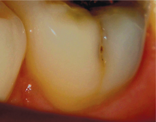
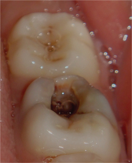
i. Pit and fissure caries (also called type-1 caries): Caries occurring on anatomical pits and fissures of all the teeth. The specific areas or surfaces involved include occlusal surfaces of molars (Figure 2) and premolars, buccal and lingual surfaces of molars (Figure 3) and lingual surfaces of maxillary incisors.
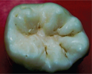
    The lesions are smaller in the beginning but become wider as they spread toward the dentin due to orientation of enamel rods.

    In these places there can be entrapment of food leading to fermentation of carbohydrates with lack of neutralization of acid produced by salivary buffers which leads to destruction of enamel and dentin. The enamel bordering the pit and fissure may appear opaque and bluish-white as it becomes undermined.

    Clinically these lesions appear brown or black, with little softening and opaqueness of the surface. When the lesion is examined by a fine explorer tip, a ‘catch point’ is often felt, where the explorer teeth catches the area.

    When the lesion reaches the dentinoenamel junction (DEJ), they spread laterally to cause undermining of the enamel.

ii. Smooth surface caries (also known as type-2 caries): These carious lesions occur on the smooth surfaces of the teeth (e.g. proximal surfaces or gingival areas of the buccal and lingual aspect of tooth).

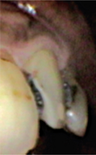
    Proximal caries usually begins just below the contact point, and appears in the early stage as a well demarcated faint white opacity of the enamel without apparent loss of continuity of enamel.

    The white spot lesion becomes pigmented yellow or brown and it often extends buccally and lingually.

    The surrounding enamel becomes bluish white as the lesion continues to progress (Figure 4). The surface of the affected enamel becomes rough and later on, there is formation of a cavity (Figure 5).
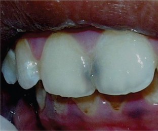
iii. Root caries: Caries occurring at the cementoenamel junction or cementum. This occurs predominantly in the older age when there is gingival recession.

iv. Linear enamel caries: Caries occurring on the labial surfaces of anterior teeth. This is also known as ‘odontoclasia’. The caries occurs at neonatal zone because of trauma at birth or metabolic disturbances.

According to Severity and Progress

i. Incipient caries: Initial carious lesion limited to the teeth is called incipient caries and is characterized by a virtually intact surface but a porous subsurface (subsurface demineralization).

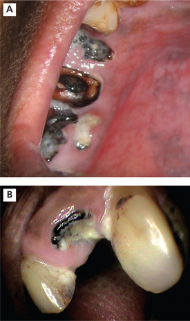
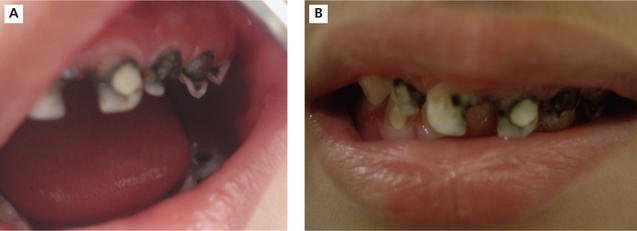
ii. Rampant caries: This is an acute fulminating type of carious process, which is characterized by simultaneous involvement of multiple number of teeth (may be all teeth) in multiple surfaces (Figure 6A, B).
iii. Arrested caries

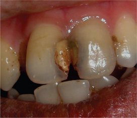
iv. Recurrent caries: It occurs at the interface of tooth and restorative material because of many factors such as defective cavity preparation, microleakage and combination of these (Figure 7).
v. Radiation caries: One of the complications of radiotherapy of oral cancer lesions is xerostomia, which leads to an early development of widespread caries.

According to Age Pattern

Nursing bottle caries: A type of acute carious lesion, which occurs among those children who take milk or fruit juice by the nursing bottle, for a considerably longer duration of time, preferably during sleep.

1. It has been variously attributed to prolonged use of

2. Invariably there is a prolonged habitual use of one of the above after 1 year of age, usually as an aid for sleeping at night or naptime.

3. Clinically presents as widespread destruction of deciduous teeth, most commonly the four maxillary incisors, followed by the first molars and then the cuspids if the habit is prolonged.

4. The lower teeth are not usually affected as they remain under the cover of the tongue, so the absence of caries in the mandibular incisors distinguishes this disease from ordinary rampant caries.

5. Both the nursing bottle and rampant cause early pulp involvement.

Because they spread at a very rapid pace and as a result, the pulp hardly gets any time to protect itself by forming secondary dentin.

According to Rapidity

i. Acute dental caries

ii. Chronic dental caries

Microbiology of Dental Caries

The predominant group of microorganism is streptococci. Among these strains S. mutans is responsible. These are gram-positive organisms which are round or ovoid. These may appear rod shaped, non-sporulating and non-motile. These can be cultured on blood agar with formation of refractory colonies measuring 0.5–1.5 mm at 37°C. These are pathogenic to human beings.

The three common organisms seen to be associated with secondary caries are S. mutans, lactobacilli and Actinomyces viscosus. Fontanna et al (1996) observed a definite relationship of S. mutans and secondary caries. S. mutans is also present in saliva and in dental plaque in individuals with rampant caries due to xerostomia as well as in children nourished with bottle milk. S. mutans and lactobacilli have been found to increase in significant numbers in the plaque as well as dentin of teeth restored with amalgams having marginal defects wider than 40 μm.

Fitzergerald et al (1994) were of the view that in association with these three major microorganisms, others also played a role in secondary caries. They found S. mutans, S. sanguis and S. salivarius in 35%, 24% and 14% of growth positive samples respectively. Other isolates like S. gordonii, S. milleri, S. oralis and S. mitis were also recognized. Certain organisms, which occurred very frequently were Propionibacterium, Bifidobacterium, Eubacterium and Peptococcus. Actinomyces were found in 46% of the samples. A. viscosus and A. naeslundii were most prevalent followed by A. israelii and A. odontolyticus.

S. mutans can adhere to the tooth surface by glucan which is produced by utilization of dietary sucrose. These organisms ferment mannitol and lactose with the production of acid. These can take up dietary sucrose and breakdown into glucose and fructose by means of the enzyme, invertase. Finally glucose and fructose is broken down to lactic acid. These have the ability to store glucose and fructose from degradation for the synthesis of acids in the absence of dietary sucrose.

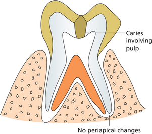
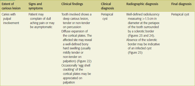
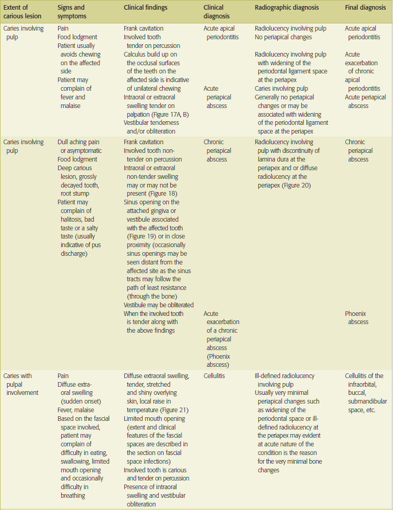
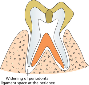
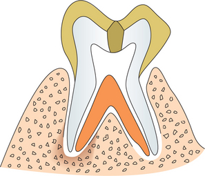
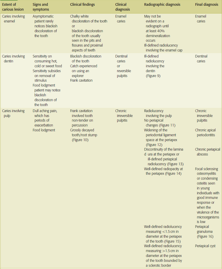
Clinical features of dental caries, pulp and periapical lesions are given in Table 1.
Stay updated, free dental videos. Join our Telegram channel

Jan 12, 2015 | Posted by in Oral and Maxillofacial Radiology | Comments Off on 15: Dental Caries

VIDEdental - Online dental courses
Get VIDEdental app for watching clinical videos