14
Local risk factors for periodontal diseases
Figure 14.1 Section VIII.A from the classification of periodontal diseases and conditions.
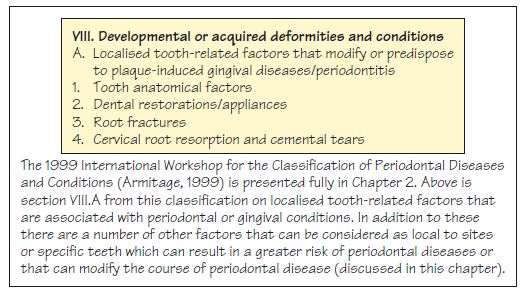
Figure 14.2 An enamel pearl.
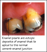
Figure 14.3 Overhanging restoration margins.
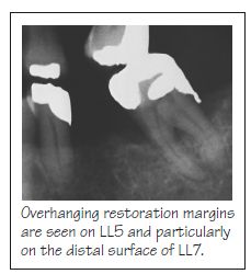
Figure 14.4 Subgingival crown margins.
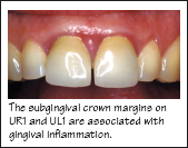
Figure 14.5 An overcontoured crown and subgingival calclus.
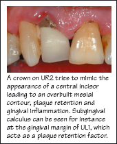
Figure 14.6 Minimising the periodontal consequences of restorative treatment.
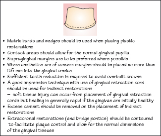
Figure 14.7 (a) Poorly designed acrylic partial denture with ging val coverage (shown out of the mouth). (b) Partial denture in situ; interproximal plaque is visible. (c) Following removal of the denture, the gingivae can be seen to be reddened and inlamed.
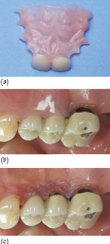
Figure 14.8 Partial denture with extensive gingival coverage.
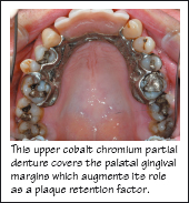
Figure 14.9 Minimising the periodontal consequences of providing a partial denture.
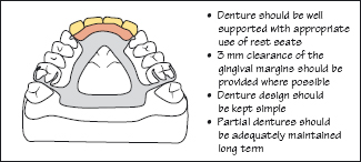
Figure 14.10 Orthodontic appliance acting as a plaque retention factor.
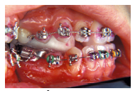
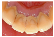
Figure 14.11 Supragingival calculus acting as a local plaque retention factor.
Local risk factors can increase the risk of development and progression of periodontal disease by acting as plaque retention factors. Local factors should be identified when carrying out a thorough dental examination of the patient and their removal or modification where possible should be included in the patient’s management.
Figure 14.1 shows an extract from the 1999 International Workshop Classification o/>
Stay updated, free dental videos. Join our Telegram channel
VIDEdental - Online dental courses


