CHAPTER 13
Treatment of Class II Malocclusions
Class II malocclusion is not a single entity but results from numerous combinations of both skeletal and dental alveolar components. The earliest description, solely a dental description, was provided by Edward Angle when he defined a Class II malocclusion as characterized by the lower molar in distal position relative to the upper 6-year molar. He further subdivided Class II malocclusions into Class II division 1, characterized by the anterior maxillary teeth being protrusive; and Class II division 2, characterized by two or more maxillary anterior teeth being retroclined. The Class II division 1 was later shown to also be characterized by a retrognathic mandible or a prognathic maxilla with variable vertical dimensions. The Class II division 2 patient was shown to exhibit an orthognathic maxilla, a short and retrognathic mandible, brachyfacial growth pattern, retroclined maxillary central incisors, and a relatively prominent chin, as well as dental deep bite. In later years further assessment of the dental Class II provided information regarding the underlying skeletal components.< ?xml:namespace prefix = "mbp" />
1 What are the components of a Class II malocclusion?
Utilizing cephalometrics and computer-based statistical evaluation, Moyers et al.
TABLE 13-1 The Six Horizontal Class II Types Determined by Moyers et al.
| DIAGRAMMATIC REPRESENTATION | DESCRIPTION |
|---|---|
| Normal | |
 |
Type A: Maxillary dental protraction |
 |
Type B: Maxillary prognathism, dental protraction |
 |
Type C: Maxillary retrognathism with flared or upright incisors, mandibular severe retrognathism with flared lower incisors |
 |
Type D: Maxillary retrognathism with dental protraction, severe mandibular retrognathism |
 |
Type E: Maxillary prognathism and dental protraction + mandibular dental flaring |
 |
Type F: Mandibular retrognathism |
Adapted from Moyers RE, Riolo MS, Guire KE, et al: Am J Orthod 1980;78(5):477-494.
The five vertical types (
TABLE 13-2 The Five Vertical Class II Types Determined by Moyers et al.
| DIAGRAMMATIC REPRESENTATION | DESCRIPTION |
|---|---|
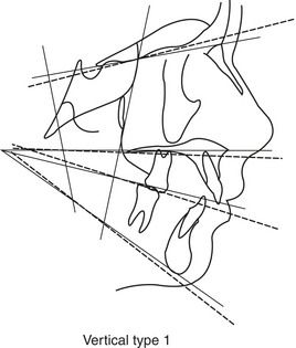 |
Type 1: Mandibular plane steeper than normal, steeper functional occlusal plane, palate tipped somewhat downward, anterior cranial base tipped upward |
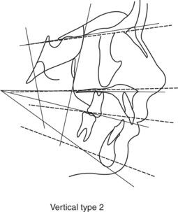 |
Type 2: Mandibular plane, functional occlusal plane and palatal plane are all flatter than normal and are nearly parallel |
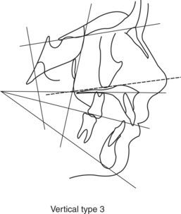 |
Type 3: Palatal plane tipped upward anteriorly |
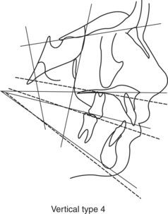 |
Type 4: Mandibular plane, the functional occlusal plane, and the palatal plane are all tipped markedly downward |
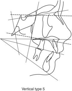 |
Type 5: Palatal plane is tipped downward, cranial base tipped downward |
Adapted from Moyers RE, Riolo MS, Guire KE, et al: Am J Orthod 1980;78(5):477-494.
In the transverse dimension, the buccal segments of Class II patients often appear normal. A 3 to 4 mm transverse discrepancy, however, usually exists at the level of the first molar due to a narrow maxillary arch. This is readily observable if the mandible is moved into the Class I relationship at the molar. Further assessment of components of Class II malocclusion in an adolescent population indicated that in a sample of 277 children with a Class II malocclusion, mandibular skeletal retrusion was the most common characteristic. The maxilla was generally either retrusive or well positioned.
2 How can Moyers’ differential diagnosis of Class II horizontal and vertical types be used to help us with treatment planning of Class II patients?
Moyers’ differential diagnosis of Class II malocclusions allows us to more easily determine the components of the Class II malocclusion problem. It identifies the skeletal problem and the dentoalveolar problem and thus directs our treatment thinking to these specific areas. Treatment planning considerations using Moyers’ differential Class II horizontal analysis is summarized, at least in part, in
TABLE 13-3 Summarization of Moyers’ Differential Class II Horizontal Analysis
| TYPE | TREATMENT CONSIDERATIONS |
|---|---|
Type A  |
|
Type B  |
|
Type C  |
|
Type D  |
|
Type E 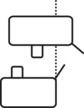 |
|
Type F  |
Adapted from Moyers RE, Riolo MS, Guire KE, et al: Am J Orthod 1980;78(5):477-494.
In addition to the horizontal considerations addressed in
3 What is the prevalence of Class II malocclusions?
According to the NHANES III Study, 15% of the U.S. population have an overjet of greater than 4 mm, 38% an overjet of 3 to 4 mm, and 33% Class II occlusal discrepancies. The same frequency for Class II dental characteristics was found in Caucasians, African-Americans and Hispanics. According to McNamara1, 75% of Class II skeletal discrepancies are the result of mandibular retrognathia.
4 What is the etiology of Class II malocclusion?
Class II malocclusion is usually an aberration of normal development and not caused by a pathologic process. It is usually the result of multiple factors that influence growth and development and not from one specific factor. The development of Class II malocclusion, however, may be related to some specific causes, genetic influences, and environmental factors. Such specific causes as the effect of teratogens on mandibular growth, mandibular deficient syndromes (Pierre-Robin and Treacher-Collins), fetal molding, trauma to the transmandibular junction (TMJ) area during the birth process, childhood fractures of the jaw, and mandibular arthritic problems may all contribute to the development of a Class II skeletal pattern. Less than 1% of orthodontic patients, however, have a disruption in embryological development that can be attributed as the major cause of malocclusion. Genetic influences have been shown to be associated with Class II malocclusions.
Local and environmental factors may also be an issue in the development of Class II malocclusions because of their alteration of the normal physiologic pressures and forces associated with craniofacial growth. These pressures and forces may be disrupted or imbalanced by the effects of abnormal function of the soft tissues. Disruption of normal lip balance such as that associated with lip incompetency may lead to flaring of the upper incisors from an imbalance of labial and lingual musculature. The need to achieve lip–tongue contact for an oral seal during swallowing can cause the lip to retrocline lower incisors and the protruding tongue to flare upper incisors, thus increasing overjet. It has also been speculated that mouth-breathing can cause the opening muscles to place a distal force on the mandible, retarding its growth and rotating the mandible clockwise. In addition, it is thought that finger-sucking habits can produce a Class II division 1 incisal relationship within a Class II or Class I skeletal pattern (
5 What treatment protocols are used to correct Class II malocclusions?
Treatment for Class II malocclusions may involve the following:
The appropriate protocol for each patient depends upon patient desires and doctor assessment of the exact nature of the Class II problem as well as the orthodontist’s treatment protocol preferences.
6 What is extra-oral traction?
Extra-oral traction is the application of force to the dentition and maxilla through the use of headgears (cervical, occipital, combination) fitted to the skull and attached facebows through which force is directly applied to the dentition, usually through the permanent maxillary first molars. Headgear wear is required for at least 12 to 14 hours per day for 6 to 18 months at a force level of 12 to 16 ounces per side for skeletal modification. Three types of headgear are commonly used depending upon the vertical craniofacial growth pattern: cervical pull headgear for low vertical dimension (Frankfort mandibular plane angle [FMA] ≤25), occipital pull headgear (FMA >30) and combination (combi) headgear for cases in which the vector of force application needs to be altered depending upon the desired effect for a specific patient. The various types of headgear and attached facebows are useful in applying forces of appropriate magnitude and direction to the maxilla via the maxillary dentition. The effect of the applied force has an orthopedic effect. It modifies maxillary position by altering its normal downward and forward growth pattern, thus normalizing the Class II to a Class I skeletal pattern (
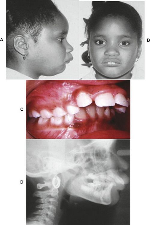
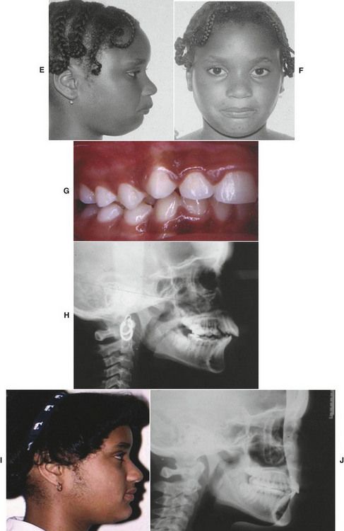
FIG 13-2 Headgear treatment of Class II division 1 malocclusion. A-D, Initial photos: facial photos (A and B), intraoral photo (C), and cephalometric radiograph (D). E-H, Post-cervical headgear: facial photos (E and F), intraoral photo (G), and cephalometric radiograph (H). I and J, Orthodontic treatment with extractions: facial photo (I) and cephalometric radiograph (J).
7 What is the distalizing protocol for the correction of Class II?
Molar distalization is used when the Class II problem is dentoalveolar in nature. Molar distalization corrects a dental Class II by moving the maxillary first molar distally into a Class I relationship. Such treatment has no orthopedic effect. D/>
Stay updated, free dental videos. Join our Telegram channel
VIDEdental - Online dental courses


