13: Canines

Canines

MAXILLARY AND MANDIBULAR PERMANENT CANINES

The four maxillary and mandibular permanent canines, one on each side of each jaw, are the longest teeth in the mouth. Located at the corners of the mouth, they are well anchored in the bone by their extremely long roots. Their location requires extra anchorage, which is furnished by the length and the shape of their roots and a special projection of bone called the canine eminence. The term canine brings to mind the fanglike teeth of dogs, which are members of the animal family Canidae.

In function the canines act as holding and tearing tools and assist both the incisors and premolars. In addition, their V shape at the corner of the mouth allows for dissipation of pressures that can force the premolars to protrude out of the mouth or the incisors farther into the mouth. The self-cleaning qualities of the canines; their smooth, pointed shape; the thickness of their crowns; and their strong anchorage make the canines the most stable teeth in the mouth.

Maxillary Canines

Evidence of calcification: 4 months

Enamel completed: 6 to 7 years

Eruption: 11 to 12 years

Root completed: 13 to 15 years

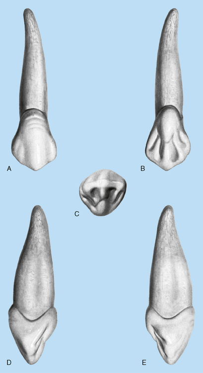
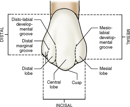
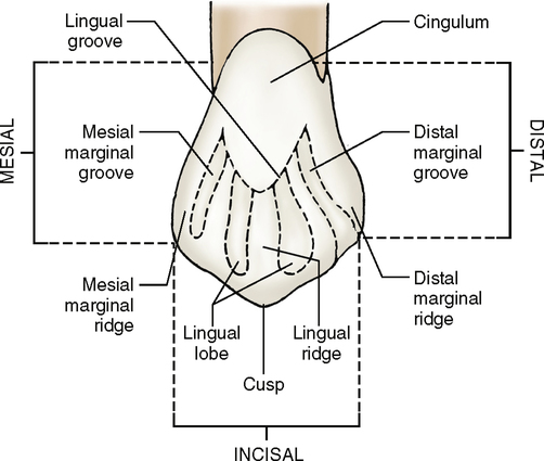
A maxillary canine (Fig. 13-1) resembles an incisor in its composition of four developmental lobes—three facial and one lingual. The three facial lobes resemble the facial lobes of the incisors except that the middle facial lobe extends farther incisally when the tooth is viewed from the labial or lingual aspect. This middle lobe extension results in the formation of a single cusp. The cusp tip is formed by the junction of four ridges. One of the ridges extends along the middle lobe of the tooth on its most facial part; another extends along the lingual part. The other two ridges run from the mesioincisal and distoincisal corners. All four ridges converge to form the cusp tip.

The lingual lobe of a canine is much larger and thicker than the lingual lobe of an incisor, which results in the canine being much wider labiolingually than a maxillary incisor is. The cingulum of a maxillary canine also shows greater development in that it is larger and bulkier than the cingulum on any of the other anterior teeth.

Labial aspect (Fig. 13-2; see Fig. 13-1, A)

The crown and root of a maxillary canine are narrower mesiodistally than those of a maxillary central incisor. The cervicoincisal length of the crown is much larger on a maxillary canine than on any other anterior tooth, except the maxillary central incisor and the mandibular canine. Although these teeth usually have longer crowns than a maxillary canine has, the roots of the maxillary canines are longer and thus make them the longest teeth in the mouth.

Mesially, the outline of the crown is straighter than laterally with a slight convexity at the contact area. The center of the mesial contact area is approximately at the junction of the middle and incisal thirds of the crown.

Distally, the outline of the crown is rounded in appearance because the distal contact area is usually at the center of the middle third of the crown. This position makes the distal convexity appear larger and more uniform. How does this differ from the location of the mesial contact area? Which contact area is located more incisally: mesial or distal?

The labial surface of the crown is smooth. The developmental lines are two shallow depressions dividing the three labial lobes. The middle lobe is much larger and has greater development than the other lobes, resulting in a ridge on the labial surface of the crown. This ridge ends incisally at the cusp tip, which is centered in the middle of the tooth (from the facial view).

The cervical line crests slightly mesial to the center of the tooth.

The root of a maxillary canine is slender in comparison with the crown and is conical in shape with a blunt root apex. It is not unusual for the root to turn sharply to the distal or mesial side in the apical third. A general rule is that most roots, if they do have an apical curvature, point toward the distal side. Although this rule applies to almost all single-rooted teeth, exceptions are noted. If an apical curvature is not present, the root itself has a tendency to point more often toward the distal than to the mesial side.

Lingual aspect (Fig. 13-3; also see Fig. 13-1, B)

The root of a maxillary canine tapers toward the lingual surface. The lingual sides of both the crown and root are narrower than the labial sides.

Compared with the facial line, the cervical line shows a more even curvature, and the crest is straighter and centered over the middle of the tooth.

The most obvious structure on the lingual surface of a maxillary canine is the well-developed cingulum. It is huge in comparison with those of all the other anterior teeth.

Confluent to the cingulum and running from the cusp tip is a well-developed lingual ridge. This ridge runs from the cusp tip on the lingual side to the cingulum. Unlike other anterior teeth that have a lingual fossa, this area on a maxillary canine is occupied by a lingual ridge, which divides the lingual side of the three facial lobes, creating two separate lingual fossae, one on the mesial and one on the distal side of the lingual ridge. These fossae are bordered by a mesial and a distal marginal ridge, respectively. When present, these fossae are called the mesial and distal lingual fossae. The borders of the lingual fossae are the incisal ridge, lingual ridge, and mesial or distal marginal ridge.

Sometimes the lingual surface of a canine crown is so smooth that no concavities or fossae are present. Usually the cingulum and marginal ridges are less developed in these instances, with little evidence of developmental grooves.

The lingual side of the root is narrower than the labial side. A cross-sectional view of the root appears triangular, with the lingual portion more tapered than the labial.

Mesial aspect

The functional form of a maxillary canine is evident on the mesial view (Fig. 13-4; see Fig. 13-1, D). The wedge-shaped outline of the crown shows the canine to have greater labiolingual bulk than any other anterior tooth. The greatest measurement labiolingually is at the cervical third. This is because of the huge cingulum of the lingual side and the more convex labial outline of the canine. The entire labial surface is more convex from the cervical line to the cusp tip than any other maxillary anterior tooth. The cervical line curves toward the cusp an average of 2.5 mm.
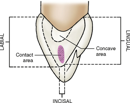
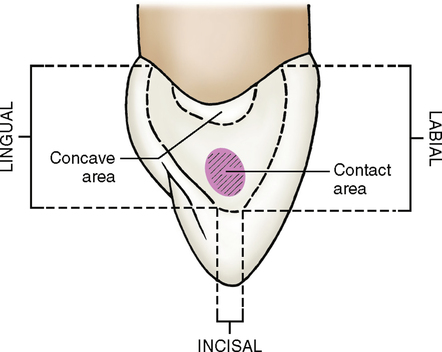
The root of a canine is broad labiolingually and is usually extremely long. The end of the root apex is blunt and may often curve to the lingual or distal lingual side. The mesial surface of the root shows much labiolingual development, with a shallow developmental depression extending from the cervical line halfway to the apex of the root. This developmental depression appears to almost divide the single root into two roots. In extremely well-developed roots, it helps anchor a canine in the bone and prevents root rotation.

The mesial surface of a canine crown is entirely convex throughout except for a small area between the contact area and the cervical line, which may be flat.

Distal aspect

The distal aspect (Fig. 13-5; see also Fig. 13-1, E) of a maxillary canine shows the same form and outline as the mesial view does. However, the cervical line shows less curvature toward the cusp tip. The distal marginal ridge is more developed and heavier in outline than the mesial marginal ridge. Although both the mesial and the distal surfaces show a slightly flat or concave area above the contact area, the distal surface displays much more concavity. The root surface on the distal aspect may show a more pronounced developmental depression than that on the mesial aspect.

Stay updated, free dental videos. Join our Telegram channel

Jan 4, 2015 | Posted by in General Dentistry | Comments Off on 13: Canines

VIDEdental - Online dental courses

Get VIDEdental app for watching clinical videos