12: Reconstructive Oral and Maxillofacial Surgery

Reconstructive Oral and Maxillofacial Surgery

The term reconstructive maxillofacial surgery refers to the wide range of procedures designed to rebuild or enhance soft or hard tissue structures of the maxillofacial region. Ablative tumor surgery (benign or malignant) and traumatic injuries (especially avulsive) commonly demand reconstructive procedures to restore the functional and cosmetic deficit. Loss of soft or hard tissue secondary to infectious processes (e.g., osteomyelitis), or tissue injury due to irradiation (e.g., osteoradionecrosis) may also require reconstructive measures. In addition, the decrease in the quantity and quality of maxillomandibular structures with age (which may be accelerated by other processes, such as early loss of teeth) can be addressed with reconstructive measures to augment the tissue for restoration using dental implants.

In the past two decades, four developments have revolutionized the reconstruction of the maxillofacial structures. First, gains in our understanding of bone biology have allowed advanced bone grafting procedures in a variety of circumstances (e.g., sinus lift procedures, mandibular augmentation/reconstruction). Second, the advent of microvascular free flap techniques has enabled the transfer of tissue to reconstruct large soft and/or hard tissue defects (e.g., radial forearm fasciocutaneous or fibula osteocutaneous free flaps). Third, the development of and advances in dental implant techniques have allowed successful dental rehabilitation. Fourth, the introduction of computer-aided surgery, using CT-generated images and virtual surgery, has significantly changed the practice of reconstructive surgery. Future research may reveal improved methods of regenerating bone, neural, and muscle tissue. Future molecular biology techniques using gene therapy may provide knowledge that can be applied to maxillofacial reconstruction.

In this chapter, we present important teaching cases that describe some of the main issues in maxillofacial reconstruction. Mandibular reconstruction remains one of the greatest challenges in maxillofacial surgery. We present cases of mandibular reconstruction using corticocancellous bone grafts with implants and other cases using the free fibula osteocutaneous flap.

Posterior Mandibular Augmentation

Examination

General. The patient is a well-nourished, well-developed 69-year-old Caucasian male in no apparent distress.

Vital signs. Blood pressure is 156/87 mm Hg, heart rate 69 bpm, respiratory rate 16 per minute, and temperature 37°C.

Maxillofacial. Normocephalic. Skin is dry and intact, pupils equal, round, and reactive to light and accommodation (PERRLA), no scleral icterus, visual acuity grossly intact, external auditory canals clear bilaterally, tympanic membranes intact, nares patent, cranial nerves II through XII grossly intact bilaterally. Neck is supple and without lymphadenopathy.

Intraoral. Mucosa is moist and pink. No ulcers, masses, or discolorations of the oral cavity noted. Teeth #3, #11 through #15, #19, #20, and #30 are absent. Generalized periodontal disease is noted with root exposure on teeth #2, #4, #18, #21, #28, and #30. A buccal horizontal defect is present at teeth #19, #20, and #30.

Assessment

Caucasian male, 69 years old, with a history of hypercholesterolemia, prostatic hypertrophy, GERD, chronic neck pain, and chronic rhinitis/sinusitis presents for an evaluation for dental implant placement after extraction of periodontally involved teeth. The physical exam reveals vertical insufficiency of the right mandibular and bilateral maxillary posterior alveolar ridges, in addition to a horizontal alveolar deficiency of the right mandibular posterior alveolar ridge (Figure 12-1).
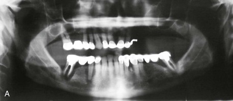
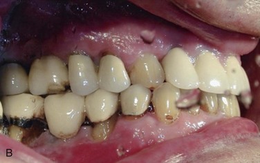
Treatment

Alveolar deficiencies of the posterior mandible present unique surgical challenges. Defects must be accurately assessed for the horizontal and vertical deficiencies of bone and the amount of keratinized tissue available to support the final prosthesis. A host of reconstructive techniques and materials must be considered, and the most appropriate method selected to maximize the individual patient’s outcome (Figure 12-2). The success of endosteal implant restorations and prostheses has made augmentation of the posterior mandible a necessary skill for oral and maxillofacial surgeons. The use of titanium mesh, autogenous bone, allogeneic/xenogeneic bone, and inlay bone grafting is discussed later.
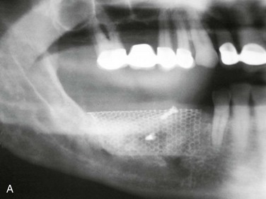
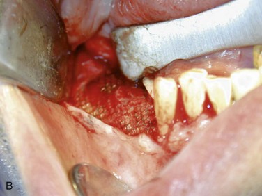
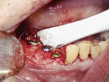
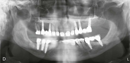
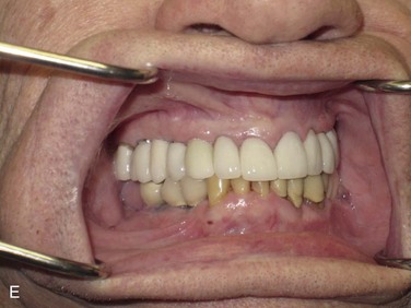
Horizontal Defects

A horizontal defect is defined as an inadequate buccolingual dimension of bone with an adequate superoinferior dimension. Horizontal defects most commonly occur on the facial aspect of the mandible. Generally, implants 5 mm in diameter are placed in the molar region; this requires 7 to 8 mm of horizontal bone to ensure 1 to 1.5 mm of bone buccal and lingual to the implant.

Block Grafts

The autogenous block graft has been widely used for horizontal defects. The harvest site of the cortical bone depends largely on the length of bone required. For smaller defects, harvest from the mandibular symphysis or mandibular ramus allows for easy access with low long-term morbidity. However, temporary V3 paresthesia has been reported in 10% to 50% of symphysis grafts, and 0 to 5% of ramus grafts. For larger defects, distant harvest sites of bone are necessary. The calvarial graft, taken from the parietal bone, provides dense cortical bone that is resistant to resorption. Harvesting a split-thickness graft from this region provides an approximately 3-mm-thick segment of bone, and the harvest site has few complications. The ilium may also be used; however, the cortical bone is thinner and less resistant to resorption due to the endochondral origin of the ilium, compared with the intramembranous origin of the parietal bone. However, a large amount of bone may be harvested from the iliac crest. The main complications of iliac crest bone grafts include gait disturbances, paresthesia, hematoma/seroma, and fracture of the hip.

The use of block allografts has been presented in case series. Nissen and colleagues placed 29 cancellous block allografts in 21 patients with posterior mandibular atrophy; the graft failure rate was 20.7%, and the implant survival rate in the remaining grafts was 95.2% at 37-month follow-up. However, long-term outcomes for allogeneic block grafts from prospective, randomized clinical trials are lacking. A systematic review by Waasdorp and Reynolds found only nine articles that met inclusion criteria, and eight of the articles were case reports or case series. The authors concluded that, although the case reports demonstrated potential for allogeneic block grafts for alveolar ridge augmentation, there is insufficient evidence to establish treatment efficacy with regard to graft stability and long-term implant survival.

Particulate Bone

Particulate bone graft material is available from a wide array of sources, including autografts (from the patient), allografts (from a human donor), xenografts (from an animal donor), and alloplastic material (synthetic material). Autogenous particulate bone may be harvested from intraoral sites, including cortical shaving from the symphysis, ramus, or zygoma, and cancellous bone can be harvested from the ilium or tibia. Autogenous bone is often combined with banked particulate bone; this increases the volume of graft material while maintaining the osteogenic and osteoinductive properties of the autogenous bone. The choice of graft material depends on the amount of particulate bone needed, osteoinductive versus osteoconductive properties, and the desires of the patient. Whether the graft is mineralized or demineralized determines the type of membrane required for graft stabilization. In general, mineralized particulate bone is able to better withstand the forces exerted on the surgical site during healing, requiring only a nonrigid membrane at the time of graft placement. Demineralized bone, however, requires a rigid membrane during the healing phase. Titanium mesh is well suited to protecting demineralized bone and can tolerate exposure to the oral cavity without a significant rate of graft failure. The titanium mesh is contoured and adapted to the alveolar ridge and secured with titanium screws.

Inlay Bone Graft

First described by Simion and colleagues in 1992, the alveolar split osteotomy technique for horizontal bone defects and subsequent implant placement has shown predictable results. The surgery is usually performed in a two-stage fashion in the mandible due to the dense cortical buccal plate. At least 3 mm of horizontal width is preferred for a controlled fracture; however, widths as narrow as 2 mm have been reported. The goal of the technique is to produce a vascularized bone flap through controlled fracture of the buccal plate. The gap produced by the fracture at the second stage can then be grafted with block or particulate bone, or implants can be placed along with a particulate graft. A membrane is generally used to protect the graft and implants during healing.

Procedure.

The initial surgery requires a full-thickness flap to expose the buccal cortical plate. A crestal incision with releasing incisions away from the planned corticotomy sites is used. Crestal, apical, and two vertical corticotomies are performed and connected to create an outline of the intended bone flap. A piezoelectric drill is often used to preserve bone. The mucosal flap is then sutured. Stage 2 is performed after approximately 4 weeks; with this interval, the periosteal blood supply to the bone is restored, but callus is still present at the corticotomy sites. The crestal incision is made along the crestal corticotomy, with care taken to reflect as little periosteum as possible. Osteotomes are used to gently out-fracture the bone flap. The bone graft and/or implants can then be placed. Primary closure can be attempted using a periosteal releasing incision, but primary closure is often difficult, requiring the use of a membrane. If implants are not placed, 4 to 6 months of healing is allowed before implant placement.

Vertical Defects

Vertical defects of the posterior mandible refer to inadequate height of alveolar bone in relation to the inferior alveolar nerve. Vertical defects can be challenging to treat due to a small surface area of crestal mandibular bone for onlay grafting, difficulty with exposure of the graft due to tension of the soft tissue after augmentation, and resorption of graft material. Techniques described include onlay grafting, particulate bone grafts, inlay grafts, and distraction osteogenesis.

In 2009, Esposito and colleagues performed a Cochrane systematic review of randomized controlled trials (RCTs) for horizontal and vertical ridge augmentation. Of the 13 trials that met the inclusion criteria, 10, enrolling 218 patients, addressed vertical ridge augmentation. Analysis of the trials found that vertical augmentation resulted in a high complication rate (20% to 60%) and graft failure rates of 10% to 15%. Interestingly, two split-mouth trials compared alloplastic grafting (anorganic bovine bone and Regenaform, respectively) with autogenous bone grafting (iliac crest and particulate bone) and showed no statistical difference in outcomes. Although both studies had small sample sizes (10 and 5 patients), the reduction in operative time, cost, and patient discomfort certainly justify further investigation. The review also included a meta-analysis of two RCTs examining mandibular ridge augmentation (iliac crest inlay graft and anorganic bovine inlay) versus short implant placement without augmentation. The meta-analysis showed an increased implant failure rate (borderline significance; p = 0.06), and a statistically significant increase in the complication rate in the augmented group. The additional time, cost (e.g., general anesthesia, hospitalization), and patient discomfort are also important factors. However, the long-term outcomes of short implants in the posterior mandible have not been adequately evaluated to date.

Particulate Bone

Particulate bone grafting for vertical deficiency has been shown to be effective when used with a rigid membrane or titanium mesh. The use of titanium mesh has been shown to be effective in the reconstruction of alveolar ridge defects, regardless of the particulate bone source. The titanium mesh acts as a permanent, rigid barrier that is biocompatible and easily molded to the desired shape. Several studies have demonstrated successful vertical augmentation (maxilla and mandible) using titanium mesh, with average vertical gains of 3.71 to 14 mm and implant success rates in the grafted area of 93% to 100%. Exposure of the titanium mesh during the healing phase is commonly reported, with a rate ranging from 5% to 52%. However, the rates of infection and graft failure remain low compared to other nonresorbable barriers. Watzinger and colleagues demonstrated that the timing of the mesh exposure is critical to the final outcome. If exposure occurred within 4 to 6 weeks of the grafting procedure, graft take was poor. However, if exposure occurred after 4 to 6 weeks, these sites had outcomes similar to those for grafts that did not have exposure. Most areas of late exposure of titanium mesh (after 4 to 6 weeks) can be managed with local wound care; removal is required only if signs of infection are present.

Inlay Graft

Inlay grafting, or “sandwich” osteotomy, provides the advantage of having a pedicled segment of alveolar bone overlying the graft material. Felice and colleagues demonstrated significantly less bone resorption of the inlay graft, compared to onlay grafting of anterior iliac crest bone, in 20 patients. In another study by Felice, no statistical difference in outcomes was found for inlay grafts with anorganic bovine bone (Geistlich Bio-Oss [Geistlich Pharma North America, Inc., Princeton, N. J.]), compared with iliac crest bone, for vertical augmentation in the posterior mandible. The disadvantages of inlay grafting are the inability to address horizontal defects with the procedure and limitation of the amount of vertical augmentation by the lingual soft tissue pedicle to the mobilized alveolar segment. Additionally, there must be at least 4 mm of bone above the mandibular canal to preserve viability of the mobilized segment while avoiding damage to the nerve.

Procedure.

A vestibular incision is made, and a subperiosteal flap is raised to expose the buccal surface of the alveolar ridge. The crestal and lingual tissue is not reflected. A reciprocating saw and/or piezoelectric handpiece is used to create the horizontal and two vertical oblique osteotomies, with care taken not to damage the lingual tissue. The horizontal osteotomy should be at least 2 mm superior to the mandibular canal, and the alveolar segment should ideally be at least 3 mm tall to tolerate the placement of titanium screws without fracturing. The bone graft is placed, and the transported and basal mandibular segments are plated with titanium miniplates and screws, thus stabilizing the graft. Gaps are filled with particulate bone, and the vestibular incision is closed. A healing period of 3 to 4 months is allowed before the hardware is removed and implants are placed.

Discussion

With the growing popularity of implant restorations, mandibular ridge augmentation has become a necessary skill for oral and maxillofacial surgeons. The posterior mandible can be a particularly difficult area to successfully augment due to the unique anatomy of the area. A thorough understanding of surgical techniques and bone grafting options is vital to maximizing the final functional and esthetic outcomes for the patient (Figure 12-3).
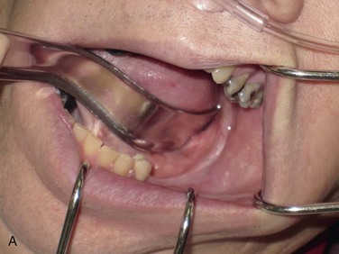
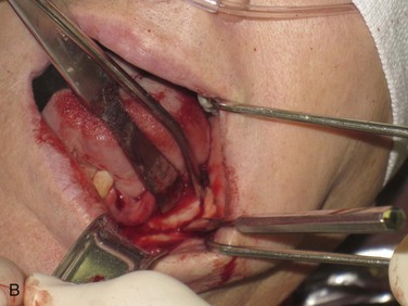
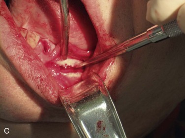
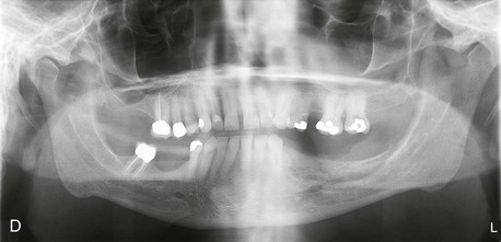
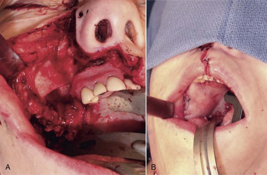
Bibliography

Bell, RB, Blakey, GH, White, RP, et al. Stages reconstruction of the severely atrophic mandible with autogenous bone graft and endosteal implants. J Oral Maxillofac Surg. 2002; 60:1135.

Chiapasco, M, Abati, S, Romeo, E, et al. Clinical outcome of autogenous bone blocks or guided bone regeneration with e-PTFE membranes for the reconstruction of narrow edentulous ridges. Clin Oral Implants Res. 1999; 10(4):278–288.

Clavero, J, Lundgren, S. Ramus or chin grafts for maxillary sinus inlay and local onlay augmentation: comparison of donor site morbidity and complications. Clin Implant Dent Relat Res. 2003; 5(3):154–160.

Cordaro, L, Amade, DS, Cordaro, M. Clinical results of alveolar ridge augmentation with mandibular block bone grafts in partially edentulous patients prior to implant placement. Clin Oral Implants Res. 2002; 13:103.

Corinaldesi, G, Pieri, F, Sapigni, L, et al. Evaluation of survival and success rates of dental implants placed at the time of or after alveolar ridge augmentation with an autogenous mandibular bone graft and titanium mesh: a 3- to 8-year retrospective study. Int J Oral Maxillofac Implants. 2009; 24:1119–1128.

Esposito, M, Grusovin, MG, Felice, P, et al. The efficacy of horizontal and vertical bone augmentation procedures for dental implants: a Cochrane systematic review. Eur J Oral Implantol. 2009; 2(3):167–184.

Felice, P, Pistilli, R, Lizio, G, et al. Inlay versus onlay iliac bone grafting in atrophic posterior mandible: a prospective controlled clinical trial for the comparison of two techniques. Clin Implant Dent Relat Res. 2009; 11(Suppl 1):e69–82.

Felice, P, Marchetti, C, Iezzi, G, et al. Vertical ridge augmentation of the atrophic posterior mandible with interpositional bloc grafts: bone from the iliac crest vs bovine anorganic bone—clinical and histological results up to one year after loading from a randomized-controlled clinical trial. Clin Oral Implants Res. 2009; 20:1386–1393.

Her, S. Titanium mesh as an alternative to a membrane for ridge augmentation. J Oral Maxillofac Surg. 2012; 70:803–810.

Louis, P, Gutta, R, Said-Al Naief, N, et al. Reconstruction of the maxilla and mandible with particulate bone graft and titanium mesh for implant placement. J Oral Maxillofac Surg. 2008; 66:235–245.

Louis, P. Bone grafting the mandible. Oral Maxillofac Surg Clin North Am. 2011; 23:209–227.

Mertens, C, Decker, C, Seeberger, R, et al. Early bone resorption after vertical bone augmentation: a comparison of calvarial and iliac grafts. Clin Oral Implants Res. 2013; 24:820–825.

Nissen, J, Ghelfan, O, Mardinger, O, et al. Efficacy of cancellous block allograft augmentation prior to implant placement in the posterior atrophic mandible. Clin Implant Dent Relat Res. 2011; 13(4):279–285.

Pieri, F, Corinaldesi, G, Fini, M, et al. Alveolar ridge augmentation with titanium mesh and a combination of autogenous bone and anorganic bovine bone: a 2-year prospective study. J Periodontol. 2008; 79:2093–2103.

Proussaefs, P, Lozada, J. The use of intraorally harvested autogenous block grafts for vertical alveolar ridge augmentation: a human study. Int J Periodontics Restorative Dent. 2005; 25:351.

Proussaefs, P, Lozada, J, Kleinman, A, et al. The use of ramus autogenous block grafts for vertical alveolar ridge augmentation and implant placement: a pilot study. Int J Oral Maxillofac Implants. 2002; 17:238.

Roccuzzo, M, Ramieri, G, Spada, MC, et al. Vertical alveolar ridge augmentation by means of a titanium mesh and autogenous bone grafts. Clin Oral Implants Res. 2004; 15:73–81.

Roccuzzo, M, Ramieri, G, Bunino, M, et al. Autogenous bone graft alone or associated with titanium mesh for vertical alveolar ridge augmentation: a controlled clinical trial. Clin Oral Implants Res. 2007; 18:286–294.

Simion, M, Baldoni, M, Zaffe, D. Jawbone enlargement using immediate implant placement associated with a split-crest technique and guided tissue regeneration. Int J Periodontics Restorative Dent. 1992; 12:462–473.

Tolstunov, L, Hicke, B. Horizontal augmentation through the ridge-split procedure: a predictable surgical modality in implant reconstruction. J Oral Implantol. 2013; 39(1):59–68.

Waasdorp, J, Reynolds, MA. Allogeneic bone onlay grafts for alveolar ridge augmentation: a systematic review. Int J Oral Maxillofac Implants. 2010; 25(3):525–531.

Watzinger, F, Luksch, J, Millesi, W, et al. Guided bone regeneration with titanium membranes: a clinical study. Br J Oral Maxillofac Surg. 2000; 38:312–315.

Radial Forearm Free Flap

Examination

General. The patient is a slim, pleasant white man who appears his stated age.

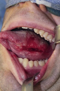
Intraoral. The patient is edentulous and has a prominent ulceration at the left posterior lateral tongue that measures 3 cm in length and 2 cm in width. The red and white lesion is tender and has a necrotic center with firm, everted edges along its periphery (Figure 12-4). The tongue is freely mobile. There appears to be no extension into the floor of the mouth.

Neck. There is no palpable lymphadenopathy.

Nasal fiber endoscopy. The lesion does not extend into the base of tongue; the bilateral tonsillar pillars, epiglottis, valleculae, arytenoids, piriform sinuses, and glottis are without obvious lesions.

Extremity. Peripheral pulses are 2+ for all extremities, and there is no cyanosis, clubbing, or edema. Bilateral Allen’s tests revealed good collateral circulation to the hands.

Allen’s test is used to assess the circulatory blood flow of the hand. The main blood supply to the hand is via the ulnar and radial artery. The ulnar artery supplies the superficial palmer branch, and the radial artery supplies the deep palmer branch. (Communication between the superficial and deep systems allows perfusion of the hand if there is interruption of one of the two main arteries to the hand, such as with the radial forearm free flap harvest.) Allen’s test determines the perfusion of the hand by simulating complete interruption of the radial artery. This is to ensure that the hand remains viable upon harvesting of the radial forearm free flap. Allen’s test is performed by elevating the intended hand and digitally occluding both the ulnar and the radial arteries. The patient is asked to clench and release a fist to cause blanching of the hand. Next, the pressure over the ulnar artery is released, and the capillary refill of the hand is evaluated. A wide range of values for hand reperfusion have been noted, ranging from 3 to 15 seconds. Additional techniques to qualify hand perfusion include the use of finger oximetry and Doppler assessment in conjunction with the Allen’s test. If hand perfusion is predominately based from the radial artery, use of the contralateral forearm or of an ulnar fasciocutaneous free flap should be considered.

Imaging

In general, if the patient has a normal result on Allen’s test, no imaging is necessary prior to radial forearm free flap harvest.

The work-up of squamous cell carcinoma of the tongue includes CT scanning of the head and neck with intravenous contrast (for improved delineation of soft tissue) and a chest radiograph (see the section Squamous Cell Carcinoma in Chapter 11).

In the current patient, axial and coronal CT scans demonstrated a 3 × 2 × 1.5-cm mass with poorly defined margins in the area of the tongue. No cervical lymphadenopathy was noted. The results of the chest radiograph were within normal limits.

Physical examination of the neck has variable reliability, with a sensitivity of 74%, specificity of 81%, and accuracy of 77%. As recommended by the National Comprehensive Cancer Network (NCCN) guidelines, the work-up for cancer of the oral cavity can include CT scans with contrast and/or MRI studies with contrast of the primary tumor location and the neck. CT imaging has a sensitivity of 83%, specificity of 83%, and accuracy of 83% for the detection of cervical metastasis. MRI, which has an effectiveness similar to that of CT, has been described as more sensitive in the identification of small metastatic cervical nodes. The combination of a physical examination with CT imaging increases the detection rate to 91%, whereas the detection rate for a physical examination alone is 75%.

Treatment

Surgery is the primary treatment modality for oral squamous cell carcinoma. Radiation therapy is an alternative with a comparable survival outcome; however, the treatment duration and likely complications of radiation-induced fibrosis and xerostomia make this option less appealing. Most patients opt for surgery, although medically compromised patients who are not suitable candidates for surgery may opt for radiation therapy.

Access to the lesion is considered first. In the current patient, the resection can be completed via a transoral approach. For larger oral squamous cell carcinomas of the oral cavity, it is not uncommon to gain access via a lip split mandibulotomy or “pull through” approach (Figure 12-5). Along with resection of the tumor, reconstruction of the defect is planned preoperatively. In general, reconstruction is based on the concept of the reconstructive ladder. The methods of reconstruction are ranked by complexity; most descriptions of the reconstructive ladder start with closure by secondary intention, primary closure, local flaps, regional pedicled flaps, and microvascular free flaps. In the current patient, the surgical defect after resection would result in a significant loss of tissue, because oncologic clearance generally incorporates 1 to 1.5 cm for the margins and, depending on frozen sections, may incorporate another 3 to 5 mm circumferentially.
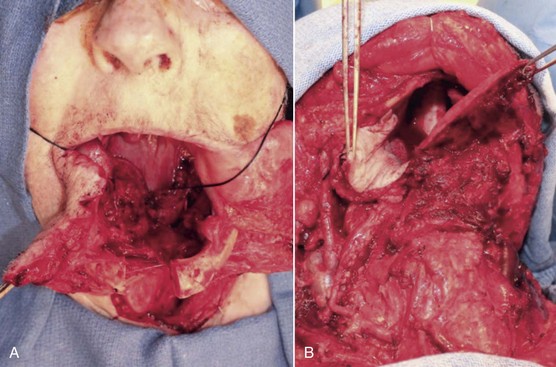
With respect to tongue defects, reconstruction should consider preserving the patient’s functions of speech and swallow. Resections incorporating the anterior tongue require the tongue tip for articulation and for propelling a food bolus posteriorly; the posterior tongue is largely involved with swallowing. Primary closure is an acceptable means of reconstruction if closure will not restrict tongue mobility. For the current patient, the radial forearm free flap was selected to provide bulk and to prevent restriction in tongue mobility that would affect speech and swallowing functions. Because tongue defects vary, a variety of free flaps can be used to restore form and function. When a glossectomy leaves more than 33% to 50% of the tongue, emphasis should be placed on maintaining mobility of the remaining tongue through the use of a thin, pliable flap, such as a radial or an ulnar fasciocutaneous free flap. When the defect leaves less than 33% of the original tongue, reconstruction shifts to the restoration of bulk to direct secretions toward the oropharynx and to provide contact of the neotongue with the palate for deglutition. For greater tissue bulk, the anterior lateral thigh free flap is an effective choice for reconstruction.

The radial forearm fasciocutaneous flap is the soft tissue flap of choice for reconstructing small to medium-sized oral and oropharyngeal defects. Based on the radial artery and cephalic vein and/or venae comitantes, it consists of thin, pliable skin and a very long pedicle, which make it well suited for use in the oral cavity. It can be designed to include tendons, muscle, or a segment of bone up to 12 cm in length, making it also useful for composite maxillary and mandibular defects.

The current patient was placed under general anesthesia and underwent a tracheostomy to secure his airway. A marking pen was used to delineate the planned resection edges, and a paper template was used to approximate the size and shape of the resection. A left hemiglossectomy was performed, and the margins of the resection were confirmed to be adequate with frozen sections (Figure 12-6, A and B).
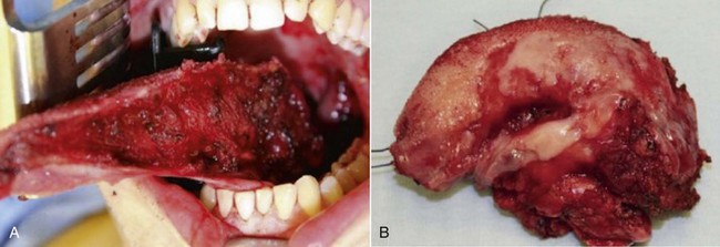
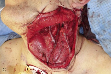
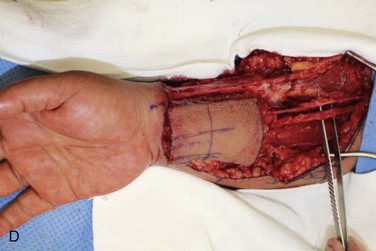
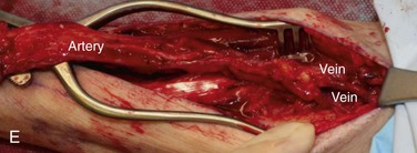
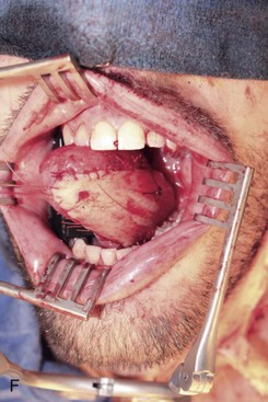
A left selective neck dissection was performed to sample lymph nodes from levels I through III and to expose and preserve vessels for microvascular reconstruction (Figure 12-6, C). These vessels included the internal jugular vein and the internal and external carotid arteries and their associated branches.

Simultaneous harvest of the radial forearm free flap was performed on the nondominant hand (Figure 12-6, D). The template was used to approximate the area of skin needed for the fasciocutaneous flap harvest. Generally, the skin of the entire volar forearm may be harvested, extending from the antecubital fossae to the flexor crease of the wrist. The skin and subcutaneous tissue are thinner in the distal forearm than in the proximal portion of the forearm. Also of note, the area of the distal forearm is thinner in males than in females.

Clinical landmarks that assist in the harvest of a radial forearm free flap include the distal extent of the flap within a flexor crease of the wrist, the antecubital fossae, and the brachioradialis, flexor carpi radialis, and palmaris longus, in addition to the outline of the planned flap. The flap is centered to overlap the region of the vascular pedicle located between the brachioradialis and flexor carpi radialis (Figure 12-6, E).

In the current patient, a tourniquet was placed proximal to the elbow and inflated to 250 mm Hg to facilitate dissection. An incision was made along the distal margin of the planned flap to expose and identify the vascular pedicle, cephalic vein, and superficial branch of the radial nerve. (After identification of the vascular pedicle, a confirmatory Allen’s test can be performed with a bulldog clamp, the tourniquet let down and perfusion checked in the hand. A subfascial dissection is completed to the extent of the planned flap, with care taken to incorporate the vascular pedicle and to travel along the brachioradialis and flexor carpi radialis and to incorporate the cephalic vein. At the proximal margin of the flap, the vascular pedicle is dissected free to the antebrachial fossae. Along the medial aspect of the skin flap, care is taken to avoid injury to the ulnar artery during dissection [deep between the flexor digitorum superficialis and flexor carpi ulnaris muscles.])

The radial artery was clamped and divided at its most proximal extent at the branching of the brachial artery to form the radial and ulnar arteries. (The cephalic vein is generally dissected free into the antebrachial fossae. The superficial branch of the radial nerve of the forearm runs with the radial artery beneath the distal belly of the brachioradialis. At approximately the midpoint of the brachioradialis, the nerve continues laterally and the radial artery courses medially. For this reason, it is often not included in the raised pedicle; therefore, the antebrachial cutaneous nerve is relied upon to supply the paddle.)

Once the radial forearm flap had been harvested, it was inset into the intraoral defect (Figure 12-6, F). Because of the vessel geometry, the pedicle was delivered medial to the left mandibular body and placed alongside the great vessels of the neck. Excess skin was removed to achieve the desired bulk, and the flap was secured to the resection wound edge prior to the microvascular vessel anastomosis. The term “ischemia time of the flap” refers to the time from harvesting of the flap to the moment the artery is reestablished. Free flap reconstructions require a period of tolerance to tissue ischemia. Studies have demonstrated that this period of tolerance varies with the tissue type. Most flaps incorporate skin, fascia, muscle, and bone tissue. Skin and fascia are the most resistant to ischemia; they have a tolerance period of 4 to 6 hours before irreversible cellular injury occurs that could jeopardize the viability of the free flap. Muscle is generally the most sensitive to ischemia time; it has a tolerance period of less than 3 hours.

In the current patient, after flap harvest, the tourniquet was released, hemostasis was confirmed, and a split-thickness skin graft was harvested from the left thigh and grafted to the radial forearm free flap donor site. The arterial anastomosis was performed in an end-to-end fashion into the facial artery with 9-0 nylon sutures in an interrupted fashion, and the venous anastomosis was performed in an end-to-side fashion into the internal jugular vein. (During the microvascular anastomosis, heparinized saline [100 µ/ml] is used to mute the process of thrombosis in the lumen of the vessels. Inherent to the anatomy of blood vessels, arteries generally have a thicker media, whereas veins have thinner vessel walls; hence, anastomosis for the artery is commonly completed with sutures, and the veins are coupled together, because their thin walls make them amenable to coupling.) Adequate perfusion of the flap was confirmed clinically by assessment of color, texture, capillary refill, and temperature. (Doppler probes are also regularly used to assess blood flow through the free flap.)

Closure of the donor site was completed in a layered fashion, with dermal sutures followed by staples for the skin. The skin paddle donor site can be closed primarily if it is small, and techniques have been described for using local flaps (Z-plasty) to facilitate closure. Most defects are repaired with either a split-thickness or full-thickness skin graft. Unlike microvascular flaps, these grafts are dependent on nutrition from the recipient bed via plasmatic circulation or imbibition for 48 to 72 hours, until capillary in-growth occurs. During the initial wound healing phase, it is necessary to bolster the graft on the recipient bed to provide stability for the healing process. The split-thickness skin graft incorporates the layers of the epidermis and a portion of the dermis. Its advantages include the ability to cover large areas with a higher rate of successful graft take. On the other hand, split-thickness skin grafts are less esthetic, are susceptible to more contracture, and require an additional donor site. Full-thickness skin grafts incorporate layers of the epidermis and dermis and provide a more esthetic result with less late wound contracture. The donor site for the full-thickness skin graft can also be closed primarily, and the graft can be harvested from inconspicuous areas of the body, such as the supraclavicular area or, more commonly, the medial aspect of the upper arm.

In the current patient, the harvested split-thickness skin graft was bolstered with a wound vacuum-assisted closure system, and the wrist was immobilized for 5 to 7 days with a volar splint to prevent shearing of the skin graft. The flap remained viable at the recipient site and healed without complications.

Complications

The surgical complication rate among patients undergoing microvascular reconstruction after head and neck surgery is 19% to 22%. The majority of these surgical complications are related to the reconstructive effort and manifest as flap loss, partial loss, and wound healing complications at either the donor or recipient site. Preoperative risk factors that have been described to contribute to the incidence of surgical complications include American Society of Anesthesiologists (ASA) status of III or IV, low preoperative hemoglobin, prolonged operative time (longer than 10 hours), and prior radiation and/or surgery. Factors specific for free flap failure have been associated with the surgeon’s experience, flap selection, and the patient’s nutritional status. Free flap failure rates have improved significantly since this technique moved into mainstream use. Initial free flap survival rates, during the first decade of popularity in the 1980s, ranged from 85% to 89%; recent studies show that they have improved to more than 95%. A review of 248 consecutive microvascular free flap reconstructions at Legacy Emanuel Hospital in Portland, Oregon, found a 95.5% success rate. The radial forearm fasciocutaneous free flap was used in 52% of cases, reflecting its popularity for reconstruction of defects in the oral cavity. Specifically, the radial forearm free flap demonstrated excellent success, with flap survival rates of 96% to 100% in multiple studies.

The main disadvantage of the radial forearm fasciocutaneous free flap is the morbidity associated with the donor site. Inherent to the harvest of the graft is the manipulation of the superficial branch of the radial nerve, because this nerve is closely associated with the cephalic vein. The superficial branch of the radial nerve provides sensory input for the dorsal surface of the thumb and the second and third digits. Abnormal sensation (hypoesthesia, hypesthesia, or paresthesia) in the donor hand occurred in 82% of patients at 3 months after surgery; however, this improved to 26% during a mean follow-up period of 14 months.

Wound healing accounts for most of the complications associated with the donor site. Subfascial radial forearm fasciocutaneous free flap harvests demonstrated a higher incidence of skin graft loss, ranging from 16% to 28%; in this same subgroup, tendon exposure and delayed healing accounted for 13% to 28% and 22% to 28%, respectively. An alternative to harvesting of the radial forearm free flap involves suprafascial dissection, which tends to leave the exposed tendons of the forearm with investing fascia and some deep subcutaneous tissue to allow for a better foundation for the split-thickness or full-thickness graft. Skin graft losses in suprafascial donor sites are reported to be 4% to 6%. The incidence of skin graft loss has been associated with the likelihood of decreased grip strength. It is reported that subjective grip strength was significantly decreased in 5% to 10% of patients who demonstrated partial skin graft failure.

Use of the radial forearm flap as an osteocutaneous flap can be associated with the risk of radial fracture. A segment of vascularized bone can be harvested, and the length is limited by the attachments of the pronator teres muscle proximally and the brachioradialis muscle distally. On average, 10 to 12 cm in length and less than half of the radius can be harvested. Prophylactic plating of the radius is recommended to decrease the likelihood of radius fracture. Prior studies in which the radius was not plated after harvesting reported a fracture incidence of 15% to 67% in donor arms.

Discussion

Since the original description of the radial forearm fasciocutaneous free flap, more than 30 years ago, this technique has evolved into a versatile and reliable flap for head and neck reconstruction. First described by Taylor in 1976 and publicized in oral cavity reconstruction by Soutar and colleagues in 1983, this flap continues to be a popular soft tissue flap despite the challenges with donor site morbidity. Alternative flaps, such as the cutaneous lateral arm flap, fasciocutaneous ulnar flap, and anterior lateral thigh perforator flap, have limited advantages, which focus on less donor site morbidity.

There are many applications for the radial forearm free flap in head and neck reconstruction. As demonstrated in this case presentation, tongue reconstruction with the radial forearm free flap is a common indication. Use of this type of flap has been described for reconstructing the ablative defects of the soft palate and mandible, restoring soft tissue deficiencies, and repairing tracheoesophageal fistulas. Dual skin flaps have been used to reconstruct through-and-through defects in the cheek and lip, and with two-stage procedures, prefabricated grafts can be prepared for reconstruction of complex structures such as the nose (Figure 12-7).

The surgical anatomy of the radial forearm flap is consistent and readily assessed. As previously mentioned, the viability of a donor site is based on the results of Allen’s test. The blood supply is based on the radial artery, which is reliably found between the brachioradialis and flexor carpi radialis. On average, the length of the radial artery, from the antecubital fossa to the wrist, is 18 to 20 cm, and the vessel has a diameter of 2 to 2.5 mm. It is relatively superficial, and dissection is completed within a subfascial plane guided by the brachioradialis and flexor carpi radialis. The radial artery is intimately associated with the overlying fascia and supplies perforating vessels that pass through to form subfascial, intrafascial, and suprafascial vascular plexuses. These networks establish the extensive subcutaneous vascular plexuses that supply the skin. The fascia provides a degree of additional protection to the pedicle and overall integrity of the flap.

Multiple variations of this flap can be developed from the incorporation of accessible anatomic resources. Additional bulk can be obtained from the brachioradialis, tendon and ligaments can be obtained from the palmaris longus when necessary, and innervation through the antebrachial cutaneous nerve and bone can be obtained through harvest of the radius.

Venous return for the free forearm flap is classified as a deep and superficial venous supply. The deep supply is composed of the venae comitantes, which run in the intermuscular septum along with the radial artery. The superficial venous supply consists chiefly of the cephalic vein. Both systems provide venous outflow for the tissue supplied by the radial artery. The harvest of both venous systems allows for anastomosis of two veins. A study of 492 head and neck free flap procedures performed at the University of Toronto compared two-vein anastomosis with single-vein anastomosis and demonstrated an improved success rate for the former (98.6% versus 93.6%). This finding was also illustrated in an evaluation of microvascular couplers in head and neck reconstruction; in this study, dual vein–coupled anastomosis showed a trend for improved success over single vein–coupled anastomosis. When only a single vein is used for reconstruction, there are no differences in using the superficial over the deep venous system as the sole outflow for the flap.

In a vessel-depleted neck, a radial forearm hybrid flap can be used to compensate for the lack of vascular options. This scenario is most common in previously operated and irradiated fields. The radial forearm hybrid flap uses a single arterial anastomosis that is carried out between the radial artery of the flap and a recipient artery and the venous drainage (the cephalic vein), to remain in continuity with the systemic venous system. Technically, the cephalic vein is dissected up to the deltopectoral groove, pedicled over the clavicle, and delivered into the neck in the subcutaneous plane.

The radius is situated lateral to the ulna and articulates with the humerus and ulna proximally and with the ulna, scaphoid, and lunate bones distally. It averages 23 cm in length and in cross section the radial shaft appears triangular. Functionally the radius plays a minor role in the stability of the elbow; however, at the radiocarpal joint, it is an integral structure of the wrist. The proximal head of the radius allows the hand to pronate, and the distal head plays a role in hand flexion, extension, adduction, and abduction.

The brachioradialis functions to flex the arm at the elbow and for pronation and supination of the forearm. Its origin is at the lateral supracondylar ridge of the humerus, and it attaches to the distal styloid process of the radius by way of the brachioradialis tendon. It is located in the posterior compartment of the forearm and is innervated by the radial nerve. In reconstruction, the brachioradialis can be used to provide bulk, such as in a total glossectomy defect.

The skin available to be harvested on the forearm is defined by the radial artery angiosome. It extends from the flexor crease of the wrist to the antecubital fossa and from the medial third of the ventral surface of the forearm to the lateral third of the dorsal surface of the forearm. If an innervated flap is considered, the somatosome of the lateral antebrachial cutaneous nerve is used to guide the location of skin flap harvesting. The somatosome of the lateral antebrachial cutaneous nerve extends from the midline of the ventral forearm to the lateral third of the dorsal surface of the forearm.

Innervated tongue flaps are accomplished by neurorrhaphy of the lateral antebrachial cutaneous nerve to a recipient nerve. Sensation recovery of the innervated radial for/>

Stay updated, free dental videos. Join our Telegram channel

Jan 12, 2015 | Posted by in Oral and Maxillofacial Surgery | Comments Off on 12: Reconstructive Oral and Maxillofacial Surgery

VIDEdental - Online dental courses

Get VIDEdental app for watching clinical videos