12
John Hunter and the London Tooth Museum
The heart of Leicester Square in the West End of London is pedestrianised and contains a small park, Leicester Square Park. In its centre are statues of two of England’s most famous entertainers, one ancient and one more modern. The larger statue is of the playwright William Shakespeare, while next to it is a statue of the silent screen comedian, Charlie Chaplin. At each corner entrance to the park is a bust representing a famous Englishman from the eighteenth century. Three are well known, namely Sir Isaac Newton, mathematician and astronomer (1643–1727); Sir Joshua Reynolds, portrait painter and one of the founders and first president of the Royal Academy of Arts (1723–1792) and William Hogarth, painter, engraver and satirist (1697–1764). The statue on the remaining corner is of John Hunter (Figure 12.1), surgeon and scientist, who is less well known and the subject of this chapter.
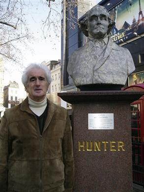
Figure 12.1 Author by statue of John Hunter in Leicester Square Park. The park is undergoing reconstruction (as of April 2012).
Source: Photographed by Mr G. Fox.
John Hunter and His Contribution to Medicine and Science
John Hunter was born in 1728 in Scotland, the 10th and youngest child of the family. Brought up on a small farm in East Kilbride and showing little promise at school, his career prospects did not look good. Realising the need for change if he were to achieve anything, when he was 20 years old he wrote to his elder brother, William, offering his services in any capacity.
William Hunter, 10 years older than John, had studied medicine in Edinburgh before moving to London in 1741 as a young trainee surgeon. In those days, physicians enjoyed high status and rarely deigned to examine patients physically. Surgeons, on the other hand, did not have such a high standing. Before the days of anaesthetics and antisepsis, and with little detailed knowledge of anatomy, only minor surgical procedures had any chance of success. Surgical work such as bloodletting or tooth pulling was often left to the barber-surgeons who themselves had little, if any, training.
Determined to transfer to the more lucrative field of obstetrics, the ambitious William Hunter realised the importance of knowledge of human anatomy both in medical practise and in helping to improve success rates in surgery. With this in mind, in 1746 he established a private school of anatomy in Covent Garden. The key to success for his venture was to guarantee every student would have the opportunity to dissect human bodies. This meant that he had to overcome the problem of legally obtaining sufficient numbers of bodies.
John’s arrival in London in 1748 fortuitously coincided with the beginning of anatomy classes for a new intake of medical students at his brother’s fledgling anatomy school. Help was needed in providing students with guidance in human dissection, and William offered his young brother employment as a dissector. The uncomfortable conditions under which John found himself working, particularly regarding the smell of the corpses and the very cold surroundings, can only be imagined, not to mention his total ignorance of the subject. As the science of preserving cadavers was still in its infancy, dissection had to be carried out in the cold winter months in order to delay the rate of decomposition of the bodies.
In a relatively short time, it became clear that John had a remarkable aptitude for dissection. He also possessed a scientific curiosity and talent for developing methods of preserving and displaying teaching material, especially of diseased structures. John demonstrated initiative in procuring sufficient corpses, even if this meant undertaking grave-robbing himself and dealing with the ‘resurrectionists’, who sold bodies without too many questions being asked about their origins.
Within a short space of time, it was apparent there was little that William could teach John, who was soon in charge of the dissecting room. From that moment on, John Hunter found his purpose in life. His hidden and considerable intellect began to flourish. From humble beginnings and lack of education, he eventually rose to become the most famous anatomist and surgeon in England.
John Hunter’s reputation as an anatomist quickly grew. His skill in dissection was greatly admired, and he started his own research into the workings of the human body, little of which was known at the time. Soon, medical students from around the world were coming to London to learn anatomy under his tutelage. When these students returned home, they took with them Hunter’s ideas and, most importantly, his meticulous scientific approach.
Among John Hunter’s early major scientific discoveries, two can be highlighted, either of which would have assured him a place in the history of anatomy. First was his demonstration that the lymphatic system was separate from the blood system. Second was his discovery that the blood circulations of a human foetus and its mother were separate and not joined directly together, as was the general opinion at the time.
John undertook dissections of human foetuses at the behest of his brother William, who was planning a book containing a small number of illustrations showing the development of the human foetus. This he hoped would establish his reputation in the field of obstetrics. When William saw his brother’s beautiful dissections, he decided to postpone publication until he had a larger, complete series of specimens. This project was not finished for 25 years when his atlas The Anatomy of the Human Gravid Uterus Exhibited in Figures was finally published in 1774 (Figure 12.2). This book is considered one of the most important in the history of medicine. However, John Hunter felt that he was not given sufficient credit, particularly for establishing the separateness of the foetal circulation. A bitter disagreement erupted into the public domain, and, sadly, the two brothers barely spoke to each other again. The artist, Jan van Rymsdyk, who produced all the wonderful engravings, was given no recognition at all.
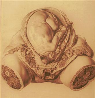
Figure 12.2 Illustration of a human foetus dissected by John Hunter and illustrated by Jan van Rymsdyk in William Hunter’s book: The Anatomy of the Human Gravid Uterus Exhibited in Figures.
Source: Reproduced by kind permission of the Hunterian Museum at the Royal College of Surgeons.
Having acquired more anatomical knowledge than anyone else in the country, John Hunter was aware that this alone would not help his advancement. He chose to pursue a career in surgery and left his brother’s anatomy school. He spent the next 3 years as an army surgeon, which required little previous experience to enlist. This took him to France and Portugal, where he gained experience in treating many war casualties. He returned to London in 1763, where his army service enabled him to establish a practice as a surgeon. Although the next few years were financially difficult, Hunter’s surgical and scientific reputation spread and by 1768 he was sufficiently experienced to be accepted into the elite Company of Surgeons.
At this time, autopsies were being undertaken (mainly at the behest of the legal profession) to help determine not only the cause of death but also to aid the medical profession in improving diagnosis and treatment. Hunter was soon acknowledged as the most experienced person in England to carry out these post-mortems.
Hunter continued his experiments on a wide range of topics on animals while continuing to dissect and preserve human material, both normal and abnormal. His aim was to try and understand, through his own observations and experiments, how the body functioned. He was not prepared blindly to accept existing medical theories, techniques and treatments that had been passed down dogmatically from bygone ages, particularly where his own experience indicated these were incorrect.
Hunter’s career as a surgeon could only advance significantly if he obtained a post in one of the major London hospitals. This he achieved in 1768, being elected surgeon at St George’s Hospital, which meant he had an influential base from which to work. Apart from his own private patients, Hunter was now treating surgical cases in his hospital. With this new position came the added responsibility of supervising surgeons-in-training. It should be remembered that in Hunter’s time, medicine was still following the ancient doctrines of Hippocrates (BC 460–370) and Galen (129–200 AD). This stressed that good health depended on the balance of the four basic humours: black bile, yellow bile, phlegm and blood. Ill health and disease resulted from an imbalance between them that could only be counteracted by procedures such as purging the patient or bloodletting.
Hunter introduced the study of comparative anatomy, looking at a wide range of animals to observe how structure and function were related in order to understand the workings of the human body. His lectures and demonstrations were not the easiest to follow, and he was not gifted in the art of speaking. However, he used his unrivalled and ever-expanding collection of specimens to educate and inspire his growing numbers of students from both home and abroad. It was through these students that his philosophy of careful experimentation, observation and rigorous thought was spread: ‘Believe what you actually observe rather than what others tell you should happen’. The term ‘Hunterian tradition’ reflects this attitude and is still used today.
One of Hunter’s students was Edward Jenner. With encouragement from Hunter, Jenner founded the science of immunology through the successful inoculation of patients with cowpox to prevent the onset of the related, but far more dangerous disease, smallpox.
Hunter’s scientific eminence was recognised when he was elected a Fellow of the Royal Society in 1767. He built a large house at Earls Court with facilities to dissect and accommodate his ever-expanding collection of specimens, later moving to much bigger premises in Leicester Square. This was still a well-known building in the 1880s, and author Robert Louis Stevenson is thought to have used it as a model for Dr Jekyll’s house, with its laboratory, in his novel The Strange Case of Dr Jekyll and Mr Hyde’.
Hunter was continually on the lookout for unusual specimens showing abnormalities, hoping they would help him gain a deeper understanding of how organs and individuals developed. He was known to dealers as willing to pay high prices for such material. Due to the ongoing expense of obtaining, preserving and housing his ever-expanding collection, Hunter was always in need of funds, despite enjoying a considerable income as one of the major London surgeons, plus fees from students. His pre-eminence in surgery was recognised by his appointment in 1776 as Surgeon Extraordinary to King George III.
No object was too large or too small to escape Hunter’s interest. He obtained the skin and some bones of the first giraffe ever to be seen in England and dissected a bottle-nosed whale, whose skeleton he displayed. He received and described some of the first marsupials following the discovery of Australia by Captain Cook. He received the corpses of many exotic animals that died in the private zoos of the wealthy, including those from the royal menagerie at the Tower of London.
One story relating to Hunter’s desire to possess unique specimens concerned Charles Byrne, the Irish Giant, who, in 1782, caused a sensation when he appeared in London at the age of 21. At that time, there was a great demand for ‘freak shows’, which included the display of giants and dwarves. Byrne was about 2.3 m tall (7 ft 8 in), the result as we know today of a tumour in his pituitary gland that caused the production of excessive amounts of growth hormone. Unfortunately, it soon became apparent that, due to the severe side effects of the untreated tumour, Byrne’s health was rapidly deteriorating and that he would soon die. It was also no secret that Hunter, among many other competing individuals, wanted this body to dissect and add to his collection, and would be willing to pay a large sum for the privilege. Terrified of being dissected, Byrne invested all the money he had, which was no small amount, into what he regarded as a foolproof scheme that would prevent his body getting into the hands of the anatomists. His plan was to hand over his money to an undertaker with strict instructions that he be interred in a large, heavy coffin watched over by loyal friends. These friends would then transport him to the coast, transfer him to a boat and bury him deep at sea. Following Byrne’s death in 1783 at the age of just 22, this request was seen to be carried out. However, somewhere along the route Byrne’s body ‘disappeared’ (assuming it was in the coffin in the first place!) so that the coffin buried at sea did not contain his remains. Five years later, in 1788, when Hunter first opened his collection of specimens to a select group of friends and colleagues, Byrne’s skeleton took pride of place and was there for all to see, its enormous height causing a sensation. It was not clear whom Hunter bribed, or how the body was obtained, but a very large sum of money must have changed hands.
Throughout his life Hunter continued to produce a stream of major scientific papers. Among important observations were those showing how bones increased in size. To shed light on this mechanism, he designed a simple experiment whereby he inserted two small metal pellets along the shaft of the leg bone of a young chicken, initially recording their distance apart. After a period of time, when the bone had grown, he found that the two metal pellets were still the same distance apart. This simple but elegant experiment proved that the bone had increased in size by growth at the ends of the bones rather than in the central shaft (Figure 12.3). This result he further confirmed by adding a natural dye called madder to the diet of animals, which stained newly formed bone red. Never afraid to experiment upon himself, Hunter also swallowed some madder and noted that it discoloured his urine.
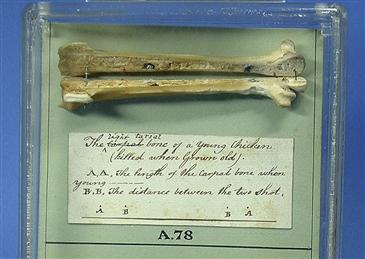
Figure 12.3 The main bone (tarso-metatarsus) in the leg of a chicken. John Hunter made two cauterised holes near the ends of the bone and filled them with metal. The chicken was later killed, and the holes were found to be of a similar distance apart, while the overall length of the bone had increased. This elegant experiment demonstrated that bone growth occurred in the ends (epiphyses), rather than in the shaft (diaphysis) of the bone. The original notes of the experiment made by Hunter’s assistant William Bell accompany the specimen.
Source: Reproduced by kind permission of the Hunterian Museum at the Royal College of Surgeons.
Hunter published two important books that furthered his scientific reputation even more, namely A Treatise on the Venereal Disease (1786) and Blood, Inflammation and Gunshot Wounds (1793). The first book was a very detailed account of the clinical features of venereal disease throughout its many stages. Hunter describes inoculating some of his own patients with diseased tissue. There is even a belief that the patient from whom many of the accurate signs and symptoms were recorded was Hunter himself, and that he had deliberately inoculated himself to obtain the information. Although this cannot be confirmed, it could account for some of the health problems that pursued him in later years and that may have contributed to his death in 1793. His second book was related to his previous experiences as an army surgeon and considered many clinical aspects of gunshot wounds.
Stay updated, free dental videos. Join our Telegram channel
VIDEdental - Online dental courses


