Chapter 12
Examples of Specific Medical Emergency Situations
Introduction
The general principles of management of medical emergencies were discussed in Chapter 11. This chapter concentrates on specific medical emergencies. The practitioner dealing with specific emergencies must not lose sight of the need to act in accordance with basic principles and be prepared to revisit them if the patient response to treatment is not satisfactory.
Specific emergencies that may arise in dental practice are listed in Box 12.1. Their signs, symptoms and management will be discussed.
Box 12.1 A summary of medical emergencies that may be encountered in a dental emergency clinic
- Vasovagal syncope (faint)
- Hyperventilation/‘panic attack’
- Acute asthma attack
- Angina/myocardial infarction
- Epileptic seizures
- Diabetic emergencies
- Allergies/hypersensitivity reactions
- Choking and aspiration
- Adrenal insufficiency
- Cardiac arrest (see Chapter 11)
Vasovagal syncope (simple faint)
The ‘simple faint’ is the most common medical emergency to be seen in dental practice and results in loss of consciousness due to inadequate cerebral perfusion. It is a reflex that is mediated by autonomic nerves leading to widespread vasodilatation in the splanchnic and skeletal vessels and bradycardia resulting in diminished cerebral perfusion. Fainting can be precipitated by pain or emotional stress, changes in posture or hypoxia. Some patients are more prone to fainting than others and it is wise to treat fainting-prone patients in the supine position.
A similar clinical picture may be seen in ‘carotid sinus syndrome’. Mild pressure on the neck in such patients (usually, the elderly) leads to a vagal reaction producing syncope. This situation may progress to bradycardia or even cardiac arrest.
Fainting – signs and symptoms
- Patient feels faint/light headed/dizzy
- Pallor, sweating
- Pulse rate slows
- Low blood pressure
- Nausea and/or vomiting
- Loss of consciousness
Fainting – treatment
- Lay the patient flat and raise the legs – recovery will normally be rapid.
- A patent airway must be maintained.
- If recovery is delayed, oxygen should be administered and other causes of loss of consciousness be considered.
Hyperventilation
Hyperventilation is a more common emergency than is often thought. When hyperventilation persists, it can become extremely distressing to the patient. Anxiety is the principal precipitating factor.
Hyperventilation – signs and symptoms
- Anxiety
- Light headedness
- Dizziness
- Weakness
- Paraesthesia
- Tetany (see below)
- Chest pain and/or palpitations
- Breathlessness
Hyperventilation – treatment
A calm and sympathetic approach from the practitioner is important. The diagnosis, particularly in the early stages, is not always as obvious as it may seem:
- Exclude other causes for the symptoms.
- Encourage the patient to rebreathe their own exhaled air to increase the amount of inhaled carbon dioxide – a paper bag placed over nose and mouth allows this.
- If no paper bag is handy, the patient’s cupped hands would be a (less satisfactory) alternative.
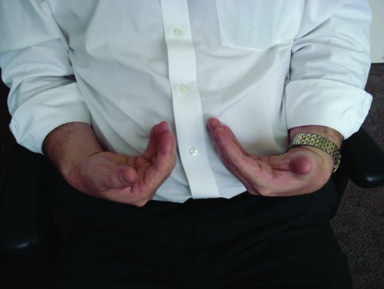
Hyperventilation leads to carbon dioxide being ‘washed out’ of the body producing an alkalosis. If hyperventilation persists, carpal (hand) and pedal (foot) spasm (tetany) may be seen (Figure 12.1). Re-breathing exhaled air increases inspired carbon dioxide levels and helps to return the situation to normal.
Figure 12.1 A demonstration of carpal spasm.
Asthma
Asthma is a potentially life-threatening condition and should always be taken seriously. An attack may be precipitated by exertion, anxiety, infection or exposure to an allergen. It is important in the history to get some idea of the severity of attacks. Clues include the precipitating factors, effectiveness of medication, hospital admissions due to asthma and the use of systemic steroids.
It is important that asthmatic patients bring their usual inhaler(s) with them – if the inhaler has not been brought it must be in the emergency kit or treatment should be deferred. If the asthma is in a particularly severe phase, elective treatment may be best postponed. Drugs that may be prescribed by dental practitioners, particularly non-steroidal anti-inflammatory drugs, may worsen asthma and, therefore, are best avoided.
Asthma – signs and symptoms
- Breathlessness (rapid respiration – more than 25 breaths per minute)
- Expiratory wheezing
- Use of accessory muscles of respiration
- Tachycardia
Signs and symptoms of life-threatening asthma
- Cyanosis or slow respiratory rate (less than 8 breaths per minute)
- Bradycardia
- Decreased level of consciousness/confusion
Asthma – treatment
- Most attacks will respond to the patient’s own inhaler, usually salbutamol (may need to repeat after 2–3 minutes).
- If no rapid response, or features of severe asthma, call an ambulance.
- A medical assessment should be arranged for patients who require additional doses of bronchodilator to end an attack.
- A spacer device may need to be used if patient has difficulty using the inhaler.
- If the patient is distressed or shows any of the signs of life-threatening asthma, urgent transport to hospital should be arranged.
- High-flow oxygen should be given whilst awaiting transfer. Four to six actuations from the salbutamol inhaler via a spacer device should be used and repeated every 10 minutes. In the British National Formulary, a technique is described for a ‘home made’ spacer device. A hole can be cut out the base of a paper or plastic cup. The mouthpiece of the inhaler is pushed through this. The open end of the cup can then be applied to the mouth when the inhaler is activated.
- If asthma is part of a more generalised anaphylactic reaction, or in extremis, an intramuscular injection of adrenaline should be given (see Section ‘Anaphylaxis’).
All patients, including those who have chronic obstructive pulmonary disease, should be given high-flow oxygen as even if these patients are dependent on ‘hypoxic drive’ to stimulate their respiration, they will come to no harm in the short-term.
Cardiac chest pain
Most patients who suffer chest pain from a cardiac cause in the dental surgery are likely to have a previous history of cardiac disease. The history is clearly important and if a patient uses medication to control known angina they should have brought this with them or it should be readily to hand in the emergency kit. Similarly, it is important that the patient has taken their normal medication on the day of their appointment.
Classically, the pain of angina is described as a crushing or band-like tightness of the chest, which may radiate to the left arm or mandible. There are many variations, however. The pain of myocardial infarction (MI) will often be similar to that of angina but more severe and, unlike angina, will not be relieved by GTN. In cases of angina, the patient should use their glyceryl trinitrate (GTN) spray, which will usually remove the symptoms. Dental treatment may be best left until another day if there is an attack, according to the practitioner’s discretion. More severe chest pain always warrants postponement of treatment and an ambulance should be called.
Features that make chest pain unlikely to be cardiac in origin are: pains that last less than 30 seconds, however severe; stabbing pains; well-localised left submammary pain and pains that continually vary in location. Chest pain that improves on stopping exertion is more likely to be cardiac in origin than one that is not related. Pleuritic pain is sharp in character, well localised and worse on inspiration.
Oesophagitis can produce a retrosternal pain that worsens on bending or lying down. A complicating factor in differentiation from cardiac chest pain is that GTN, due to action on the muscle of the oesophagus, may ease the pain.
Musculoskeletal pain is generally accompanied by tenderness to palpation in the affected region. As mentioned earlier, hyperventilation may produce chest pain. A list of possible causes of chest pain is given in Box 12.2.
Box 12.2 Chest pain – possible causes
- Angina
- Myocardial infarction
- Pleuritic, e.g. pulmonary embolism
- Musculoskeletal
- Oesophageal reflux
- Hyperventilation
- Gall bladder and pancreatic disease
It is clearly important to exclude angina and MI in the patient complaining of chest pain. If in doubt, treat as cardiac pain until proven otherwise.
Myocardial infarction – signs and symptoms
- Severe, crushing chest pain, which may radiate to the shoulders and down the arms (particularly the left arm) and into the mandible.
- Shortness of breath.
- The skin becomes pale and clammy.
- Pulse becomes weak and patient may become hypotensive.
- Often, there will be nausea and vomiting.
Not all patients fit this picture and may exhibit only some of the signs and symptoms mentioned in the preceeding text.
Myocardial infarction – treatment
- The practitioner should remain calm and be a reassuring presence.
- Call emergency number.
- Most patients will be best managed in the sitting position.
- Patients who feel faint should be laid flat.
- Give high-flow oxygen (15 L/min).
- Give sublingual GTN spray.
- Give 300 mg aspirin orally to be chewed (if no allergy) – ensure that when handing over to the receiving ambulance crew that they are made aware of this as thrombolytic therapy is given by some ambulance crews.
- A patient who has had surgical dental treatment should be highlighted to the ambulance crew as any significant risk of haemorrhage may affect the decision to use thrombolytic therapy
- If the patient becomes unresponsive, the practitioner should check for ‘signs of life’ (breathing and circulation) and start cardiac pulmonary ressusication.
Epileptic seizures
The history will usually reveal the fact that a patient has epilepsy. A history should obtain information with regard to the nature of any seizures, their frequency and degree of control. The type and efficacy of medication should be determined. Signs and symptoms vary considerably.
Epilepsy – signs and symptoms
- The patient may have an ‘aura’ or premonition that a seizure is about to occur.
- Tonic phase – loss of consciousness, patient becomes rigid and falls and becomes cyanosed.
- Clonic phase – jerking movements of the limbs, tongue may be bitten.
- Urinary incontinence, frothing at the mouth.
- The seizure often gradually abates after a few minutes but the patient may remain unconscious and may remain confused after consciousness has been regained
- Hypoglycaemia may present as a fit and should be considered (including in epileptic patients). Therefore, blood glucose measurement at an early stage is wise.
In patients with a marked bradycardia (less than 40 beats per minute), the blood pressure may drop to such an extent that it causes transient cerebral hypoxia leading to a brief fit. This is not a true fit and represents a vasovagal episode.
Epilepsy – treatment of a fit
The decision to give medication should be made if seizures are prolonged (with active convulsions for 5 minutes or more (status epilepticus) or seizures occurring in quick succession). If possible, high-flow oxygen should be administered. The possibility of the patient’s airway becoming occluded should be constantly remembered, and therefore, the airway must be protected:
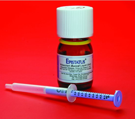
Figure 12.2 Buccal/intra-nasal midazolam. The solution in the bottle is drawn up in the syringe provided, which is calibrated into 0.1 mL increments.
- As far as possible, ensure safety of the patient and practitioner (do not attempt to restrain).
- Midazolam given via the buccal or intra-nasal route (10 mg for adults). The buccal preparation is marketed as ‘Epistatus’ (10 mg/mL) (Figure 12.2).
- For children:
- Child 1–5 years-5 mg
- Child 5–10 years-7.5 mg
- Child more than 10 years-10 mg
- The parents of some children with poorly controlled epilepsy will carry rectal diazepam. As part of pre-treatment preparation, it is wise to arrange with the parent for them to be on hand to administer this should a fit arise.
Stay updated, free dental videos. Join our Telegram channel
VIDEdental - Online dental courses


