11: Gastrointestinal Disease

Chapter 11

Gastrointestinal Disease

Gastrointestinal diseases such as peptic ulcer disease, inflammatory bowel disease, and pseudomembranous colitis are common and may affect the delivery of dental care. When a patient who has one of these conditions presents for treatment, several areas of clinical importance require consideration by the dental practitioner. The dentist must be cognizant of the patient’s condition, must monitor for symptoms indicative of initial disease or relapse, and must be aware of drugs that interact with gastrointestinal medications or that may aggravate these conditions. In addition, oral manifestations of gastrointestinal disease are not uncommon, so the dentist also must be familiar with oral patterns of disease.

Peptic Ulcer Disease

Definition

A peptic ulcer is a well-defined break in the gastrointestinal mucosa (greater than 3 mm in diameter, as defined by many industry-sponsored studies) that results from chronic acid or pepsin secretions and the destructive effects of and host response to Helicobacter pylori. Peptic ulcers develop principally in regions of the gastrointestinal tract that are proximal to acid and pepsin secretions (< ?xml:namespace prefix = "mbp" />Figure 11-1). The first portion of the duodenum is the location of most ulcers in Western populations, whereas gastric ulcers are more frequent in Asia.1 The upper jejunum rarely is involved. Peptic ulcer disease usually is chronic and focal in distribution; only approximately 10% of patients have multiple ulcers.

image

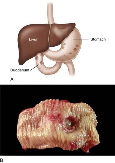
FIGURE 11-1 A, Location of peptic ulceration (shaded areas). Darker-stippled areas are higher-risk sites. B, Peptic ulcer of the duodenum.

(B, From Kumar V, Abbas A, Fausto N, editors: Robbins & Cotran pathologic basis of disease, ed 7, Philadelphia, 2005, Saunders. Courtesy Robin Foss, University of Florida–Gainesville.)

Epidemiology

Incidence and Prevalence

Peptic ulcer disease is one of the most common human ailments, once affecting up to 15% of the population in industrialized countries. Current estimates suggest that 5% to 10% of the world population is affected, and 350,000 new cases are diagnosed annually in the United States.26 The incidence of peptic ulceration peaked between 1900 and 1950 and progressively decreased thereafter. The decline in northern Europe and the United States may be the result of decreased cigarette and aspirin consumption, increased use of vegetable cooking oils (a rich source of raw materials for synthesis of prostaglandins, which have cytoprotective properties), and better sanitation leading to fewer H. pylori infections.7 The disease affects 5% to 7% of northern Europeans and accounts for about 200,000 hospitalizations annually in the United States. Peptic ulcers are rare in Greenlander Eskimos, southwestern Native Americans, Australian aborigines, and Indonesians.2

Peak prevalence of peptic ulceration has shifted to the elderly population.5 Until the 1980s, the male-to-female ratio in the United States was 2:1, but current data approximate this ratio at 1:1.8 First-degree relatives have a three-fold greater risk of developing the disease.5 Persons who smoke and are heavy drinkers of alcohol are more prone to development of the disease. An association with blood type O also is recognized. A higher prevalence is seen among patients with hyperparathyroidism and hypersecretory states (e.g., renal dialysis, Zollinger-Ellison syndrome, mastocytosis). Use of nonsteroidal antiinflammatory drugs (NSAIDs), including aspirin, for longer than 1 month is associated with an annual rate of 2% to 4% for gastrointestinal bleeding or ulcer complications in patients who ingest these drugs.9,10

The disease is rare in children, with only 1 in 2500 pediatric hospital admissions attributable to peptic ulceration.11 When a peptic ulcer is diagnosed in a child younger than 10 years of age, the condition most often is associated with an underlying systemic illness, such as severe burn injury or other major trauma.12 Most deaths that result from peptic ulcer disease occur in patients older than 65 years of age. An average dental practice of 2000 adult patients is predicted to serve about 100 patients with peptic ulcer disease.

Etiology

Peptic ulcers result when the balance between aggressive factors that are potentially destructive to the gastrointestinal mucosa and defensive factors that usually are protective of the mucosa is disrupted (Figure 11-2). The primary aggressive factor is H. pylori (formerly Campylobacter pylori). This organism is present in 60% to 90% of duodenal ulcers and in 50% to 70% of gastric ulcers.1,13 Use of NSAIDs is the second most common cause of peptic ulcer disease. Other aggressive factors include acid hypersecretion, cigarette smoking, and psychological and physical stress.14,15 Cytomegalovirus infection is a rare cause noted in human immunodeficiency virus (HIV)-positive patients.16,17 Non-NSAID, non–H. pylori peptic ulcers are infrequent and occur more often in elderly persons.

image

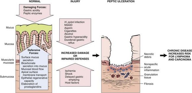
FIGURE 11-2 Complex interplay of aggressive and defensive factors involved in the formation of peptic ulcer disease.

(Modified from Kumar V, Abbas A, Fausto N, editors: Robbins & Cotran pathologic basis of disease, ed 7, Philadelphia, 2005, Saunders.)

H. pylori is a microaerophilic, gram-negative, spiral-shaped motile bacillus with 4 to 6 flagella.18 H. pylori was first reported to reside in the antral mucosa by Marshall and Warren.19 The organism is an adherent but noninvasive bacterium that resides at the interface between the surface of the gastric epithelium and the overlying mucous gel. It produces a potent urease that hydrolyzes urea to ammonia and carbon dioxide. This urease may protect bacteria from the immediate acidic environment by increasing local pH while damaging mucosa through generation of its byproduct, ammonia. Upregulation of cyclooxygenase-2 (COX-2), chemotaxis of neutrophils, and the cellular immune response are involved in the local tissue damage that subsequently occurs.

Humans are the only known hosts of H. pylori. This bacterium infects 0.5% of adults annually—a rate that has been declining since the early 1990’s.20,21 H. pylori is acquired primarily during childhood, possibly as a result of entry from the oral cavity via contaminated food and poor sanitary habits. The organism resides in the oral cavity,22 from which it probably descends to colonize the gastric mucosa. H. pylori can persist in the stomach indefinitely, and infection with the bacterium remains clinically silent in most affected persons.23 The rate of H. pylori acquisition is higher in developing than in developed countries. In developing countries, 80% of the population carries the bacterium by the age of 20 years, whereas in the United States, only 20% of 20-year-olds are infected. The prevalence of infection among African Americans and Hispanics is twice that for whites in the United States.24 Infection is correlated with lower socioeconomic status, contaminated drinking water, and familial overcrowding, especially during childhood. Approximately 20% of infected persons go on to develop peptic ulcer disease,15 suggesting that other physiologic and psychological (stress) factors are required for presentation of this disease.25

Use of NSAIDs is an etiologic factor in 15% to 20% of cases of peptic ulcer.1,10 These drugs directly damage mucosa, reduce mucosal prostaglandin production, and inhibit mucus secretion. Ulcers caused by NSAIDs are located more often in the stomach than in the duodenum. Risk with NSAID use increases with age older than 60 years, high-dosage long-term therapy, use of NSAIDs with long plasma half-lives (e.g., piroxicam) rather than those with short half-lives (i.e., ibuprofen), and concomitant use of alcohol, corticosteroids, anticoagulants, or aspirin.26 Use of orally administered nitrogen-containing bisphosphonate drugs (aledronate, risedronate) for the treatment of osteoporosis and immunosuppressive medications such as mycophenolate is associated with development of esophageal and gastric ulcers.1,27

Pathophysiology and Complications

Ulcer formation is the result of a complex interplay of aggressive and defensive factors (see Figure 11-2). Resistance to acidic breakdown normally is provided by mucosal resistance, mucus and prostaglandin production, blood flow, bicarbonate secretion, and ion carrier exchange. Additional resistance is gained from the actions of antibacterial proteins such as lysozyme, lactoferrin, interferon, and α-defensin, or cryptdin.

Under normal circumstances, food stimulates gastrin release, gastrin stimulates histamine release by enterochromaffin-like cells in the stomach, and parietal cells secrete hydrogen ions and chloride ions (hydrochloric acid). Vagal nerve stimulation, caffeine, and histamine also are stimulants of parietal cell secretion of hydrochloric acid.28 Aggressive factors include vagal overactivity and agents and events that enhance the release of pepsin, gastrin, and histamine. Physical and emotional stress, obsessive-compulsive behavior, parasitic infections, and drugs such as caffeine, high-dose corticosteroids, and phenylbutazone enhance hypersecretion of stomach acid. Alcohol and NSAIDs are directly injurious to gastric mucosa. Alcohol alters cell permeability and can cause cell death. NSAIDs including aspirin disrupt mucosal resistance by impairing prostaglandin production and denaturing mucous glycoproteins. Hyperparathyroidism enhances gastrin secretion, and renal dialysis does not adequately remove circulating gastrin. Smoking tobacco and family history are risk factors independent of gastric acid secretion for peptic ulcer disease.8,29 Tobacco smoke, similar to other aggressive factors, can affect gastric mucosa by reducing levels of nitric oxide,30 which is important for stimulating mucus secretion and maintaining mucosal blood flow.31

H. pylori is strongly associated with peptic ulcer disease32; however, the mechanism whereby infection with H. pylori results in peptic ulcer disease is not completely understood. Current evidence suggests that H. pylori causes inflammation of the gastric mucosa by producing proteases and increasing gastrin release by G cells, which leads to increased gastric acid production, acute gastritis, and eventually ulcer formation.3335 Complications associated with peptic ulcer disease vary with the degree of destruction of the gastrointestinal epithelium and supporting tissues. Superficial ulcers are characterized by the presence of necrotic debris, fibrin and subjacent inflammatory infiltrate, granulation tissue, and fibrosis. Ulcers that penetrate through the fibrotic tissue into the muscularis layer (muscularis mucosae) can perforate into the peritoneal cavity (peritonitis) or into the head of the pancreas. Arteries or veins in the muscularis layer may be eroded by ulcers, giving rise to hemorrhage (a bleeding ulcer), anemia, and potential shock. Untreated ulcers often heal by fibrosis, which can lead to pyloric stenosis, gastric outlet obstruction, dehydration, and alkalosis. Complications are more common in elderly people, and approximately 5% of those with duodenal ulcers die annually as a result of such complications.36

H. pylori is associated with the development of a low-grade gastric mucosa–associated lymphoid tissue lymphoma.37 Accordingly, H. pylori has been classified by the World Health Organization (WHO) as a definite (class I) human carcinogen.38

Peptic ulcers rarely undergo carcinomatous transformation. Ulcers of the greater curvature of the stomach have a greater propensity for malignant degeneration than do those of the duodenum. Evidence that long-term use of proton pump inhibitors (PPIs) increases the risk for atrophic gastritis and stomach cancer remains controversial; however, the eradication of H. pylori halts progression of atrophic gastritis and may lead to regression of atrophy.3941

Clinical Presentation

Signs and Symptoms

Although many patients with active peptic ulcer report no ulcer symptoms, most experience epigastric pain that is long-standing (several hours) and sharply localized. The pain is described as “burning” or “gnawing” but may be “ill-defined” or “aching.” The discomfort of a duodenal ulcer manifests most commonly on an empty stomach, usually 90 minutes to 3 hours after eating, and frequently awakens the patient in the middle of the night. Ingestion of food, milk, or antacids provides rapid relief in most cases. In contrast, patients with gastric ulcers are unpredictable in their response to food; in fact, eating may precipitate abdominal pain. Symptoms associated with peptic ulceration tend to be episodic and recurrent. Epigastric tenderness often accompanies the condition.

Changes in the character of pain may indicate the development of complications. For example, increased discomfort, loss of antacid relief, or pain radiating to the back may signal deeper penetration or perforation of the ulcer. Protracted vomiting a few hours after a meal is a sign of gastric outlet (pyloric) obstruction. Melena (bloody stools) or black tarry stools indicate blood loss due to gastrointestinal hemorrhage.

Laboratory Findings

A peptic ulcer is diagnosed primarily by fiberoptic endoscopy and laboratory testing for H. pylori. Endoscopy affords the opportunity for visualization, access for biopsy, and therapeutic procedures if bleeding is present. During endoscopy, a biopsy of the marginal mucosa adjacent to the ulcer is performed to confirm the diagnosis and rule out malignancy. A rapid urease test is then performed to detect the bacterial product urease in the mucosal biopsy specimen. Microscopic analysis of biopsied tissue prepared with Giemsa, acridine orange, and Warthin-Starry stains is effective in the microscopic detection of H. pylori (Figure 11-3). Culture of the organism is reserved for cases in which antimicrobial resistance is suspected, because the technique is tedious, difficult, and no more sensitive than routine histologic analysis.

image

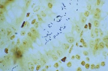
FIGURE 11-3 Helicobacter pylori organisms (dark rods) evident in the lumen of the intestine. (Warthin-Starry stain.)

(Courtesy Eun Lee, Lexington, Kentucky.)

Nonendoscopic laboratory tests include urea breath tests (UBTs), serologic tests, and, less commonly, H. pylori stool antigen tests. A UBT is a highly sensitive, noninvasive test that involves the ingestion of urea labeled with carbon-13 (13C) or carbon-14 (14C). Degradation of urea by the bacillus releases 13C or 14C in expired carbon dioxide.42 These tests are advantageous because they indirectly measure the presence of H. pylori before treatment and its eradication after treatment. Serologic testing is useful in determining current or past infection but is limited in documenting the eradication of H. pylori, because antibody titers persist after the organism has been eliminated. Upper gastrointestinal imaging is infrequently performed, because it lacks the sensitivity of biopsy.

Medical Management

Most patients with peptic ulcer disease suffer for several weeks before going to a doctor for treatment. If the peptic ulcer is confined and uncomplicated and H. pylori is not present, antisecretory drugs are administered (Table 11-1). If the patient is infected with H. pylori, inhibitors of gastric acid secretion and antimicrobial agents are recommended.43 Combination therapy is recommended because antisecretory drugs, such as histamine H2 receptor antagonists and PPIs, provide rapid relief of pain and accelerate healing, and antibiotics are effective in eradicating H. pylori. Combination treatment accelerates healing and produces an ulcer-free state in 92% to 99% of treated patients.39,40

TABLE 11-1 Antisecretory Drugs

image

Four-drug treatment regimens, including a PPI plus three antimicrobials (clarithromycin, metronidazole or tinidazole, and amoxicillin), or a PPI plus a bismuth plus tetracycline and metronidazole, provide the best results for persons with a peptic ulcer and H. pylori infection (Box 11-1). This four-drug regimen is recommended over the previously standard triple therapy (a PPI, amoxicillin, and clarithromycin) because of increasing antibiotic resistance.44 Therapy is given for 10 to 14 days, and eradication of infection should be confirmed afterward.

image

Box 11-1

Antimicrobial Regimens for the Treatment of Helicobacter Pylori Infection in Peptic Ulcer Disease

First-Line Therapy*

Proton pump inhibitor (or ranitidine bismuth citrate) twice daily plus 3 antimicrobials:

    Clarithromycin 500 mg twice daily plus
    Metronidazole 500 mg twice daily or tinidazole 500 mg twice daily plus
    Amoxicillin 1000 mg twice daily

Alternative First-Line Therapy*

Quadruple therapy: proton pump inhibitor twice daily, bismuth subsalicylate/subcitrate 120 mg four times daily, plus metronidazole 500 mg twice daily, tetracycline 500 mg four times daily

Data from Rimbara E, Fischbach LA, Graham DY: Optimal therapy for Helicobacter pylori, Nat Rev Gastroenterol Hepatol 8:79-88, 2011.

* Medications are given for 10 to 14 days. Clinicians are advised to select drugs on the basis of the presence or absence of antibiotic resistance.

image

Prior to 2000, more than 50% of patients with peptic ulcer disease experienced recurrences after treatment. Such recurrence was likely because regimens consisting solely of antisecretory drugs were the treatment of choice; however, these drugs alone do not eradicate H. pylori infection and are noncurative of peptic ulcer disease. Eradication of H. pylori with antibiotic treatment reduces the rate of recurrence of peptic ulceration by 85% to 100%.43,45 Reemergence of an ulcer usually is traced to the persistence of H. pylori after treatment because of inappropriate drug choice, discontinuance of drug therapy, lack of behavior modification, or bacterial resistance.1

In all patients who undergo peptic ulcer therapy, ulcerogenic factors (e.g., use of alcohol, aspirin or other NSAIDs, and corticosteroids; consumption of foods that aggravate symptoms and stimulate gastric acid secretion; persistent stress) should be eliminated to accelerate healing and limit the occurrence of relapse. Patients benefit from smoking cessation, in that perforation rates are higher in smokers, and continued smoking results in a higher relapse rate after treatment and lower rates of eradication of H. pylori.46 When H. pylori is successfully eradicated, cigarette smoking does not appear to increase the risk of recurrence.47

Elective surgical intervention (e.g., dissection of the vagus nerves from the gastric fundus) largely has been abandoned in the management of peptic ulcer disease. Today, surgery is reserved primarily for complications of peptic ulcer disease such as significant bleeding (when unresponsive to coagulant endoscopic procedures), perforation, and gastric outlet obstruction. On occasions when peptic ulcer disease is associated with hyperparathyroidism and parathyroid adenoma, surgical removal of the affected gland is the treatment of choice. Resolution of gastrointestinal disease occurs after abnormal endocrine function is terminated. Prototype protein-based vaccines against H. pylori continue to be investigated.48

Dental Management

Medical Considerations

The dentist must identify intestinal symptoms through a careful history that is taken before dental treatment is initiated, because many gastrointestinal diseases, although they are chronic and recurrent, remain undetected for long periods. This history includes a careful review of medications (e.g., aspirin and other NSAIDs, oral anticoagulants) and level of alcohol consumption that may result in gastrointestinal bleeding. If gastrointestinal symptoms are suggestive of active disease, a medical referral is needed. Once the patient returns from the physician and the condition is under control, the dentist should update current medications in the dental record, including the type and dosage, and should follow physician guidelines. Further, periodic physician visits should be encouraged to afford early diagnosis and cancer screenings for at-risk patients.

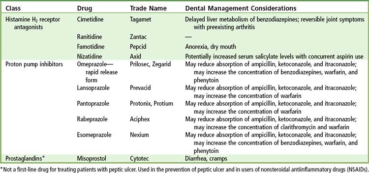
Of primary importance are the impact and interactions of certain drugs prescribed to patients with peptic ulcer disease (Box 11-2). In general, the dentist should avoid prescribing aspirin, aspirin-containing compounds, and other NSAIDs to patients with a history of peptic ulcer disease because of the irritative effects of these drugs on the gastrointestinal epithelium. Acetaminophen and compounded acetaminophen products are recommended instead. If NSAIDs are used, a cyclooxygenase-2–selective inhibitor (e.g., celecoxib [Celebrex]) given in combination with a PPI or misoprostol (Cytotec), 200 µg 4 times per day—a prostaglandin E1 analogue—is advised for short-term use to reduce the risk of gastrointestinal bleeding.4951 Analgesic selection should be based on patient risk factors (previous gastrointestinal bleeding, advanced age, use of alcohol, anticoagulants, or steroids), and the lowest dose for the shortest period to achieve the desired effect should be prescribed. Histamine H2 receptor antagonists and sucralfate are not beneficial selections because they do not appear to protect patients from NSAID-induced complications.52

image

Box 11-2 Dental Management

Considerations in Patients with Gastrointestinal (GI) Disease

P

Patient Evaluation/Risk Assessment (see Box 1-1)

Evaluate and determine whether GI disease or comorbid conditions exist.

Obtain medical consultation if patient’s disease is poorly controlled, if signs or symptoms appear suggesting an undiagnosed condition, or if the diagnosis is uncertain.

Potential Issues/Factors of Concern

A
Analgesics Avoid prescribing aspirin, aspirin-containing compounds, and other nonsteroidal antiinflammatory drugs (NSAIDs) for patients with a history of peptic ulcer disease or inflammatory bowel disease (IBD). Use acetaminophen-containing products or celecoxib (Celebrex) in combination with a proton pump inhibitor or misoprostol (Cytotec).
Antibiotics Selection of antibiotics for oral infections may be influenced by recent use of antibiotics for peptic ulcer disease; certain drugs can increase the risk of intestinal flareup in a patient with inflammatory bowel disease. Avoid long-term use of antibiotics, especially in elderly and debilitated persons, to minimize risk of pseudomembranous colitis. Monitor for signs or symptoms (diarrhea, GI distress) suggestive of pseudomembranous colitis or disease worsening. Contact patient’s physician if GI symptoms worsen while patient is on antibiotics, so that alternative therapies can be initiated.
Anesthesia No issues.
Anxiety Intraoperative sedation can be provided by an oral, inhalation, or intravenous route.
Allergy No issues.
B
Bleeding Use of acid-blocking drugs and proton pump inhibitors can enhance the blood levels of warfarin (Coumadin). Obtain complete blood count if medication profile increases patient risk for anemia, leu/>

Only gold members can continue reading. Log In or Register to continue

Stay updated, free dental videos. Join our Telegram channel

Jan 4, 2015 | Posted by in General Dentistry | Comments Off on 11: Gastrointestinal Disease

VIDEdental - Online dental courses

Get VIDEdental app for watching clinical videos