11: Diseases of Salivary Glands

Diseases of Salivary Glands

•. Developmental Disturbances

•. Saliva, Xerostomia, Hyposalivation, and Sialorrhea

•. Inflammatory Conditions of Salivary Glands

•. Viral-induced Salivary Gland Pathology

•. Non-inflammatory Conditions of Salivary Glands

•. Salivary Gland Tumors

•. Benign Tumors

•. Malignant Tumors

Salivary glands and saliva play a critical role in maintenance of oral and systemic health. Salivary glands are frequently involved in a wide array of conditions which result in glandular dysfunction. These may be subdivided into five categories which include:

Many of these conditions result in salivary gland hypofunction, enlargement, pain and facial nerve paresthesia that will prompt the patient to seek evaluation and treatment from the dentist. This chapter will address each of these five subgroups focusing on the etiology, clinical features, diagnosis and treatment of these conditions.

Developmental Disturbances

Aplasia/Agenesis and Related Aberrancy of Salivary Glands

Congenital aplasia or agenesis of the major salivary glands is an uncommon finding, characterized by partial or total lack of development of the gland. Agenesis may involve one gland, pairs, or multiple glands, and be unilateral or bilateral. It may be a single independent finding or be associated with other developmental anomalies.

Congenital agenesis of major salivary glands was first reported by Gruber in 1985. A recent search of the world literature reported 30 documented cases. Three cases of unilateral aplasia of the parotid have been reported. As it may be asymptomatic and go unnoticed, the true incidence is unknown. Bilateral agenesis is more common, with 10 cases reported in the English literature. Sometimes congenital absence of one parotid or submandibular gland is associated with hypertrophy of the contralateral gland.

Aplasia may occur as a single event or with autosomal dominant inherited developmental conditions such as first branchial arch anomalies, hemifacial microsomia and mandibulofacial dysostosis. Parotid gland aplasia may be associated with malformations of the lacrimal apparatus as well. A combination of multiple developmental anomalies is found in the lacrimo-auriculo-dento-digital (LADD) syndrome (Levy–Hollister syndrome). This is an autosomal-dominant multiple congenital anomaly disorder characterized by hypoplasia, aplasia or atresia of the lacrimal and salivary systems, ear anomalies, hearing loss, digital malformations and dental alterations. As the parotid gland develops during the 4th week of uterine life, and the submandibular and sublingual, and minor glands develop between 6th and 12th weeks, association of salivary gland aplasia with other congenital abnormalities is easily understood. Bilateral aplasia of the parotid gland has also been reported in a patient with Down’s syndrome. In ectodermal dysplasia, aplasia of the submandibular glands and alterations in salivary gland function have been reported as well.

Clinical features and diagnosis

Salivary gland agenesis may be asymptomatic if only partial agenesis occurs or if the condition is isolated to one gland. If more extensive involvement occurs, agenesis can produce profound hyposalivation in children resulting in advanced dental caries, candidiasis, ascending sialadenitis, and even laryngitis and pharyngitis. A child with subjective xerostomia and functional hyposalivation should be carefully examined for salivary gland dysfunction to include partial or complete salivary gland agenesis. Familial parotid gland aplasia has been reported, hence examination of the siblings of a child with agenesis, might be prudent. Sometimes, congenital absence of salivary glands is not noticed until adulthood resulting in unfortunate sequelae. However, clinical suspicion of salivary gland agenesis should be heightened in the setting of the ‘non-drooling baby’. Lack of complete development of the submandibular gland duct has been reported in two infants which presented as unilateral cystic swellings in the floor of the mouth. Both cases responded to simple incision and decompression of the fluid-filled ducts. Early treatment is important to avoid feeding difficulties and possible later complications such as ranula or sialadenitis.

Hyperplasia of Minor Salivary Glands

Clinical features

Adenomatoid hyperplasia of the minor salivary glands is a rare lesion characterized by localized swelling that clinically mimics a neoplasm. The lesion most often develops on the hard or soft palate, although it has been reported in other oral minor salivary gland sites. The most common presentation is during the 4th to 6th decades of life. The typical presentation is a painless, indolent palatal swelling which may be soft or firm to palpation. The overlying mucosa is usually normal in color, although some lesions are red or bluish in color. The etiology is uncertain. In one report, the occurrence of adenomatoid hyperplasia on the palate occurred in patients who were tobacco smokers or denture wearers or both. The authors, Barrett and Speight suggested that chronic, local trauma could be a factor in the development of the condition.

Saliva, Xerostomia, Hyposalivation and Sialorrhea

Saliva

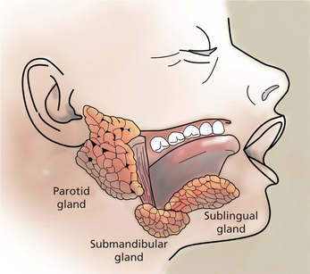
Saliva is an essential fluid for maintaining oral and systemic health and a satisfactory quality of life. While saliva is sometimes considered bothersome during restorative procedures, the alteration in quality and/or quantity of saliva has serious sequelae for the patient. Saliva is the product of three paired major salivary glands (the parotid, submandibular, and sublingual), and minor glands found in the hard and soft palate, lips, tongue, and buccal mucosa (Figure 1). Major salivary glands are composed of ductal and acinar structures. The acinar cells are the secretory components and ductal cells are the branching network that transports saliva into the oral cavity. The acinar cells of the parotid gland are purely serous elements and upon stimulation, parotid secretions account for at least half of the volume of whole saliva. Serous secretions are ‘watery’ and contain digestive enzymes, water, salts, and ions. They aid in mastication, clearing agents from the oral cavity, and facilitate swallowing and speech. Fluid from the parotid gland enters the oral cavity through the main excretory duct, Stensen’s duct, located in the buccal mucosa next to the maxillary second molars. In the submandibular gland, the acinar components are mixed mucous-serous, but predominantly serous. At rest, the submandibular gland secretions account for approximately two-thirds of the unstimulated saliva production. Wharton’s duct is the main duct of the submandibular gland and enters the floor of the mouth on either side of the lingual frenum. The sublingual gland, the smallest of the three major glands, is located above the mylohyoid muscle. Acinar elements are mixed, but predominantly mucous. The mucin produced facilitates lubrication and swallowing. The main excretory duct of the sublingual gland, Bartholin’s duct, may join Wharton’s duct resulting in a blending of secretions, or open into the oral cavity with a separate sublingual papilla. Numerous sublingual ducts may join the submandibular gland duct or open separately into the floor of the mouth. Minor salivary glands are named according to their location—lingual (anterior and posterior), labial, buccal, palatine and glossopalatine. Table 1 summarizes the nomenclature for the major and minor salivary glands and their acinar components.

Table 1

Major and minor salivary glands and secretion

Glands Type of secretion
Parotid Purely serous
Submandibular Mixed, however predominantly serous
Sublingual Mixed, however predominantly mucous
Lingual minor salivary glands  
Anterior lingual (glands of Blandin and Nuhn) Primarily mucous Purely serous
Posterior lingual (glands of von Ebner) Purely mucous
Labial and buccal minor salivary glands Mixed
Glossopalatine and palatine minor salivary glands (Weber’s glands) Purely mucous

In health, approximately 750 ml of saliva is produced in a day. The principal control of salivary secretion is mediated by sympathetic and parasympathetic innervation. The adrenergic (α and β) receptors are regulated by the sympathetic nervous system and the muscarinic receptors by the parasympathetic nervous system. At least five muscarinic-cholinergic receptor subtypes exist and it has been determined that the muscarinic receptor that medicates secretion of saliva is the M3 subtype. The parasympathetic nerve stimulation leads to an increased volume of saliva, whereas the sympathetic stimulation has an effect on protein content and salivary composition.

Saliva plays a major role in the local and systemic protection of the oral cavity, oropharyngeal region, and the upper gastrointestinal tract. Normal quantity and protein composition is essential for buffering the acidity of the oral cavity, lubrication of tissues, maintaining the integrity of the oral tissues, providing antimicrobial proteins and digestive enzymes, and remineralization of the teeth. Lack of saliva, hyposalivation, may increase a patient’s susceptibility to dental decay, oral ulcers, fungal infections, and dysphagia or difficulty in swallowing.

Saliva contains many antimicrobial proteins that help maintain a normal oral flora. The histatins provide antifungal properties, and proline-rich proteins help reduce bacterial colonization by modifying the ability of organisms to attach to tissues. Mucins are salivary glycoproteins that play a role in mucosal lubrication and assist in aggregation of oral microorganisms. Statherin and proline-rich proteins have calcium-binding properties to assist remineralization. These proteins combined with the cleansing and flushing ability of saliva constitute significant protective mechanisms for the oral cavity and upper gastrointestinal system. Of special importance to the dentition is the presence of buffers to help maintain a neutral pH and the remineralizing capacity. The pH range of saliva will vary from 6.7 to 7.4 in health. Of concern is pH below 5.5, which will promote enamel dissolution and increase caries susceptibility.

Hypofunction and Xerostomia

Altered saliva production, both qualitatively and quantitatively, creates significant oral health concerns for the patient and challenges the dentist to provide appropriate patient education and treatment. Patients frequently present with the complaint of a ‘dry mouth’. Xerostomia is the subjective sensation of dry mouth and may be determined by questioning individuals about their perceptions of oral dryness. The need to sip water while eating dry foods, difficulty in swallowing food without liquids, and a feeling of ‘too little’ saliva in the mouth have been correlated with salivary gland hypofunction. Hyposalivation is the production of inadequate or less than normal amount of saliva, usually from gland hypofunction. A few simple chairside assessments could be performed to quickly assess oral dryness. The lack of salivary pooling in the floor of the mouth is a basic clinical indicator of hyposalivation. Another indicator is the ‘cracker sign’. If a patient states that they cannot eat a cracker without drinking fluids, xerostomia and hyposalivation should be suspected. Another indicator is the ‘lipstick sign’. Patients with hyposalivation may have lipstick adhere to the incisal edges of the anterior teeth because they lack saliva that would normally prevent adherence. Another clinical clue to hypofunction is the ‘mouth mirror test’. The clinician places the mirrored surface of the mouth mirror over the buccal mucosa and attempts to move it. If the mirror glides effortlessly over the tissue, one can deduce that salivation is adequate.

The prevalence of xerostomia in the population varies widely due to the lack of a clear definition for xerostomia. Some clinicians use the terms ‘xerostomia’ and ‘hyposalivation’ interchangeably. Hyposalivation or salivary gland hypofunction results when the salivary gland flow rate is lower than normal. Defining ‘normal’ is problematic due to variations in the literature on collection methods, time of day collections were performed, and the inability to control for medications. Unstimulated whole salivary flow rates of less than 0.15 ml/min might provide a good reference for hyposalivation. The determination of hyposalivation and xerostomia do not consistently correlate as one might expect. Patients with salivary gland hypofunction are sometimes without complaints of oral dryness, or xerostomia. Conversely, patients with complaints of ‘dry mouth’ may not have reduced salivary flow rates. Xerostomia and salivary hypofunction affect the oral health-related quality of life. Reported reasons include the negative impact of oral dryness on speaking, eating, and wearing dental prostheses. Both xerostomia and salivary gland hypofunction require a careful systematic evaluation to determine causative factors.

Dry mouth has a variety of causes as indicated in Box 1. Common causes of salivary gland dysfunction leading to hyposalivation include medications, irradiation to the head and neck, and autoimmune diseases such as systemic lupus erythematosus (SLE) and Sjogren’s syndrome (SS). Other systemic conditions affecting salivary glands include sarcoidosis, sialadenosis, and viral infections such as HIV and hepatitis C.

Mouth-breathing, tobacco smoking, alcohol use, and consumption of beverages containing caffeine can contribute to oral dryness as well. Age-related hyposalivation has been reported due to structural changes in salivary glands with increasing age. Other studies have reported no age-dependent decrease in saliva flow rate in healthy elderly populations. The incidence of xerostomia will increase and the oral manifestations will continue to be a challenge for the clinician.

Treatment of xerostomia is symptomatic and supportive, but should be aggressively proactive in prevention of dental decay associated with salivary hypofunction. Patient education and patient empowerment so they become participants in their oral healthcare are key to successful outcomes. Hydration, dietary modifications and meticulous oral hygiene are especially helpful. Symptomatic treatment is aimed at minimizing the subjective complaints, and includes drinking water or finely crushed ice chips and sugar-free fluids. Patients should be advised that caffeine can contribute to the feeling of oral dryness and consequently, caffeine-free and sugar-free drinks should be strongly encouraged. Avoiding alcohol, including alcohol-based mouthrinses, spicy foods, and strong flavorings, such as cinnamon and mint, should also minimize oral discomfort. Artificial saliva is helpful for many patients due to the coating action, but does not provide long-lasting relief. Some patients find relief by chewing sugar-free gum or sucking on sugar-free candies. Lip balms and moisturizers may be helpful as well.

Because patients with hyposalivation are at increased risk for dental decay, oral applications of topical fluorides should be initiated. The fluoride is incorporated into the enamel of the teeth to increase resistance to demineralization and decay. Fluoride products are available over-the-counter as rinses and as more highly concentrated brush-on gels (0.4% stannous fluoride gel). Prescription products are available which provide 1.1% neutral sodium fluoride treatment in convenient brush-on gels or rinses. These should be used after regular toothbrushing in the evening just before retiring. Patients should be reminded to avoid rinsing, eating or drinking for 30 minutes following the fluoride application. Stannous fluoride or neutral sodium fluoride treatments can be provided using fluoride carriers, or custom trays fabricated from a cast of the patient’s mouth. The carriers are particularly helpful in treating severe hyposalivation resulting from head and neck radiation therapy. Preventive fluoride programs should be customized by the dentist to meet the specific needs and conditions of the patient.

Xerostomia and Salivary Gland Hypofunction due to Medications

Jacobsen and Chavez in their article describe that individual above 65 years of age comprise 13% of the population, but consume approximately one-third of all drugs prescribed. Loesche et al and Foc have reported many studies that have demonstrated a link between dry mouth in the elderly and polypharmacy. Shinkai et al reported that in a group of 1,163 adults (age range 32–81 years), 57% reported dry mouth to be the most frequent side effect of their medications. Hundreds of medications can cause subjective complaints of dry mouth and many induce hyposalivation. The drugs most frequently implicated in dry mouth include the tricyclic antidepressants, antipsychotics, atropinics, beta blockers and antihistamines. In another study as reported by Thomson, the unstimulated (resting) salivary flow rate was reduced among individuals who were older, female, or taking antidepressants, and higher among smokers or people who were taking hypolipidemic drugs. Other studies have correlated dry mouth with the number of medications taken, rather than specific types of medications. Field et al have demonstrated that medication is a better predictor of risk status for dry mouth than either age or gender. Medications that induce salivary hypofunction are a concern among the elderly, but xerostomia can also be a concern for young adults. Thomson et al in his study involving a large cohort of 32-year-old subjects, reported that the prevalence of xerostomia was found to be 10%, with no apparent gender difference. There was a strong association between xerostomia and diminished oral health-related quality of life. Xerostomia was significantly higher among those taking antidepressants, iron supplements, or narcotic analgesics, and those subjects taking antidepressants at both 26 and 32 years of age demonstrated 22 times the odds of reporting xerostomia.

A study of medications and caries among older individuals by Thomson et al revealed that an adjusted coronal caries increment (AdjCI) was higher among males and those taking a beta blocker or an anti-asthma drug for the previous 5 years. Several classes of drugs have been associated with dry mouth (Box 2). The most common groups include those with: (i) anticholinergic effects such as tricyclic antidepressants, drugs for urinary retention and over-active bladder, antipsychotics, diuretics, and antihistamines; (ii) sympathomimetic drugs such as antihypertensives, anti-depressants, decongestants, and bronchodilators; (iii) skeletal muscle relaxants; (iv) benzodiazepines; (v) proton pump inhibitors; and (vi) anti-migraine agents. Dry mouth has been reported to be a consequence of cytotoxic drugs such as 5-fluorouracil. Medications used for treatment of HIV have also been associated with dry mouth. These include didanosine and protease inhibitors.

It is important for the dentist to inquire about all medications, both prescription and over-the-counter agents, during the health history review. Sometimes the dentist can provide assistance by sharing concerns of medication-induced hyposalivation with the primary care physician. In some situations, the dentist might suggest medications such as pilocarpine or cevimeline to promote salivation. These medications will be discussed in the section on Sjögren’s syndrome.

Xerostomia and Salivary Gland Hypofunction due to Radiation Therapy

The role of saliva in oral health and function relates both to its fluid characteristics and to its specific components. The water (fluid) phase accounts for 99% of the volume and the remainder is salts and proteins. Essential functions of these components include flushing, buffering the acidity, and mucosal coating to maintain tissue integrity. Mucosal coating and tissue lubrication are essential for speech, taste perception, mastication, and swallowing. Cancer patients undergoing treatment for head and neck disease often experience severe difficulties maintaining such functions. Salivary function can also be diminished by radioiodine therapy for thyroid carcinoma, especially if multiple doses are administered. Deterioration of oral health and radiation-induced oral dryness has a significant influence on patients’ overall quality of life during and after treatment.

Together with surgery, radiation therapy is the main treatment for head and neck tumors. All or part of the major and minor salivary glands may be included within the radiation field due to the site and extension of the tumor and the lymphatic spread. Exposing the glands to radiation often results in severe salivary gland hypofunction and changes in saliva composition. A profound decrease in salivary flow occurs during the 1st week of radiation therapy and continues throughout the course of therapy. In general, fully irradiated parotid glands exposed to doses exceeding 60 Gy sustain permanent damage resulting in severe hypofunction, and there is no recovery of gland function over time. Partial salivary flow recovery may occur at lower doses.

Radiation mucositis may begin during the 2nd week of therapy. Primary sites are intraoral mucosal surfaces within the direct portals of radiation. A whitish discoloration will appear from keratin accumulation, which is followed by sloughing, revealing atrophic erythematous, and friable mucosa. Ulceration then develops producing burning and pain which is exacerbated by eating and oral hygiene. Symptomatic support for radiation mucositis is important to assist the patient in maintaining adequate nutrition.

The mucositis will slowly resolve 2–3 weeks after cessation of the treatment. A loss of taste has been reported which generally is recovered after 6 months.

Minimizing the probability of osteoradionecrosis (ORN) includes a dental examination at least 2 weeks prior to the initiation of radiation therapy to address or remove teeth that have a hopeless long-term prognosis. Daily fluoride treatments in custom carriers and close follow-up aid in reducing the incidence of xerostomia-induced dental caries. Due in part to more efficient radiation techniques, the incidence of ORN has been declining in radiation patients over the last two decades. Advances in radiation techniques, including the use of fractionated radiation doses, have minimized the incidental damage to adjacent tissues. Furthermore, use of three-dimensional dosimetric intensity-modulated radiation therapy (IMRT) has been shown to reduce late salivary toxicity, since the portion of tissue exposed to low radiation doses has a potential for repair. Based on recent publications, the prevention of ORN remains controversial. A recent report compiled by Chang et al after reviewing a large series of ORN studies stated that extraction of teeth with poor prognosis before radiation therapy did not appear to reduce the risk of ORN. The investigation of IMRT by Wu et al to achieve sparing of the parotids and yet achieve higher tumor control appears to show promise. Until additional evidence is available to define guidelines, a pre-radiation referral for a dental evaluation is necessary. To facilitate prevention of ORN, irradiated dental patients should maintain a high level of oral health. Pre – and post-therapy close collaboration by a multidisciplinary team can be invaluable for patients receiving head and neck radiation therapy.

Sjögren’s Syndrome

Sjögren’s syndrome (SS) is a chronic autoimmune disease affecting the exocrine glands, primarily the salivary and lacrimal glands. Patients most commonly complain of a subjective persistent feeling of dry mouth (xerostomia) and of dry eyes (keratoconjunctivitis sicca). This is due to lymphocytic infiltrates and destruction of salivary and lacrimal glands and systemic production of autoantibodies. In 1933, Henrik Sjögren, a Swedish ophthalmologist, presented his doctoral thesis entitled ‘Zur Kenntnis der Keratoconjunctivitis Sicca’, and described the clinical and histopathological aspects of the disease.

Sjögren’s syndrome occurs worldwide and while it may occur at any age, the peak incidence is between 40 and 50 years. Sjögren’s syndrome has one of the highest female-to-male ratio (9:1) of any autoimmune rheumatic disease. In addition to ocular and oral dryness, a wide spectrum of extraglandular manifestations may occur as well. The musculoskeletal, hematological, vascular, pulmonary, gastrointestinal, dermatological, renal and nervous systems may be involved. Patients with SS have an increased risk of developing lymphoma. Early reports estimated that patients with SS had up to 44 times increased risk of developing lymphoma compared with the general population. Chronic fatigue, depression, and a diminished quality of life are also common components of SS.

Classification

Two forms of the disease are recognized. Primary SS is the presence of sicca syndrome, xerostomia, or ‘dry mouth’, and xerophthalmia, or ‘dry eyes’ together, with no other autoimmune disease. Secondary SS is sicca syndrome plus another associated autoimmune disease such as rheumatoid arthritis (RA), SLE, or scleroderma. Based upon the classification criteria applied, the prevalence of SS may range from 0.5 to 3.0% of the population. All classification systems use a combination of both subjective and objective findings in the diagnostic process. The most recent 2002 criteria include subjective symptoms of dry mouth and dry eyes, and the following objective tests: ocular signs by Schirmer’s test and/or Rose Bengal score; focal sialadenitis by histopathology; salivary gland involvement by either salivary scintigraphy, parotid sialography or unstimulated salivary flow rate; and autoantibodies of SS-A/Ro and/or SS-B/La specificity. Box 3 presents the 2002 American-European Consensus Group Criteria for SS.

Box 3   European American Consensus Group criteria for Sjögren’s syndrome

Ocular symptoms (must have 1 of 3)

Oral symptoms (must have 1 of 3)

Ocular signs (1 of 2)

Histopathology in minor salivary gland biopsy

Salivary gland involvement (1 of 3)

Autoantibodies

For primary SS

For secondary SS

Exclusion criteria

Source: Vitali et al. Annals of Rheumatic Diseases 2002;61:554-58.

Etiopathogenesis

Currently, the etiology of SS is not clearly understood, but appears to be multifactorial. It has been suggested that environmental agents may trigger SS in genetically predisposed individuals. Experimental and clinical evidence suggest that immune reactivity is modulated by gender. Immune reactivity is higher in females than males, and lymphocytes and monocytes from female subjects show higher antigen presenting activity and mitogenic responses. Taiym et al have found higher prolactin levels in SS patients than controls. Estrogens might be pro – or anti-inflammatory, based on dose-related metabolite conversions. The role of sex hormones in rheumatic autoimmune diseases has yet to be clarified. Potential mechanisms underlying SS include disturbances in apoptosis, circulating autoantibodies against the ribonucleoproteins Ro and La or cholinergic muscarinic receptors in salivary and lacrimal glands or cytokines. These processes interfere with normal glandular function; and as the mucosal surfaces become sites of chronic inflammation, the disease appears to enter a self-perpetuating inflammatory cycle.

While a genetic predisposition to SS appears to exist, no simple Mendelian inheritance pattern has been demonstrated. Cases of two or more individuals with SS per family and SS in twins have been described. However, the level of genetic contribution is not known. Because large twin studies in SS are lacking, the twin concordance rate cannot be estimated. Familial clustering of different autoimmune diseases and co-association of multiple autoimmune diseases has been reported by Becker et al. Reports have also indicated that a SS proband may have relatives with other autoimmune diseases in approximately 30–35% of the cases. Assessing human leukocyte antigen (HLA)-DR and HLA-DQ gene segments in patients with SS reveals an increased use of haplotypes B, Drw52 and DR3. Correlations have been found between presence of HLA-DR haplotype and the presence of Ro/LA in SS. Gene polymorphisms have been analyzed, but no clear-cut relationship between these and primary SS have been identified. Clustering of non-major histocompatibility complex (MHC) susceptibility candidate loci in human autoimmune diseases supports a hypothesis that, in some cases, clinically distinct autoimmune diseases may be controlled by a common set of susceptibility genes.

Clinical features and diagnosis

Dry mouth and dry eyes are the most common complaints of patients with SS. Patients’ ocular concerns may include a history of dry eyes, a sensation of sand or gravel in the eyes, and/or frequent (> 3 times per day) use of tear substitutes. Objective ocular tests for dry eyes include a Schirmer’s test for tear production. Using sterile strips of filter paper placed just inside the lower lid, tear production can be assessed by measuring the length of wetness on the filter paper. Less than 5 mm of wetness in 5 minutes, without local anesthesia is considered a positive test, meeting the 2002 Sjögren’s consensus criteria. A second ocular test utilizes Rose Bengal staining of the cornea, and measures areas of increased dye intensity. With the use of a slit-lamp, the stain will detect devitalized tissue. The ocular dryness is sufficient to produce disruption in the integrity of corneal and conjunctival epithelium and dye will accumulate in these areas. The total areas identified are used to determine the extent of ocular damage. Patients with complaints of ocular dryness, especially if combined with other symptoms of SS, should be referred to the ophthalmologist for evaluation. Untreated keratoconjunctivitis sicca can progress to corneal ulcerations and even blindness.

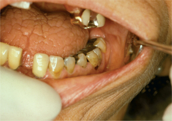
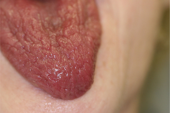
The most frequent oral signs and symptoms a dentist will encounter are xerostomia, a subjective sensation of oral dryness, and hyposalivation, or a diminished salivary flow rate. These will vary between patients, and the subjective dryness may not directly correlate with objective measures of hyposalivation. Initial indications of a diminished salivary output would be a lack of pooling in the floor of the mouth, thick or frothy saliva, and observing examination gloves sticking to the tongue or buccal mucosa. Patients may complain of difficulty chewing and swallowing, difficulty wearing their dentures, and altered taste. They will relate the necessity for drinking liquids to aid in swallowing food or to enhance their ability to speak. They may admit to keeping water by their bedside at night or frequently waking with a dry mouth. Patients with SS frequently carry water with them and often need to sip every 10–15 minutes during a consultation appointment. Upon intraoral examination, the tongue may appear fissured, slightly erythematous, and sometimes depapillated. If the patient complains of a ‘burning tongue’ and dysgeusia, oral candidiasis should be suspected (Figure 2). Salivary flow rate is diminished in primary SS patients compared with controls. In addition to quantitative changes, the protein content of the saliva is altered as well. Proteins necessary for buffering the oral acidity, countering fungal and microbial organisms may be altered. The lack of adequate salivary flow and qualitative changes in protein content may predispose the patient to dental decay, particularly in the cervical area, tooth loss, candidiasis and oral ulcerations (Figure 3). The change in saliva is linked to the lymphocytic infiltrate in the glands and subsequent damage to the functional units.
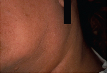
From one-fourth to two-thirds of patients with primary SS will have a diffuse enlargement of the major salivary glands during the course of their disease (Figure 4). This swelling may be unilateral or bilateral, intermittent or constant in nature. If the parotid swelling initially is unilateral, it often becomes bilateral with time. It commonly produces mild to no discomfort. However, diminished flow will enhance a patient’s susceptibility to bacterial infection in the glands and recurrent sialadenitis, which will produce pain. Although SS is considered primarily a disease of middle-aged females, it has also been reported in children and adolescents. Recurr ent bilateral parotitis in children and adolescents should include the possibility of SS in the differential diagnosis.

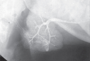
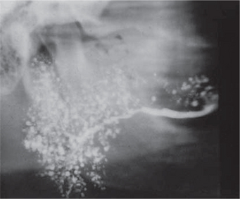
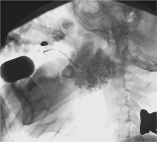
Conventional sialography renders useful information about the gland architecture and changes within it. Water-based radiopaque dye is injected into the major gland ducts followed by conventional imaging. Peripheral ducts within the glands are usually affected first with the inner ductal structure relatively well preserved. However, punctuate collections of the contrast material may be visible in the early stages of the disease followed by globular or larger collections as the condition progresses as shown in Figures 57. Once extensive intraglandular destruction has taken place and infection has become established, dilatation of central ducts is noted. Abscesses within the gland may be noted with a uniform distribution, unlike focal abscesses caused by other types of infections. However, sialography is an invasive procedure and other imaging modalities may be considered.
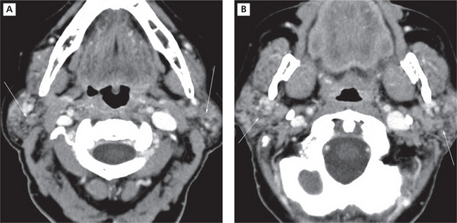
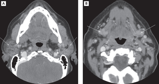
Imaging with CT and MRI is common. The glands enlarge with time, assuming a denser appearance on CT. A honeycomb appearance is not infrequent but this is also seen with granulomatous conditions. Bilateral enlargement with cystic and solid lesions is noted (Figure 8A, B). In the early stage, the parotids appear normal. Multiple cysts appear during the intermediate stage. These eventually grow with time. If the mass assumes an invasive appearance, malignant transformation may be suspected. Foci of calcification within the glands are not uncommon. Heterogeneous enhancement may therefore be expected (Figure 9A, B). The glands could eventually appear smaller in size. In MRI, numerous punctuate areas appear within the glands with low signal intensity on T1 and T2 weighted images as the disease progresses. This is considered diagnostic of the condition. But, a lymphoma arising in a benign lymphoepithelial lesion (BLEL) does not have characteristic features that would help differentiate it from other tumors. However, it may be considered in patients who present with a parotid mass. Function of the glands is directly correlated with the amount of fat deposition, thereby indicating that a monitoring of this feature may be useful in diagnosing the condition. Another imaging technique that is useful is magnetic resonance sialography. It has been shown to be highly accurate with excellent sensitivity and specificity. Globular, punctuate or a lytic appearance is typical of the condition. If cysts develop within BLEL, these are detected using CT or MR. The absence of lymphadenopathy helps exclude HIV-related lymphoepithelial cyst formation.
Another test to confirm altered salivary gland function is whole unstimulated sialometry. Sialometry is the measurement of salivary flow rate. It is instrumental in reaching a diagnosis of hyposalivation (below normal salivary flow rate), a common finding in patients with SS. In an ideal situation, patients should not eat, drink, smoke, or brush their teeth for 90 minutes before the sialometric assessment. The patient is asked to expectorate into a preweighed container, while sitting upright for 15 minutes. After re-weighing the tube post-collection, dividing by 15 and applying the conversion factor (1 g = 1 ml), a flow rate can be determined. Unstimulated salivary flow ≤ 1.5 ml/15 min or < 0.1 ml/min is considered consistent with Sjögren’s diagnosis. After an SS diagnosis has been established, periodic flow rates (every 3–6 months) may provide meaningful information to assess disease progression. Subsequent salivary assessments should be collected at the same time each sampling, preferably either 9:00 AM-noon or 1:00 PM–3:00 PM, to avoid fluctuations due to circadian rhythm of salivary secretion and composition. Often, an SS patient will be prescribed a sialogogue to enhance salivary flow. Periodic flow rates are helpful to track medication efficacy as well.

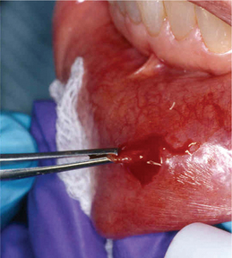
Lymphocytic infiltration in the lacrimal and salivary glands is a major feature of SS. One of the criteria for classification of SS includes the biopsy of the labial salivary glands. A 1.5-cm incision is made on normal appearing mucosa of the lower lip lateral to the midline (Figure 10). Five or more accessory salivary gland lobules are examined histopathologically for the presence of focal chronic inflammatory aggregates. A focus is 50 or more lymphocytes and some plasma cells within a 4 mm2 field in the salivary gland biopsy specimen. As noted in Figure 11A, B, these aggregates are adjacent to normal-appearing acini and are found throughout the glands. A finding of more than one focus within a 4 mm2 area of glandular tissue is supportive of a SS diagnosis. Manthorpe et al have reported an interesting fact that the focus scores are lower in SS patients who are cigarette smokers.

Stay updated, free dental videos. Join our Telegram channel

Jan 12, 2015 | Posted by in Oral and Maxillofacial Radiology | Comments Off on 11: Diseases of Salivary Glands

VIDEdental - Online dental courses
Get VIDEdental app for watching clinical videos