10: Temporomandibular Joint Disorders

Temporomandibular Joint Disorders

Care of the patient with temporomandibular joint (TMJ) disorders is difficult. At present there is no formal TMJ specialty. There are TMJ interest groups, but many of the members of these groups are not surgeons. Although the majority of patients with temporomandibular joint dysfunction (TMD) are treated nonsurgically, it is clear that oral and maxillofacial surgeons are in a unique position to treat TMJ disorders; they can provide the full scope of treatments, ranging from occlusal guards to total TMJ replacement.

Disorders of the TMJ can result in debilitating pain and limited function. Although TMD includes a broad spectrum of disease states, it can be classified into two general categories: intracapsular disease (internal derangement or ankylosis) and extracapsular disease (myofascial pain dysfunction). Most cases of TMD can be managed nonsurgically with conservative therapy. Accurate diagnosis of the etiology of TMD is paramount for avoiding unwarranted invasive treatment.

The teaching cases in this chapter cover identification and management strategies for internal derangement, myofascial pain disorder, degenerative joint disease, and ankylosis of the temporomandibular joint. Arthrocentesis and arthroscopy also are discussed. The distinction between intracapsular and extracapsular TMD is emphasized. As in many complex disorders, the majority of information is obtained from the patient’s presenting complaint and history of symptoms. In these cases the key features of the chief complaint (CC) and the history of the present illness (HPI) are emphasized. The significant findings in the physical examination are highlighted, along with explanations of these findings.

Although nonsurgical management strategies are more consistent between individual practitioners, various surgical strategies have been used based on surgeons’ preferences and the clinical presentation. Some of the advantages and disadvantages of different treatment modalities are outlined. Surgical options are discussed, along with the rationale for treatment and the relative success rates. Reconstructive strategies for advanced disease states are also presented.

Myofascial Pain Dysfunction

HPI

The patient reports that the pain is dull and, while often present upon awakening, it continues to worsen throughout the day (characteristic of MPD). When asked to point to the regions of pain, she readily identifies the areas over her temporalis and masseter muscles. She has difficulty falling asleep and wakes up frequently throughout the night. The pain is worse when eating food, especially when chewing tough foods, such as steak (increasing pain and muscle fatigue during mastication are typical findings in MPD). She has modified her diet by excluding hard and chewy foods to reduce the pain and discomfort associated with eating. She reports no history of migraines but admits to a high level of stress at work (work- or home-related stress can exacerbate MPD). She denies any prior history of temporomandibular joint dysfunction and is not aware of any parafunctional habits such as bruxism. (Bruxism is grinding of the teeth. Many patients may be unaware of nocturnal bruxism, unless reported by their bed partner. Also, patients with nocturnal bruxism are characteristically worse on waking and improve over the course of the day).

Examination

General. The patient is a well-developed and well-nourished, anxious woman.

Maxillofacial. There is no facial swelling or asymmetry. On palpation, there is tenderness of the temporalis, masseter, and sternocleidomastoid muscles bilaterally (temporalis and masseter muscles are most commonly involved in MPD). There is no TMJ capsular tenderness and no clicks or crepitus (making an intracapsular source of pain less likely). The patient has a maximal incisal opening of 28 mm (less than normal) with a soft end feel, which can be stretched to 39 mm with pain (limited opening due to muscle guarding that can be slowly stretched to a normal opening is consistent with MPD). Her left and right lateral excursions are 9 and 8 mm, respectively (normal condylar translation makes TMJ internal derangement less likely). The remainder of her physical examination is noncontributory.

Treatment

The treatment of MPD begins with the correct diagnosis. The etiology of MPD is multifactorial; therefore, the management of MPD requires a multimodal approach. Initially, the patient should be reassured that the pain is purely myofascial and likely to be the result of increased muscle activity secondary to any of a number of entities. These may include stress, anxiety, bruxism, clenching, malocclusion, parafunctional oral habits, internal derangement of the TMJ, rheumatologic diseases (polymyalgia rheumatica), fibromyalgia, and vasculitis (e.g., temporal arteritis). Treatment must address the etiology; however, given the difficulty associated with correct diagnosis, the approach is often generic.

Conservative therapy is generally the first-line treatment unless other identifiable associated diagnoses (tumors, infection, severe internal derangements, degenerative joint disease) are present that are thought to exacerbate the symptoms of MPD. Treatment options include reassurance, stress management (relaxation exercises, biofeedback), occlusal splint therapy, physical therapy, application of heat to affected muscles, nonsteroidal antiinflammatory drugs (NSAIDs), muscle relaxants, and anxiolytics (anxiolytics should be prescribed with caution due to abuse potential). Conservative treatment often results in significant improvement in or resolution of the MPD.

Patients who do respond to conservative therapy with an occlusal splint and have a significant malocclusion may be considered for orthodontic treatment or orthognathic surgery (there is some evidence that malocclusion may be associated with MPD). These modalities may offer a long-term solution to MPD, but they are invasive and not without complications.

Trigger point injections may be beneficial in a select group of patients with MPD who are refractory to all conservative approaches. Typically, a local anesthetic (with or without a steroid) is injected directly into tender areas in the muscles. This can be repeated as often as necessary. It may also be possible to improve MPD with injection of botulinum toxin into the muscle to reduce muscle activity. This may need to be repeated every 3 to 6 months, due to the temporary effect of the botulinum toxin. Regeneration of the nerve endings at the motor end plate of the neuromuscular junction is responsible for cessation of the clinical effects. Excessive muscle activity alone may not explain the majority of cases of MPD, and the response to botulinum toxin is not predictable. Intraarticular procedures, including arthrocentesis, arthroscopy, and arthroplasty, have no place in the management of isolated MPD.

The current patient was encouraged to manage her life stressors more effectively by taking stress management classes and using biofeedback to reduce muscle tension. She was instructed to avoid hard-to-chew foods and to apply moist heat (using a warm, moist towel) to the affected muscles as often as necessary for symptomatic relief. A short course of ibuprofen 800 mg three times a day was prescribed (a muscle relaxant can be added to this regimen). A hard, flat-plane maxillary occlusal splint was constructed, and the patient wore this at all times, except while eating or brushing her teeth. The splint was adjusted weekly to ensure good contacts in centric relation and no interferences during lateral excursions. Splint adjustments became less frequent as the occlusion stabilized, and by 2 months the patient was wearing the splint only at night and was able to open to 44 mm, with complete resolution of her pain.

Complications

With conservative (nonsurgical) approaches to the treatment of MPD, complications are relatively uncommon and are mostly related to the failure of available treatments to alleviate pain, the side effects of medications, or difficulties with occlusal splint therapy.

NSAIDs are often helpful and carry no risk of physiologic dependence, although gastrointestinal irritation and/or bleeding, platelet dysfunction, and decreased renal function are potential complications. The use of some muscle relaxants and anxiolytics can be associated with dependence and abuse, which are compounded by the frequently chronic and recurrent nature of MPD.

Occlusal splint therapy is not without complications (especially when the splint is inappropriately designed). Several different types of splints are used by prescribing clinicians, and unfortunately, there are no clear evidence-based guidelines for splint therapy. Different splints include maxillary, mandibular, flat-plane, anterior repositioning, and pivotal splints. Flat-plane occlusal splints, whether maxillary or mandibular, are the most popular and technically the least demanding. Although complications related to conservative splint therapy are uncommon, an incorrectly adjusted splint can result in exacerbation of the preexisting TMJ dysfunction, tooth movement, and/or the development of new symptoms. Anterior repositioning splints are occasionally useful in patients with Class II malocclusions and function by holding the mandible in a forward position; this unloads the richly innervated retrodiscal tissue within the TMJ and helps to reestablish a more normal disk-condyle relationship. These splints are likely to be associated with permanent occlusal changes, and considerable clinician experience is required in their use. Pivotal splints are rarely used and are thought to function by decreasing masticatory muscle forces (via periodontally mediated biofeedback).

After splint therapy, changes in the occlusion are not uncommon. Before splint therapy, most patients have a centric occlusion–centric relation discrepancy. A flat-plane occlusal splint may eliminate this discrepancy over time, resulting in a less than ideal occlusion when the splint is removed or discontinued. This may necessitate continued splint therapy, occlusal adjustment, orthodontics, or orthognathic surgery.

Discussion

Myofascial pain disorder of the masticatory muscle system is the most common of all temporomandibular joint disorders. The main muscles of mastication are the temporalis, masseter, lateral pterygoid, and medial pterygoid muscles. They all function harmoniously during speech and deglutination. As with any group of muscles, they are susceptible to inflammation, which may in turn cause pain. This is commonly due to excessive activity of these muscles, but the exact pathophysiology has not been clearly defined. The causes of muscle hyperactivity are many and may include malocclusion, parafunctional habits, TMJ internal derangement, cervical pain, and psychological stressors. Management of the acute symptoms of MPD is generally similar, regardless of the etiology, but long-term treatment and success need to address any known precipitating or etiologic factors. As is often the case, no definitive factors can be identified; consequently, a generic approach using several modalities must be adopted.

Bibliography

Giannakopoulos, NN, Keller, L, Rammelsberg, P, et al. Anxiety and depression in patients with chronic temporomandibular pain and in controls. J Dent. 2010; 38:369–376.

Graff-Radford, SB. Temporomandibular disorders and headache. Dent Clin North Am. 2007; 51:129–144.

Hersh, E, Balasubramaniam, R, Pinto, A. Pharmacologic management of temporomandibular disorders. Oral Maxillofac Surg Clin North Am. 2008; 20:197–210.

Klasser, G, Greene, C. Oral appliances in the management of temporomandibular disorders. Oral Surg Oral Med Oral Pathol Oral Radiol Endod. 2009; 107:212–223.

Kurtoglu, C, Gur, OH, Kurkcu, M, et al. Effect of botulinum toxin-A in myofascial pain patients with or without functional disc displacement. J Oral Maxillofac Surg. 2008; 66:1644–1651.

Okeson, JP, Leeuw, RD. Differential diagnosis of Temporomandibular disorders and other orofacial pain disorders. Dent Clin North Am. 2011; 55:105–120.

Schmitter, M, Kress, B, Leckel, M, et al. Validity of temporomandibular disorder examination procedures for assessment of temporomandibular joint status. Am J Orthod Dentofacial Orthop. 2008; 133:796–803.

Scrivani, SJ, Keith, DA, Kaban, LB. Temporomandibular disorders. N Engl J Med. 2008; 359:2693–2705.

Vedolin, G, Lobato, V, Conti, P, et al. The impact of stress and anxiety on the pressure pain threshold of myofascial pain patients. J Oral Rehabil. 2009; 36:313–321.

Internal Derangement of the Temporomandibular Joint

Examination

General. The patient is a well-developed and well-nourished woman in no apparent distress.

Maxillofacial. The right external auditory meatus is patent, without evidence of erythema or exudate. The tympanic membrane is normal. There is no TMJ capsular tenderness. An opening click (caused by the condyle translating and recapturing a normal position beneath the disk) and a reciprocal click (a second click that occurs during closure of the mandible with anterior displacement of the disk) are evident within the right TMJ to both lateral capsular and endaural palpation. Auscultation over the TMJ reveals a harsh opening click and a softer closing click. No crepitation is present (crepitus would be suggestive of disk perforation with degeneration of the condyle and glenoid fossa). The left TMJ clinical examination is within normal limits. There is no evidence of masticatory muscle tenderness (masseter and temporalis muscles). The patient has an initial interincisal opening of 22 mm with a right-sided deviation (due to restricted right condylar translation), followed by a right TMJ click and correction of the deviation (as the anteriorly displaced right disk is recaptured). The maximum interincisal opening is 44 mm. The Mahan test is bilaterally negative (biting on a tongue depressor on one side, eliciting pain in the contralateral TMJ, is a positive test result that suggests intracapsular pathology). The patient is noted to have a Class II division II malocclusion (may be associated with an increased incidence of TMD). The remainder of her clinical examination is unremarkable.

Imaging

The panoramic radiograph is the initial screening study of choice for assessment of TMDs, especially when pain is present (to assess pain of odontogenic origin). It provides a general overview of the bony morphology of the mandible and condyle. Magnetic resonance imaging (MRI), in open and closed mouth positions, is considered the standard when evaluating for TMJ internal derangement. It provides the most information regarding the soft tissue structures and disk position. A TMJ arthrogram (fluoroscopy with dye injected into the superior joint space) is an invasive procedure that shows the disk in dynamic function and is the only study that can readily identify disk perforations. Arthrograms can also be used to evaluate disk position, but the study is technique sensitive and is not readily available in most institutions. Computed tomography (CT) scans are indicated when bony or fibrous ankylosis of the TMJ or other bony pathology is suspected.

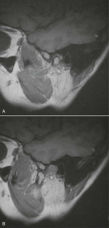
In the current patient, no osseous or dental abnormalities were seen on the panoramic radiograph. Sagittal and coronal MRI scans showed an anteromedially displaced (most frequent location of a dislocated disk) right TMJ disk in the closed mouth position (Figure 10-1, A), which reduced to a normal anatomic relationship in the open mouth position (Figure 10-1, B). The disk demonstrated a normal morphology (anatomy is best seen with T1-weighted images). No joint effusion was seen (inflammation and effusions are best evaluated with T2-weighted images).

Assessment

Internal derangement of the right TMJ; a nonpainful anterior disk displacement (ADD) with reduction of the right TMJ.

Patients with ADD with or without reduction may present with or without pain originating from the joint itself or from the muscles of mastication (i.e., myofascial pain dysfunction [MPD]). ADD without reduction presents with different clinical findings, including no opening or closing click, and potentially with restricted condylar translation on the affected side (reduced lateral excursion to the contralateral side). The MRI scan would demonstrate anterior displacement of the disk with no evidence of disk recapture during opening. It is not uncommon for MPD to accompany a painful internal derangement of the TMJ. It is important to distinguish between internal derangement and MPD, because their treatment is very different. MPD may also present as the sole source of pain, which warrants proper diagnosis to avoid unnecessary and inappropriate surgical management (see the section Myofascial Pain Dysfunction earlier in this chapter).

The Wilkes classification system for internal derangement of the TMJ characterizes progression of the disease as having five stages, based on the clinical, radiographic, anatomic, and pathologic features (Table 10-1).

Table 10-1

Wilkes Classification System for Internal Derangement of the TMJ

image

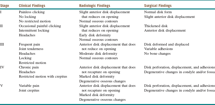
Modified from Wilkes CH: Internal derangement of the temporomandibular joint: pathological variations, Arch Otolaryngol Head Neck Surg 115:469-477, 1989.

Treatment

Treatment of internal derangement is generally guided by the presence of pain and/or limited function. In the absence of symptoms, active treatment may be avoided or minimized, as long as adequate patient education and reassurance are provided.

Conservative (reversible or nonsurgical) treatment is generally the first line of therapy in symptomatic patients. Such treatment includes splint therapy; a soft, nonchewing diet; elimination of parafunctional habits (bruxism); warm, moist compresses; physical therapy; nonsteroidal antiinflammatory drugs (NSAIDs); and muscle relaxants. However, protocol-driven treatment should be avoided, and individualized patient assessment is necessary. Undue delay with conservative therapy, when an effective surgical solution is indicated, can be counterproductive, resulting in a delay in resolving the problem and further frustration for the patient and clinician.

Patients who are unresponsive to conservative therapy or who present with advanced disease are candidates for various invasive (surgical) interventions. Surgical options include arthrocentesis, arthroscopy, arthroplasty with disk plication, meniscectomy (with or without autogenous, allogeneic, or alloplastic graft/replacement), modified condylotomy, and total joint replacement.

Arthrocentesis is accomplished by irrigating and distending the superior joint space with lactated Ringer’s solution, removing inflammatory mediators, and improving joint mobility by lysis of immature adhesions. A steroid or hyaluronic acid injection may follow, particularly if pain is a significant component of the patient’s complaint.

Arthroscopy is a more invasive procedure, often requiring general anesthesia, but it enables the surgeon to visualize, irrigate, and lyse adhesions within the superior joint space. Arthroscopy is reported to have a success rate approaching 80%. Coblating instruments and the holmium : YAG laser can be used during arthroscopy for lysis of adhesions, discectomy, and partial synovectomy of the inflamed synovium. Arthroscopy may be expected to produce a similar reduction in pain but a greater increase in the mean interincisal opening, compared with arthrocentesis.

Open joint arthroplasty with disk plication, the most conservative open technique, involves mobilizing the anteriorly displaced disk and plicating it posteriorly (with sutures or titanium anchors) to ensure that it rests in the correct anatomic position on the mandibular condyle. Success rates approaching 90% have been reported.

Meniscectomy involves removal of the disk, and although it readily eliminates the disk displacement, it may be associated with significant degenerative joint disease (DJD) unless it is replaced with some type of graft (cartilage, fat, and dermis grafts have been used) or flap (temporalis muscle–fascia flap).

The modified condylotomy is an extraarticular procedure that spares the TMJ itself but involves allowing the mandibular condyle to reposition inferiorly and anteriorly to facilitate a more normal relationship between the condyle and disk. This procedure is associated with significant postoperative occlusal changes that can be difficult to manage in the long term, especially when the procedure is performed bilaterally.

In total joint replacement (TJR), the condyle is removed and the condyle and fossa are replaced with a prosthesis. TJR is performed in certain circumstances, such as ankylosis, degenerative joint disease, aseptic necrosis of the condyle, and systemic arthritides (rheumatoid arthritis, ankylosing spondylitis). Success after total joint replacement, in terms of pain and range of motion, is strongly influenced by the number of prior open surgical procedures.

In the current patient, although anterior disk displacement with reduction was present within the right TMJ, no symptoms were present. The patient did not require any treatment; she was reassured that her clinical findings were not uncommon and, in the absence of pain or limited function, observation was all that was necessary.

Complications

Although not complications, the sequelae of observational treatment include progression to symptomatic disease and the development of ADD without reduction or DJD. Progression of disease may warrant further noninvasive and/or invasive surgical treatment (see Discussion), each with its associated potential complications.

Complications associated with arthrocentesis are rare and mostly related to traumatic needle placement. Arthroscopy is more invasive and therefore may be associated with several complications, including facial nerve injury; penetration into the middle cranial fossa; damage to the joint structures; laceration or edema of the external auditory canal; otologic injury, resulting in hearing loss; infection; and instrument failure. Increased joint noise is common after arthrocentesis or arthroscopy, especially in patients with ADD without reduction.

Open joint procedures are the most invasive and are associated with the most potential complications, which include those mentioned for arthroscopy. Preauricular or endaural incisions are most commonly used to access the joint, which may result in sensory and motor nerve injuries. Injury to the auriculotemporal nerve (most common sensory nerve injury) usually results in altered sensation to the skin overlying the preauricular region (although this is usually temporary). Frey’s syndrome (auriculotemporal nerve syndrome or gustatory sweating) may result from injury to the auriculotemporal nerve, which carries parasympathetic fibers to the parotid gland and sympathetic fibers to the sweat glands of the skin. Misdirected nerve regeneration may cross the sympathetic and parasympathetic pathways, causing ipsilateral facial sweating when tasting or smelling food. Gustatory neuralgia (much less common) is similar to Frey’s syndrome but results in electric shock and/or pain in the preauricular region when tasting or smelling food. Injury to the temporal (frontal) branch (most common motor nerve injury) of the facial nerve (crosses the zygomatic arch 8 to 35 mm, 20 mm on average, anterior to the external auditory meatus) results in weakness or paralysis of the frontalis (resulting in eyebrow ptosis) or orbicularis oculi muscles (resulting in lagophthalmos). Other branches or the main trunk of the facial nerve may be injured when more complex reconstructive joint procedures are performed due to the need for wider access and multiple incisions. There is a higher incidence of motor nerve injury in patients who have undergone multiple TMJ operations.

Temporary postoperative malocclusion (ipsilateral posterior open bite) is common after many invasive joint procedures. This may result from anatomic changes and edema in the joint. Modified condylotomy (extracapsular procedure) is associated with temporary or permanent postoperative malocclusion (anterior open bite and increased overjet), especially when performed bilaterally. This may result from condylar sag and loss of posterior vertical height or from condylar dislocation. Most cases of malocclusion may be treated with elastics, but surgical correction may be required.

Progression to DJD may occur both in patients who have undergone surgery and those who have/>

Stay updated, free dental videos. Join our Telegram channel

Jan 12, 2015 | Posted by in Oral and Maxillofacial Surgery | Comments Off on 10: Temporomandibular Joint Disorders

VIDEdental - Online dental courses

Get VIDEdental app for watching clinical videos