10: Posterior Direct Composites

Chapter 10 Posterior Direct Composites

Section A Posterior Composites

Relevance of Posterior Composites to Esthetic Dentistry

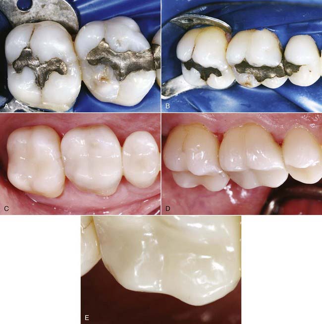
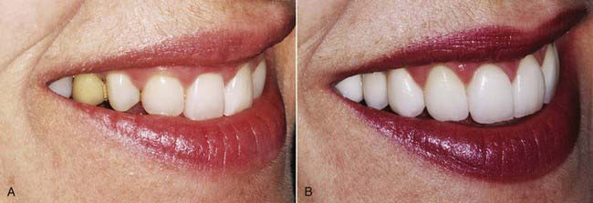
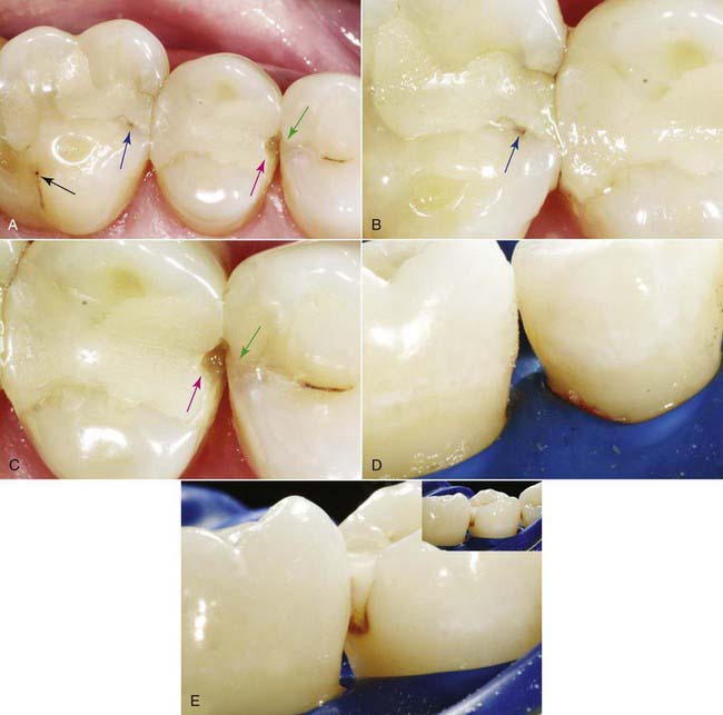
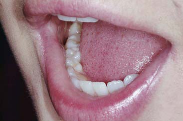
When esthetic dentistry began its evolution, the posterior teeth were considered unimportant. As patient expectations have increased, more focus has been placed on the esthetic contribution of posterior teeth (Figure 10-1). With the mechanics of mandibular function, as humans speak, laugh, and exhibit the behaviors considered human, the incisal edges of the lower anterior teeth and the occlusal surfaces of the posterior teeth are critical (Figure 10-2).

Many patients inhibit behaviors and develop a lack of confidence from a lack of pride in the anterior teeth. The same problems occur with the patient’s quality of life with regard to the posterior dentition. Many practitioners have seen these behaviors in patients with unacceptable anterior teeth. It is a valid exercise to examine the psychology of what happens when posterior tooth esthetics are not ideal. These problems have an impact on both quality of life and self-esteem (Box 10-1). Interestingly, habits such as pursing of lips and raising the hand to cover the mouth are the same regardless of whether patients dislike the appearance of their anterior or their posterior teeth.

Box 10.1 Posterior Dentition’s Impact on Self-Image and Quality of Life

A senior partner at a large, powerful accounting firm commented that she would arrange the seating for business meetings so that her colleagues were seated directly in front of her rather than to the side. Why? So that they would not see her maxillary bicuspid, which had a small interproximal display of gold. The culprit was a gold onlay placed by her previous dentist 20 years earlier. Over time a dinginess often occurs with interproximal metal restorations (see Figure 10-1, A and B).

This conversation was initiated when the patient was shown buccal corridor photographs. A traditional anterior photograph or “lips” picture does not really show the side view. Photographs where people are looking either obliquely or laterally allow the dentist and patient to discuss problems seen only from these perspectives.

This patient referred herself to the practice because she distrusted self proclaimed “cosmetic dentists”who aggressively promote their procedures. Yet a routine use of intraoral photography allowed her to enter into a course of self-discovery and served as the basis for an honest exchange of ideas and concerns. Such a course can lead to esthetic dentistry procedures without the ethical dilemma of “pushing” esthetics on the patient.

Happily, this patient chose a comprehensive esthetic reconstruction (see Figure 10-1, C to E). She now smiles and laughs and conducts meetings without rearranging the chairs in the room.

Brief History of the Clinical Development and Evolution of the Posterior Composite Procedure

For anterior composites, North American dental schools rapidly integrated both materials and updates. Cavity preparations were adapted to the material very early on. In terms of longevity, anterior composites have been deemed acceptable by both practicing dentists and dental schools. It is important to talk about perception of success with teaching institutions and outcome studies because often they are unrelated. Two factors were key in the success of anterior composites and their early integration. First was the clear Mylar matrix—the simple, single-space filling technique. Second was an early recognition by dentists and dental schools that the cavity preparation benefits from significant changes in the preparations done for silicate restoration or gold foil restorations, which the composites replaced. Thirty years later, after constant evolution, the modern anterior cavity preparation that has little or no mechanical undercuts and long infinity edge margins bears little resemblance to the silicate and gold foil preparations that anterior composite has replaced.

Unlike with anterior composites, the integration of posterior composites has had a tumultuous journey because of the lack of a regimented cavity shape and filling technique. Posterior composites have a somewhat dysfunctional relationship with dental schools. Many dental schools have discouraged the placement of posterior composites, knowing full well that on graduation most dentists would place posterior composites on a wholesale basis. In the schools’ defense, the outcome studies referenced, combined with the instructors’ personal observations of the parade of failing, leaking composite fillings, validated schools’ resistance to embracing the use of posterior composite on a wide-scale basis. It should be noted, though, that the posterior composite can be the most minimally invasive, biomimetic, and esthetically pleasing of all posterior restorations.

The Five Cs

Cracks and Fractures

Cracks and fractures are now the third leading cause of tooth loss in industrialized nations. The original hope was that because composite restorations are adhesive, the composite would be able to repair or splint the tooth together after the cavity preparation has weakened the tooth. Unfortunately, this is not the case, as recent outcome studies have shown the same number of cuspal fractures in amalgam-restored and composite-restored posterior teeth. Some immediate strengthening of the tooth can occur after placement of a composite, but two important factors must be borne in mind. First, crack initiation and propagation in the human dentition take years or even decades to accomplish. Today dentists are seeing cracks and fractures in teeth with posterior composites because enough time has elapsed. Much of the immediate strengthening of the tooth lessens dramatically over time. Many teeth that had posterior composites placed 15, 20, or 30 years ago are now fracturing. Second, it is not possible to predictably rely on composite to keep the tooth splinted long term. That challenges the dental profession to rethink the entire posterior composite approach. Unless the cavity design is changed dramatically, clinicians should consider posterior composites to act as “white amalgam” in terms of tooth fracturing.

Dental practitioners also worry about cracks and fractures in the material itself. A full explanation of composite’s characteristics is beyond the scope of this text. The most important characteristics in preventive dentistry for cracks and fractures can be divided into two different camps: characteristics during placement and polymerization, and characteristics of the composite once it is in the tooth and functional. Radiopacity, flexural strength, modulus of elasticity, fracture toughness, and total fracture work all contribute, but the most important factor from a clinician standpoint is fracture toughness. Catastrophic problems occur with posterior composites lacking fracture toughness. The fracture toughness of composite materials in general is inferior to that of gold—one of the liabilities of composite. These concerns must be overcome.

Crack initiation must be carefully evaluated and managed during cavity preparation and placement of composite and finishing. This addresses both crack initiation in the tooth and crack initiation or propagation in the composite material.

The C-Factor

The C-factor is a concept that is bandied about by many in the dental community. Although the concept has never been proven scientifically, it is the best guide to the management of polymerization shrinkage in various cavity preparations. C-factor stands for configuration factor and expresses the ratio of internal walls versus external surfaces. A second way to describe C-factor is internal surface area versus external surface area. C-factor is a fundamental flaw in traditional cavity preparations because the parallel walls for resistance and retention work against the dentist during polymerization shrinkage (Figure 10-3).

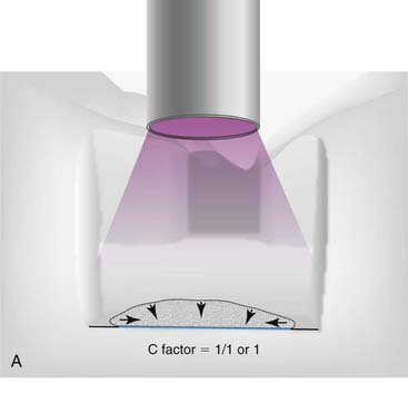
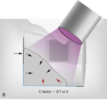
As the curing light hits the composite, it will shrink toward the center (Figure 10-4). The shrinkage can be measured as either volume or linearly. On a linear basis, most direct composites shrink 2% to 5%. All composites shrink on polymerization at this point, but the way the composite shrinks is critical and is based on the C-factor. The shape of the cavity preparation, the number of opposing walls, how they oppose one another, and the angle at which they oppose one another are extremely critical to the behavior of composite shrinkage.

image image image image image image image

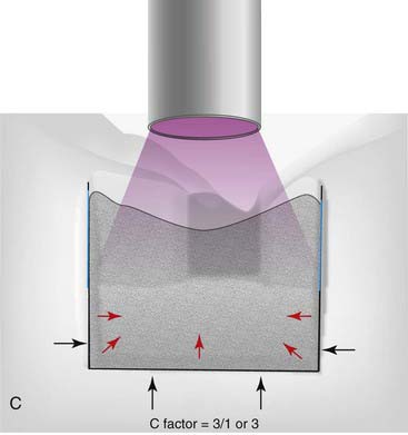
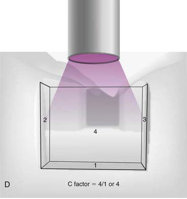
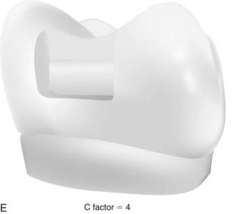
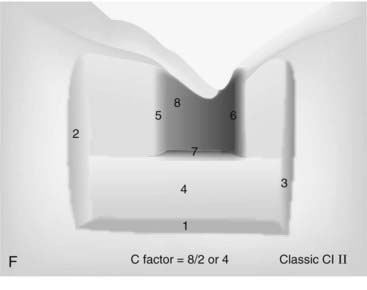
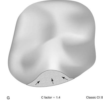
FIGURE 10-4 A, The curing light in the box area where the composite has been selectively placed on the gingival floor. The composite shrinks toward the single wall to which it has been applied. The C-factor is 1 because there is one internal wall that is being touched by the composite and one surface of the composite. A C-factor of one is very favorable. B, The light angled in. The C-factor is 2 because there are two cavity walls that the composite is touching at the same time and only one external surface to the composite, yielding a C-factor of 2. The red arrows shows where the composite is pulling toward the occlusal margin because of the shrinkage. In this example, the C-factor is 2 over 1 or 2. C, Filling the box area. The composite is engaging three walls of the cavity preparation—buccal, lingual, and gingival margin—simultaneously. The curing light is applied and shrinkage develops. The C-factor is 3. What typically happens in situations such as this is the shrinkage develops away from the gingival margin, the gingival dentin. This is extremely problematic both in post-operative sensitivity and microleakage on the gingival margin. There are three walls to the tooth cavity in this section of the box, and only one external surface of composite. This is an extremely unfavorable C-factor. The red arrows show the area where the composite pulls away from the tooth. D, When the actual wall of the cavity preparation is added to the buccal, lingual, and gingival walls, there are four cavity walls and realistically only one external surface area, which would be the interproximal area of the composite. The C-factor of 4 occurs in the classics. E, The new slot preparation, which has a C-factor of 4 for that box area. F, Classic G. V. Black class II preparation with an occlusal element. The C-factor is actually about the same as it was in the slot preparation because one traditional external surface has been added to the composite, the occlusal portion of the external surface of the composite. The ratio becomes eight internal walls over two external surfaces of composite, yielding a C-factor of 4, which is still unfavorable. G, Clark Class II preparation. Note the saucer shape. The C-factor on this is calculated at 1.4, significantly less than the 2 that was problematic. As composite is placed in this flattened cavity preparation, the C-factor is favorable enough that it is possible to injection mold the entire restoration as one without worrying about mitigating a C-factor with exotic layering techniques.

The C-factor is a ratio of the internal walls divided by the external walls, or it can be expressed in terms of surface area of the external surface. For C-factor a high number is unfavorable. Realistically a number of 2 or above is a problem when it comes to performance of the composite. Stress that is put onto the tooth and or composite can compromise the bond, cause microfracturing of the enamel, and lead to lack of adhesion in certain areas of the composite. The higher the C-factor, the worse for the situation. To simplify things, basically everything that dental school taught students about making a good preparation for amalgam with retention and resistance form are problematic for composite because of the C-factor (Figure 10-5).

Contamination

Contamination during posterior composite use occurs realistically in three ways:

1. Residual bacteria. Caries present on the tooth must be completely removed, although deep in the tooth some residual caries can be acceptable. The modern method of pulp capping is to avoid pulpal exposure if at all possible. Follow-up of teeth with indirect pulp caps has demonstrated that when small amounts of carious dentin are left over the pulp, after a few months this infected dentin heals and becomes hardened and sterile. However, this should not be misconstrued to assume that sloppy caries removal is acceptable. Within 1.0 to 1.5 mm of the margin, residual contamination in the tooth or as caries often results in recurrent decay.

2. Contamination of the infinity edge margin, or slight extension of the composite past the finish line. The long bevel or infinity edge margin combined with acid etching and bonding the composite a little past the margin is done with great success in anterior sites but has never been fully recognized with posterior placements. For the infinity edge or “Margeas margin,” the composite tends to go slightly past the finish line. Although this can be a strength in anterior restorations and achieves great esthetics, it is more difficult to clean posterior teeth. Compounding the problem is the problem that dentists unfortunately abandon protocols used on anterior teeth when preparing and filling posterior teeth, such as aggressively de-plaquing the teeth with rubber cup and coarse pumice. When the margin is not on enamel but on biofilm, no technique can provide an adequate seal.

3. Contamination that occurs during the restorative process. If fluid—water, saliva, or blood—is incorporated into the composite material, problems result. With amalgam, contamination is less detrimental.

Clinical Considerations

Contraindications

The most important contraindications to posterior composites are based on individual tooth considerations. For example, when both the mesial and the distal surfaces were previously restored with either composite or amalgam and fracture is suspected, that is not the best indication for posterior composite. When large areas of the margin are on dentin or cementum or when cast restorations are relying on dentin cementation, then an indirect restoration is more predictable than with large areas of dentin bonding on the margins. Dentin cementation is more predictable than dentin bonding when there are large areas of the margins exposed. For a severely caries-prone patient, a patient with salivary disorders, or a patient undergoing cancer or radiation therapy, amalgam or glass ionomer may be preferred. Composite has not been shown to release therapeutic levels of fluoride, and the current composites have no ability to act as a fluoride bank or to be rechargeable unless they are glass ionomers. Although research shows that significant caries resistance for therapeutic restoratives such as glass ionomer is absent, it is generally recognized that the valid approach is to use a glass ionomer. Amalgam is more bactericidal than composite and tends to accumulate less decay. If there is biofilm underneath a composite restoration, that can lead to recurrent decay. In addition, amalgam is more inert than all of the resins and will not degrade. The key here is that composite resins that are turning brown are actually degrading as if the bacteria were consuming the composite. This is most common at the gingival margin.

Material Options

The chemistry in the composite really has been unchanged for the past 25 years, but recently composites have been broken into two categories: pastes and flowable composites. Each of these can be subcategorized as a microfill, a preconglomerated microfill, or a microhybrid. The new, popular term “nanofill” is more marketing than science. Furthermore, those camps can be broken down into light cured materials, chemical cured materials, or those that have both light and chemical cures. In the paste camp, a new organic chemistry has finally been introduced to achieve low shrinkage. With this, the material actually expands as it contracts during polymerization shrinkage.

The paste composites have better polishability, but more important, as already discussed, they have the ability to maintain polish and surface integrity. Many of the studies show polishability as an asset. Many flowables allow a good polish, but that polish is very temporary. One of the problems with the literature is that studies do not look at long-term ability to retain a polish, which is more important for esthetics. In general the pastes are far superior in maintaining the polish compared with the flowables. The flowables in general tend to lose luster much more quickly than does a well-polished paste.

The advantages of flowable composite are superior handling and wetting of the cavity preparation. The research on using a flowable composite as the first layer to fill in the nooks and crannies and seal to the gingival margin reveals that it is not superior to putting paste composite directly into the cavity preparation. There is a perception that when the dentist places a flowable composite, it will fit into the nooks and crannies better in a class I or a class II preparation. Recently the American Dental Association analyzed the two composite types. One thing that research does not consider is the microgap versus the macrogap. If the dentist, during handling of a paste composite, leaves large voids in the restoration, then there will be microleakage. Flowable composite may not show a better result in research studies, but in practicality, most dentists feel they can get a better result using flowable composite.

Nearly all the physical characteristics of flowable composites are inferior to those of the paste composites. The advantages of the flowable materials are nearly all focused on ease of application as an aid to the paste composite and avoidance of mistakes. Flowable composite should never be considered as a replacement for paste composite.

In comparing a microfill and a microhybrid, microfills tend to have less wear but inferior strength. Microhybrids, conversely, have greater tendency for wearing and marginal ditching because of the clumping phenomenon as the larger particles fall out of the matrix. On the other hand, they tend to have higher compressive strength.

Current Best Approach

The traditional metal matrix and the translucent systems are completely different. For metal matrix the current best approach is layering at 2-mm increments. For the first layer a flowable composite is popular but has not been proved scientifically to be better. The goal is to use as little flowable composite as possible to avoid fracturing and weakening from the nexus of the flowable composite. The reason 2-mm increments are needed is because currently that is the deepest one can guarantee that the curing light will penetrate. The problem with the 2-mm layering, especially in a taller preparation, is that it is quite difficult and highly susceptible to developing seams and gaps between layers. The best approach using a non-metal matrix or translucent system is either the “snow plow” or the “injection-molded” technique. The former involves using a flowable and then a paste injection using the bulk loader. The author’s preferred technique is the injection-molded technique, which is a total-etch technique, with placement of resin, then flowable composite, then paste in sequence with no curing between applications.

The current best approach for large cavities or teeth with early incomplete fractures is a direct composite onlay. The best environment for posterior composite in class I or class II cavities is when all the margins can be placed and maintained on the enamel. Therefore great care should be taken during tooth preparation to preserve residual enamel along the finish line. Even if it is a tiny sliver of enamel, it should be carefully maintained.

Many dentists are going away from the total-etch technique and using self-etching resin. The author’s recommendation is still a total-etch technique because it allows the dentist to create a good bond on the infinity edge portion of the margin, which is the part of a composite that extends slightly past the finish line on the enamel. However, the self-etching resins do not provide as strong a bond on uncut (un-abraded) enamel. To obtain that last purchase, the last seal at the marginal extreme, a total-etch technique allows a light feather etch that the self-etch technique does not. The total-etch technique can be done with either a one-bottle or a two-bottle formulation. When a total-etch one-bottle technique is used, some clinicians have reported problems with sensitivity. The recommendation for the total-etch one-bottle technique is to place and dry two coats, then air thin and cure two coats of resin over the dentin before binding to the enamel to increase the dentin bond and to decrease sensitivity. Many clinicians have adapted the technique and achieved significant reduction in postoperative activity. A few new self-etching resins can be used after etching with phosphoric acid, Easy Bond (3M ESPE, St Paul, Minnesota) being the best known. Most other self-etching resins have poor dentinal bonds if the dentin is etched first.

The current best approach as far as adhesives and adhesion to enamel is still the total/>

Stay updated, free dental videos. Join our Telegram channel

Jan 3, 2015 | Posted by in Esthetic Dentristry | Comments Off on 10: Posterior Direct Composites

VIDEdental - Online dental courses
Get VIDEdental app for watching clinical videos