Oblique lateral radiography
Introduction
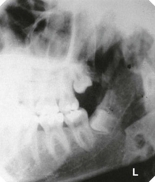
Oblique lateral radiographs are extraoral views of the jaws that can be taken using a dental X-ray set (see Fig. 10.1). Before the development of panoramic equipment they were the routine extraoral radiographs used both in hospitals and in general practice. In recent years, their popularity has waned, but the limitations of panoramic radiographs (see Ch. 12) have ensured that oblique lateral radiographs still have an important role.
Terminology
Lateral radiographs of the head and jaws are divided into:
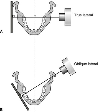
The differentiating adjectives true and oblique are used to indicate the relationship of the film, patient and X-ray beam, as shown in Fig. 10.2.
True lateral positioning
The image receptor and the sagittal plane of the patient’s head are parallel and the X-ray beam is perpendicular to both of them. This is the positioning for the true lateral skull radiograph taken in a cephalostat unit described in Chapter 11.
Main indications
The main clinical indications for oblique lateral radiographs include:
• Assessment of the presence and/or position of unerupted teeth
• Detection of fractures of the mandible
• Evaluation of lesions or conditions affecting the jaws including cysts, tumours, giant cell lesions, and osteodystrophies
• As an alternative when intraoral views are unobtainable because of severe gagging or if the patient is unable to open the mouth or is unconscious (see Ch. 6, Fig. 6.1)
• As specific views of the salivary glands or temporomandibular joints.
Equipment required
This includes (see Fig. 10.3):
• An extraoral cassette containing film and intensifying screens or a digital phosphor plate (usually 13 × 18 cm)
• A lead shield to cover half the cassette when taking bimolar views.
Specially constructed angle boards, as shown in Fig. 10.3, can be used to facilitate positioning, but are not considered necessary by the authors.
Stay updated, free dental videos. Join our Telegram channel

VIDEdental - Online dental courses


