10
Musculoskeletal disorders
Introduction
There are many ways in which disorders of the musculoskeletal system can impact on dental management. For example, cervical spine involvement may lead to poor neck extension, disorders of bones may impact on surgical management, and muscular disorders can cause difficulties with the safe provision of intravenous sedation or general anaesthesia.
Diagnosing musculoskeletal problems is achieved by recognising the pattern of disease (Table 10.1). Timing divides acute arthritis (<6 weeks duration) and chronic arthritis (≥6 weeks). Inflammation is found in varying degrees in all types of arthritis, but osteoarthritis typically has mild or short-lived inflammation, compared with the prolonged and/or severe inflammation of a true inflammatory arthritis, such as rheumatoid arthritis. The presence of deformity strongly suggests chronicity, but the type of deformity is often diagnostic (e.g. ulnar deviation of the hands in rheumatoid arthritis).
If one joint is involved (a monoarthritis), then infection or a crystal arthritis or seronegative arthritis are likely causes. Involvement of 2–4 joints (an oligoarthritis) is most commonly caused by seronegative arthritis. Polyarthritis involves ≥5 joints.
The impact on the activities of daily living of the patient are very important. Functional limitation is a significant consideration in determining treatment.
Classifying musculoskeletal problems
The key distinction is the presence of inflammation. The disorders shown in Table 10.2 all have prominent inflammatory elements. They are known collectively as the seronegative arthritides.
In contrast, the presence of inflammation in a second group of disorders is mild, or absent and mechanically driven. These disorders include osteoarthritis, back and neck pain, and sports injuries. Finally, there are connective tissue disorders, where the defect is immunological, but the main manifestations are distant from the joint. Such disorders include Sjögren’s syndrome, systemic lupus erythematosus (SLE), scleroderma and myositis.
Osteoarthritis
Osteoarthritis is a mechanically driven, but biochemically mediated disease of synovial joints. It is a group of allied diseases where there is an imbalance between cartilage synthesis and degradation. It is characterised by pain, tenderness, decreased range of movement and variable inflammation. There are changes to the cartilage and classical radiographic changes, as will be described.
In clinical practice, the presence of pain plus typical changes on radiographs is generally accepted for diagnosis.
Clinical features
Signs of generalised osteoarthritis are often found in those with one painful joint. The typical pattern is involvement of the hands, knees and hips, although foot disease may often be unrecognised.
Symptoms
Patients complain of localised pain which, in the case of the hip, is increased on weight bearing. Advanced disease causes non-weight-bearing/nocturnal pain (probably due to raised intraosseous pressure). There may be short-lived or absent early morning joint stiffness, or no symptoms.
Table 10.1 Key features for diagnosis of joint disease
| Feature | Findings |
|
Acute versus chronic |
|
Yes/no |
|
Yes/no (mild/moderate/severe) |
|
Mono/oligo/polyarthritis |
|
|
| Impairment | e.g. bent knee |
| Activity restriction | Cannot walk |
| Participation | Cannot go to the pub |
Table 10.2 The seronegative arthritides and disorders with prominent inflammatory components
|
|
|
|
|
|
|
* These are collectively known as the seronegative arthritides, remembered with the mnemonic PEAR.
Signs
In the hand, outgrowths known as nodes may be seen on the fingers. Distal interphalangeal (DIP) nodes (towards the end of the finger) are termed Heberden’s nodes, whereas proximal interphalangeal (PIP) nodes are termed Bouchard’s nodes (Fig. 10.1). There is generalised muscle wasting.
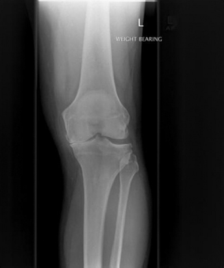
The radiographic signs of osteoarthritis are summarised in Table 10.3. A radiograph of a knee joint showing the changes of osteoarthritis is shown in Figure 10.2.
Risk factors for osteoarthritis
In primary osteoarthritis there is incomplete understanding of the aetiology. The incidence and prevalence increase with age and it is usually more common in women. Osteoarthritis is more common in occupations and populations where the load on joints is greater, e.g. in the obese and runners (knee). There are recognised genetic associations for some subgroups, explaining a minority of disease.
Secondary osteoarthritis occurs earlier with more predictable severity in damaged joints, e.g. after a cruciate ligament injury of the knee.
Table 10.3 Radiographic signs of osteoarthritis
|
|
|
|
Figure 10.1 Osteoarthritis in the hands.
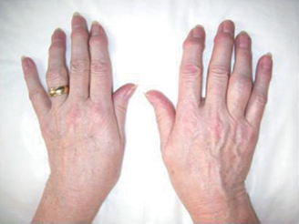
Figure 10.2 Radiographic changes of osteoarthritis (see Table 10.3).
Demographic changes
The incidence of osteoarthritis is increasing; this is mainly due to the ageing population. There is estimated to be at least a 40% increase in the numbers needing total hip replacements (THRs) in the future.
Management
The management of osteoarthritis can be divided into pharmacological and non-pharmacological methods.
The pharmacological route relies on analgesics, particularly the NSAIDs. Injections of steroid/hyaluronan may also be used.
Non-pharmacological methods include exercises to develop musculature, e.g. in the lower limb exercises to develop the quadriceps muscle, weight loss and the provision of aids. Education and support are also valuable.
Surgical treatment is discussed later. It is reserved for recalcitrant cases.
Rheumatoid arthritis
Rheumatoid arthritis is an autoimmune systemic inflammatory illness, characterised by symmetrical joint inflammation and variable extra-articular features. It may be defined according to the criteria shown in Table 10.4, derived from the American College of Rheumatology.
In clinical practice, the presence of symmetrical joint swelling with prolonged early morning stiffness (≥30 min) lasting for 6 weeks or longer, is generally accepted.
Clinical features
The typical pattern is of involvement of the peripheral joints, including the upper and lower limbs, but sparing the DIP joints.
Symptoms
There will be pain in or around the joint, sometimes with myalgia (muscle pain), which may be increased in the morning or with rest (gelling). The early morning stiffness of ≥30 min may be severe or prolonged. Patients may have dry eyes, dry mouth or other extra-articular symptoms.
It is important to recognise that rheumatoid arthritis is a multisystem disorder and may involve diverse systems. A summary is given in Table 10.5.
Table 10.4 American College of Rheumatology criteria used to define typical rheumatoid arthritis
|
|
|
|
|
|
|
Signs
On examination, there is a ‘boggy swelling’ of affected joints which may be hot, tender and sometimes red. Typical deformities seen are the swan-neck/boutonnière deformity of the fingers so-called due to their shape, or a Z-thumb. Classically there is ulnar deviation at the metacarpophalangeal (wrist) joints (Fig. 10.3).
Table 10.5 The multisystem nature of rheumatoid arthritis
| Cardiovascular |
|
|
|
| Respiratory |
|
| Renal |
|
| Liver |
|
| Skin |
|
|
| General |
|
|
|
Figure 10.3 Clinical features of rheumatoid arthritis – ulnar deviation of the hands.
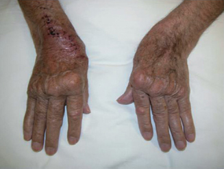
Table 10.6 Radiographic signs of rheumatoid arthritis
| Early |
|
|
| Late |
|
|
|
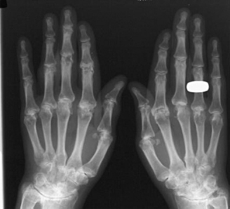
Figure 10.4 Radiographic features of rheumatoid arthritis (see Table 10.6).
Periarticular signs of rheumatoid arthritis include rheumatoid nodules (rubbery swellings, typically over elbows or other extensor surfaces, although they can be found anywhere). Bursitis/tenosynovitis may also be seen.
Extra-articular signs are seen in Sjögren’s syndrome. Osteoporosis may be seen.
Radiographic signs of rheumatoid arthritis
Radiographic signs of rheumatoid arthritis are summarised in Table 10.6 and shown in Fig. 10.4.
Burden of rheumatoid arthritis
Rheumatoid arthritis affects around 1% of the population and is twice as common in women compared with men. There is an increasing incidence and prevalence after age 50.
Table 10.7 Recent advances in treatment
|
|
|
|
|
|
|
| Commonly used |
|
|
|
|
| Less commonly used |
|
|
|
|
* Caution, due to infection risk especially dissemination/ tuberculosis; autoimmune disease such as SLE and potential for increased primary or recurrent malignancy.
DMARD = disease-modifying antirheumatic drug; TNF = tumour necrosis factor.
Management of rheumatoid arthritis
Education with regard to the nature of the disease is important. Various splints and devices can be used for joint protection. Analgesics, including NSAIDs, are used. There have been many developments in recent years of disease-modifying antirheumatic drugs (DMARDs). DMARDs aim to reduce symptoms and prevent joint damage. They require very close monitoring to check for side effects, which can be significant. They are slow in their action.
Recent advances in treatment are summarised in Table 10.7.
Reactive arthritis
Reactive arthritis is an autoimmune inflammatory condition, triggered by an infection characterised by usually self-limiting inflammation of one or more joints. It is a clinical diagnosis based on the development of a typical mono- or oligoarthritis, developing 10–14 days after been triggered by a gastrointestinal or genitourinary infection.
Clinical features
Monoarthritis or oligoarthritis of a large joint(s) occurs, most commonly the knee, therefore septic arthritis may need to be excluded by joint aspiration. A triggering infection, e.g. by Campylobacter or Salmonella, can be identified in up to 50% of cases and should be actively sought to aid effective treatment.
Reiter’s syndrome may be found. This is an association of reactive arthritis with conjunctivitis and urethritis. Reiter’s syndrome may be associated with a rash of the hands and feet, histologically indistinguishable from psoriasis (keratoderma blenorrhagica).
Viral infections, especially parvovirus B19 and Coxsackie viruses, can cause a reactive polyarthritis with a rheumatoid arthritis-like pattern, but which is self-limiting.
Signs and symptoms
The signs are consistent with the pattern of involvement. Symptoms include pain and morning stiffness in the affected joint(s). Symptoms may be improved by exercise or NSAIDs.
Radiographic signs of reactive arthritis
There are usually no radiographic changes. If radiographic changes are seen, then a reconsideration of the diagnosis should take place.
Management of reactive arthritis
Reactive arthritis is usually self-limiting. NSAIDs are usually beneficial, occasionally DMARD therapy may be needed. There is a 50% recurrence rate.
Other joint disorders
Juvenile chronic arthritis may lead to an increased incidence of caries and periodontal disease as a result of difficulty in maintaining good oral hygiene. In addition, if the temporomandibular joint (TMJ) is involved, facial growth may be disturbed.
As mentioned earlier, one of the early signs of development of rheumatoid arthritis may be stiffness of the fingers, particularly in the early morning (‘early morning stiffness’) which usually decreases during the day. In more advanced disease, the direction of the fingers appear to drift away from the thumb – ‘ulnar deviation’. The onset is often slow, but it can be acute with malaise, fever and joint pain. There is anaemia which is normocytic and normochromic – the so-called anaemia of chronic disease. Treatment in the early stages is usually with NSAIDs. Second-line treatment includes a variety of agents such as gold and the chemotherapy agent methotrexate (this may lead to folic acid deficiency with the potential for secondary oral problems). Corticosteroids have been used for treatment, as have antimalarial medications. The mainstay of physical treatment involves occupational therapy and />
Stay updated, free dental videos. Join our Telegram channel
VIDEdental - Online dental courses


