Dental Caries in Children and Adolescents
Marit Slåttelid Skeie, Anita Alm, Lill‐Kari Wendt, and Sven Poulsen
Dental caries is the most common chronic disease among children and adolescents [1], and thus the one most often affecting both oral and general health [2,3]. Untreated cavitated dentine lesions may also negatively influence children’s quality of life [4]. The impact of caries on oral and general health is associated with the age the child, when lesions appear, the depth of the lesions, and the location of the lesions in the dentition. The impact of the disease is more serious in younger children, in children with chronic health conditions, and in children living in countries with poor access to adequate dental health services.
Childhood dental caries as a health problem
“Health is a state of complete physical, mental and social wellbeing, not merely the absence of disease and infirmity.” This definition of health by World Health Organization, which dates back half a century, although criticized for its shortcomings [5] (Chapter 1), has been a starting point for today’s generally accepted holistic approach to health [3]. As oral health is an integrated part of general health, it is natural to use this concept even when describing caries and its sequelae in children and adolescents [6].
Dental caries has impacts on both oral and general health (Box 10.1), and when left untreated, often leads to pain, and consequently a reduced ability to chew and eat [2]. Limitations in choice of foods, loss of appetite, and reduced pleasure from eating [2] are some of the reasons why untreated severe caries may be associated with reduced body weight and impaired growth [6–8]. However, when restorative treatment was so extensive that general anaesthetic was indicated, some studies showed that children afterwards often gain weight rapidly [8]. Research also indicates that restorative rehabilitation of young children with severe caries experience is associated with substantial and highly significant improvements in their parent‐assessed oral‐health‐related quality of life [9,10].
An Irish study concluded that almost half of all hospital dental emergencies were due to caries sequelae [11]. Candida, osteomyelitis or sepsis due to infected teeth are reported in countries that have poor access to adequate dental or general health services [3]. Even in the Western world, for some children with chronic health conditions (Chapter 23), dental infections and to some extent the treatment of carious lesions may represent a threat to life [12].
Caries with onset early in life indicates an increased risk of future caries development [13–15], a topic that will be covered in more detail later in this chapter. Insufficient oral hygiene around a tooth with caries also often induces additional gingival problems [16]. Furthermore, deep carious lesions constitute a risk for endodontic complications and abscess development [17], which often leads to infection, abscess formation with swelling and further pain and discomfort during chewing. A high number of untreated decayed teeth is found to be related to dental sepsis as well [18]. For some children, hospitalizations and emergency dental visits are the consequences [19]. As use of antibiotics is common in dentistry [20,21], the risk for antibacterial drug resistance is another problem which should be considered. Regarding infected primary teeth, there is a risk for potential injuries to the permanent tooth such as enamel opacities, hypoplasia or developmental disturbances [12].
Extractions of primary teeth due to caries at an early age may lead to reduced dental arch length, tooth displacement, tilting, and rotations [22]. If several teeth with important phonetic functions are extracted, normal language development may be compromised [23]. Having to perform operative restorative treatment of caries in children at an early age may also affect future oral health negatively. About one‐third of fillings in primary teeth will need replacement between the ages of 7–12 years [24]. It has also been documented [25,26] that approximal cavity preparations may damage two‐thirds or more of the neighboring sound surfaces, making them more susceptible to caries. Children with caries experience at 6 years of age had three and a half times more treatments performed (i.e., new restorations, replacement of restorations, or other treatment such as disking or extraction) in primary teeth from 7 to 12 years compared to children without previous caries experience at the age of 6 years [27].
A well‐documented consequence of severe caries with pain is the reduction of the individual’s quality of life [4,6]. Disturbed sleeping, concentration problems [1,28,29] and interruption in play and schoolwork [2,30] may induce emotional stress with anger and irritability. Because of aesthetics and/or phonetic problems, there is also a risk that children will be teased, which again may negatively influence their self‐esteem, resulting in the child acquiring a silent demeanor or avoiding smiling and laughing [31].
Experiences of pain during dental treatment during childhood are well documented to increase the risk of developing dental behavioral management problems and dental anxiety later in life [32]. A study has shown that more than every third child with more than 10 carious lesions at age 5 years presented with dental anxiety 5 years later [33], indicating that painful experiences during restorative treatment are a major risk factor for development of dental anxiety. A higher prevalence of missed dental appointments among children who have had dental treatment in connection with toothache has also been documented [34]. Neglected oral health may be the consequence, and also increased economic treatment costs later in life, both for the individual and for society.
Epidemiology of dental caries in children and adolescents
Epidemiology is defined as “The study of the occurrence and distribution of health‐related states or events in specified populations, including the study of the determinants influencing such states, and the application of this knowledge to control the health problems” [35]. From this definition it is obvious that understanding and interpretation of epidemiologic data are an essential part of managing pediatric dental care. More specifically, epidemiology has two important applications in pediatric dental care: to describe the distribution of caries in the population, and to describe changes in caries prevalence over time. It is important to define certain basic epidemiologic terms in order to understand epidemiologic data on dental caries in children and adolescents (Box 10.2). Epidemiologic data can be used, for example, to provide information on:
- The prevalence of caries in the population, according to age, gender, socioeconomic and ethnic background. This is important in order to determine the magnitude of the problem and the distribution of the burden of disease in the population.
- The incidence of caries in the population. This will give information as to the future level of disease in the population.
- Oral health strategic planning for caries control, e.g., how to use the existent personnel resources efficiently, how to evaluate child dental care, and how to inform the dental health authorities responsible for the financing of health care.
- Formulation of goals and determination of whether these goals are fulfilled or not.
Thus, epidemiologic data are important in quality development and quality control of pediatric dental care.
Epidemiologic data on dental caries
During recent years increasing emphasis has been placed on the dynamics of the carious lesion as developing from a subclinical lesion through an initial, noncavitated lesion to a manifest lesion, eventually resulting in complete destruction of the tooth. It is now generally accepted that arrest and even reversal of initial, noncavitated carious lesions can occur [36]. This has resulted in the development of interceptive strategies in children and adolescents in the management of these kinds of lesions (see Chapter 12). An important aspect of these strategies is the need to inform the parents that appropriate preventive measures may allow a natural arrest of lesion progression while it is confined to the enamel. As guardians, they have the right to be informed about this. Parents should be shown the sites of enamel lesions and given advice for future optimal oral health behaviors. Dentists not taking this task seriously may be at future risk to encounter demands. The best period for such prevention and to reach and motivate caregivers, is during eruption of the primary teeth [37]. It might be expected in the future that operative intervention, not a treatment of the caries disease, but its consequences, will be chosen only as a secondary alternative only when nonoperative intervention methods have failed [38]. The previous traditional “care” concept, meaning operative treatment as restorations and extractions, is nowadays changed to also include nonoperative treatment in the disease management.
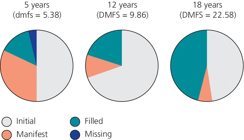
Nordic countries have, for administrative purposes, established data collection systems to monitor the level of dental caries. The data collected in some of these systems are the numbers of already placed restorations and/or the number teeth which according to the dentist’s judgment, need restorative treatment. Today’s shift to a more conservative treatment philosophy [39] may in this system compromise valid comparisons between studies from different time periods. An example of one exception is the system implemented by the National Board of Health in Denmark [40]. Caries data are collected by this institution according to written epidemiologic criteria, and noncavitated carious lesions are recorded as a separate caries diagnosis. Over the last few decades, several other epidemiologic projects in the Nordic countries have been carried out, using similarly detailed diagnosis criteria. These studies have shown that noncavitated carious lesions constitute a large proportion of the total number of carious lesions (Figure 10.1). In teenagers, it is reported that approximal noncavitated carious lesions may make up as much as 80–90% of the total number of carious lesions [14,41]. This actually underlines previous arguments from other researchers [42,43] that national reports considerably underestimate the total number of lesions. Additionally, by not including enamel caries, the need for modern nonoperative treatment will not be demonstrated. Thus, for a number of reasons it is very important to make explicit the criteria used for diagnosis, when presenting, interpreting, and making use of data on the occurrence of caries in children and adolescents (Box 10.3).

Figure 10.1 Contribution of initial and manifest lesions, filled and missing (due to caries) surfaces of the total caries index in Norwegian children.
Source: Amarante et al. 1998 [42]. Reproduced with permission of John Wiley & Sons.
However, the different caries measurement systems capable of incorporating noncavitated carious lesions vary in detail, in the way they are used, and in terminology (Box 10.4). A common trait is ambiguity and incompatibility in caries diagnostic thresholds around the dentino‐enamel border. Although detailed caries diagnostic systems should be considered as a step forward in the field of epidemiology, measures of caries activity are still relatively poorly developed [44]. However, Nyvad argues that additional information related to caries diagnostic decision can be obtained if carious lesions were dichotomized into “inactive” and “active” lesions [45].
Table 10.1 lists a number of Nordic surveys on which the description, the patterns of caries in children and adolescents in this chapter is based. Descriptive epidemiologic data from these surveys are not presented because, as previously discussed, differences in diagnostic criteria hamper comparison of data.
Table 10.1 Nordic surveys of caries in children and adolescents on which the description in the present chapter is based
| Author (year of publication) | Country | Age (month/year) |
| Seppä et al. (1989) [79] | Finland | Follow‐up (6–13 yr) |
| Wendt et al. (1991, 1992) [57,134] | Sweden | 1 yr, 3 yr |
| Grindefjord et al. (1993,1995) [13,135] | Sweden | 1 yr, 3.5 yr |
| Amarante et al. (1995) [136](136) | Norway | 5 yr, 12 yr, 18 yr |
| Petersen (1996) [113] | Denmark | 2–3 yr, 7 yr |
| Vehkalathi et al. (1997) [85] | Finland | 5 yr, 15 yr |
| Mattila et al. (1998) [51] | Finland | Follow‐up (3–5 yr) |
| Mejàre et al. (1998, 1999, 2000) [90,91,137] | Sweden | Follow‐up (6–12 yr), (11–22 yr) and (12–18 yr) |
| Wendt et al. (1999) [78] | Sweden | Follow‐up (3–6 yr) |
| Poulsen & Pedersen (2002) [138] | Denmark | 5 yr, 7 yr, 12 yr, 15 yr |
| Stenlund et al. (2002) [89] | Sweden | Follow‐up (11–22 yr) |
| Wennhall et al. (2002) [61] | Sweden | 3 yr |
| Sundby & Petersen (2003) [112] | Denmark | 3 yr, 5 yr |
| Ruottinen et al. (2004) [139] | Finland | Follow‐up (7 mo–10 yr) |
| Skeie et al. (2004, 2006) [76,40] | Norway | Follow‐up (5–10 yr) |
| Stécksen‐Blicks et al. (2004, 2008, 2014) [62,84,141] | Sweden | 4 yr |
| Hugoson et al. (2005, 2008) [73,81] | Sweden | 3 yr, 5 yr, 10 yr, 15 yr, 20 yr. |
| Mattila et al. (2005b, 2005a) [124,127] | Finland | Follow‐up (0–7 yr) and (0–10 yr) |
| Skeie et al. (2005, 2006) [86,114] | Norway | 3 yr, 5 yr |
| Wennhall et al. (2005) [142] | Sweden | Follow‐up (2–3 yr) |
| Alm et al. (2007, 2008) [14,58] | Sweden | Follow‐up (3–15 yr) |
| Wigen et al. (2009, 2011) [75,143] | Norway | 5 yr |
| Ekbäck et al. (2012) [144] | Sweden | Follow‐up (3–19 yr) |
| Socialstyrelsen (2013) [63] | Sweden | 3 yr, 6 yr, 12 yr, 19 yr |
| Isaksson et al. (2013) [59] | Sweden | Follow‐up (3–20 yr) |
| André Kramer et al. (2014) [74] | Sweden | Follow‐up (3–6 yr) |
Dental caries in the young and the late primary dentition
Dental decay in infants and toddlers has a distinctive pattern. The definitions first used to describe this condition were related to etiology, with the focus on inappropriate use of nursing practices. This generated the terms “nursing bottle mouth,” “baby bottle tooth decay,” “nursing bottle syndrome,” and “nursing caries.” The current internationally accepted definition of caries with early onset is “early childhood caries” (ECC). This terminology encompasses a broader meaning, better fitting the understanding that unfavorable feeding practice is not the only important cause of ECC. However, pediatricians have not yet succeeded in agreeing on a mutual and uniform definition of ECC, and inconsistencies in the case definitions and diagnostic criteria still exist (Box 10.5) [46,47]. The definition of ECC used in this chapter is the occurrence of any sign of dental caries on any tooth surface during the first 3 years of life [48]. A more extensive form of ECC, severe ECC (S‐ECC), is defined of the basis of age, caries prevalence, and depth of the carious lesions. Typical for S‐ECC is a high caries progression rate. Grindefjord et al. have documented that children with early caries development, before 2.5 years of age, exhibited a high caries progression rate during the period up to 3.5 years of age [49].
Stay updated, free dental videos. Join our Telegram channel

VIDEdental - Online dental courses


