10: Bilobe Flaps

Bilobe Flaps

Introduction

The original design of the bilobe flap is attributed to Esser,1 who described its use in 1918 for reconstruction of nasal tip defects. Zimany2 and others expanded the use of the flap to reconstruct defects on the trunk and soles. However, most authors now share the opinion that this flap is most useful for facial reconstruction, particularly of the nose.

Esser’s original design required that the angle of tissue transfer be 90° between each lobe of the flap, for a total pivotal movement of more than 180°. An example of this design for repair of a nasal tip defect would be the first lobe of the flap harvested from the nasal sidewall and the second lobe from the glabella. This design maximizes the distance that skin can be moved, but the wide angles between the two lobes (90°) also maximize the standing cutaneous deformities and the likelihood of creating trap-door deformities of both first and second lobes. These were common problems with the original flap design and limited the use of the bilobe flap for facial reconstruction.

Bilobe flaps are double transposition flaps that share a single base. Similar to single transposition flaps, bilobe flaps move around pivotal points located at their base and develop standing cutaneous deformities as they pivot. Because each flap or lobe moves around an independent pivotal point, each lobe develops an individual standing cutaneous deformity. The greater the arc of movement about their pivotal points, the larger are the standing cutaneous deformities.

In general, bilobe flaps are designed in such a way that the first lobe is immediately adjacent to the defect and has a surface area that is less than the surface area of the defect. Thus, part of the closure of the defect is achieved by secondary movement of surrounding skin through direct advancement. The relationship between the surface area of the first lobe and the size of the defect depends on the location of the defect on the face and the elasticity of skin surrounding the defect. On the caudal nose, for instance, the first lobe must nearly approximate the size of the defect because of the inelasticity of nasal tip skin. However, on the cheek, the first lobe may be designed considerably smaller than the size of the defect (up to 25% less in surface area) because of the general elasticity and redundancy of the skin surrounding cheek defects. The second lobe of a bilobe flap is usually designed so that its surface area is less than the surface area of the defect left by harvesting of the first lobe. Again, advancement of adjacent skin assists the second lobe in repair of the donor site of the first lobe. The defect left by the second lobe is closed primarily by direct advancement of surrounding skin. Thus, for the bilobe flap to work, there must be considerable skin laxity in the vicinity of the first and second lobes to achieve wound repair without excessive wound closure tension.

Biomechanics

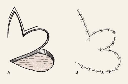
The bilobe flap would appear to be a modified rotation flap with some component of transposition. However, during transfer of the flap, there is noticeably less restriction of tissue movement than would be present with a pure rotation flap. This ease of tissue movement of the first lobe is the result, in part, of transposing the triangle-shaped peninsula of skin located between the defect and the first lobe of the flap. The transposition of this skin peninsula adjacent to the distal portion of the first lobe of the flap in essence represents a modified Z-plasty (Fig. 10-1). Likewise, there is a triangular peninsula of skin that forms between the first and second lobes that is also transposed during closure of the donor site of the second lobe. Thus, the bilobe flap in some ways represents a modified double Z-plasty. This results in repositioning of the skin adjacent to the defect and the two lobes of the flap. This in turn results in an overall reduction in wound closure tension compared with use of a single transposition or rotation flap.

Bilobe flaps expand the use of transposition flaps. The major advantage of bilobe flaps is the ability to recruit skin for construction of a flap from areas of skin redundancy that are not adjacent to the defect. On the cheek, this skin redundancy may be located at some distance from the defect and may be difficult to transfer to the recipient site by other surgical approaches. Defects that cannot be easily repaired with a single transposition flap without excessive wound closure tension causing distortion of facial structures may often be reconstructed with a bilobe flap. This is because the second lobe of the transposition flap reduces overall wound closure tension and provides additional skin to the proximity of the reconstruction. Furthermore, bilobe flaps transfer the tension of wound closure through a 90° arc, which is more than the usual 45° to 60° arc of transfer of a single transposition flap. This greater distribution of wound closure tension helps minimize distortion of structures surrounding the primary defect.

Flap Design

McGregor and Soutar3 altered the design of bilobe flaps and noted that the degree of pivotal movement could be varied greatly from the original 90° between each lobe. In 1989, Zitelli4 published his experience using the bilobe flap for nasal reconstruction. He emphasized the use of narrow angles of transfer, 45° between each lobe, so that the total pivotal movement of tissue occurs over no more than 90° to 100°. This eliminated the need to excise standing cutaneous deformities, and trap-door deformities were frequently avoided. Burget5 confirmed the excellent results with this design for reconstruction of the nose. Other surgeons also advocated a similar design for repair of skin defects of the cheek, chin, and lips.6,7 They used narrow angles between the lobes of the flap and achieved better results than with use of traditionally designed bilobe flaps that are transposed through an arc of 180°.

Limiting the angle between the axis of the first and second lobes of the bilobe flap is more important on the nose than on the cheek. When bilobe flaps are used on the cheek, the primary determinants of the position of the first and second lobes of the flap are the location of the defect and the availability of donor skin for construction of the two lobes. In such instances, the first and second lobes are designed in areas of greatest skin laxity or in areas where scar camouflage will be maximized. This may on occasion necessitate design of the second lobe along an axis that is 180° to the axis of the defect.

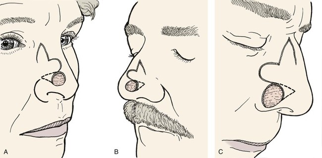
Variations of the bilobe flap are useful. On the nose, the flap may be based medially, although it works best and is most often designed with a lateral base (Fig. 10-2). The lobes of the flap may be designed with rhombic shapes for smaller defects. Bilobe flaps may be used to repair large defects located on the cheek in lieu of larger cervical-facial rotation advancement flaps. They may also be used to transfer skin from the postauricular area to cover helical rim defects that might otherwise require skin grafting.

The bilobe flap is best suited for reconstruction of circular defects on the caudal third of the nose. In this location, reconstruction with other types of nasal cutaneous flaps is difficult. The bilobe nasal flap enables the surgeon to repair defects using adjacent skin without causing nasal distortion. The aesthetic result is usually excellent and often surpasses the result with use of full-thickness skin grafts. This is especially true for deep nasal cutaneous defects. As a consequence, the bilobe flap is a preferred flap for reconstruction of the nose in the area of the tip and caudal dorsum.

The primary disadvantage of the bilobe flap is that most of the incisions necessary to create the flap produce scars that do not parallel relaxed skin tension lines (RSTLs). On the nose, incisions for the flap do not lie within boundary lines separating nasal aesthetic units. The resulting scar is also lengthy due to the need to elevate two lobes. Because bilobe flaps have curvilinear incisions, they are prone to develop a trap-door deformity. This is especially true when they are used on the nose in patients with thick skin or with sebaceous gland hyperplasia. On the nose, trap-door deformity may be minimized by extensive peripheral undermining of the nasal skin as far laterally as the cheek. Designing the lobes of the flap with angles, such as with rectangular- or rhombic-shaped lobes, may also reduce the incidence of trap-door deformity. Adjusting the thickness of the first lobe so that it matches the depth of the recipient site is accomplished by thinning the flap if necessary, and it may be helpful in preventing the deformity.

Clinical Applications

Cheek

The bilobe flap may be used to repair medium-sized (3-6 cm) skin defects of the cheek. It is particularly useful when simple rotation or transposition flaps will not provide sufficient tissue for repair. This occurs when the defect is large and located in the midcheek away from the central part of the face. In this situation, the amount of remaining adjacent cheek skin available for construction of a local flap may be insufficient to cover the cheek defect and still enable closure of the flap donor site. Instead, a bilobe flap designed to recruit upper cervical skin can be useful. Obviously, the lines of wound closure do not fall in RSTLs on the cheek, but the advantage of reducing wound closure tension outweighs the disadvantages of the curvilinear scar created by use of the flap. Like all transposition flaps, bilobed flaps take advantage of lax skin adjacent to the defect to assist in wound closure. The flap is designed to resemble a mitten (Fig. 10-3). The first lobe adjacent to the cheek defect is designed slightly smaller than the defect, and the second lobe is designed to be even smaller. The flap is raised in the subcutaneous tissue plane and transposed into position. The trick is to make sure the donor defect of the second lobe can be closed primarily. This is determined by the pinch test to see if the skin in the proposed area of the second lobe is sufficiently loose to permit primary wound closure. The test is accomplished by gathering the skin of the designed second lobe between the thumb and index finger. If sufficient skin laxity is present, the pinch test should enable the surgeon to approximate the anticipated borders of the second lobe defect.
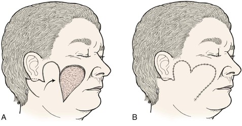
Bilobed flaps may be used anywhere on the cheek; however, care should be taken in using the flap because not all the incisions required to create the flap lie parallel to the natural lines of the face, and the aesthetic result could be disappointing. The flap is best used to repair large to moderate-sized defects of the central cheek. In such cases, the remaining lateral preauricular skin is used to construct the first lobe, and the posterior auricular or superior cervical skin is the source of the second lobe.

Nose

The nose has unique characteristics that a surgeon must consider when choosing a local flap for reconstruction. The topography is complex, with multiple adjacent convex and concave surfaces. The free margins of the alar rims are mobile and easily elevated or displaced. The skin over the caudal third of the nose has limited mobility, restricting its recruitment for local flaps. Skin texture is unique so that only nearby skin will provide an adequate match.

The bilobe flap is well suited for reconstruction of the nose. Many surgeons with experience using bilobe flaps report that it is best suited for use on the caudal third of the nose. In one review of 400 nasal reconstructions, the bilobe flap was the most commonly used flap.4 With little wound closure tension on the first lobe, there is little or no distortion when the flap is used for repair of defects located near the alar rim, provided the first lobe is made sufficiently large. The use of skin adjacent to the defect allows excellent skin color and texture match. The donor site of the second lobe is closed primarily. This is possible because the second lobe is harvested from the lax skin of the upper dorsum and nasal sidewall, where primary approximation of the donor site of the second lobe can occur.

Whenever possible, the nasal bilobe flap is based laterally. Medially based flaps are hardy, although the vascular supply is not as abundant as that of flaps based laterally. Bilobe flaps are ideally suited for repair of defects less than 1.5 cm in maximum dimension on the central or lateral nasal tip and without extension to the ala. Ideally, the defect should be at least 0.5 cm above the margin of the nostril. The flap recruits skin from the mid and upper dorsum and sidewall, where more generous skin laxity is present. Defects of the cephalic half of the nose are not well suited for reconstruction with a bilobe flap unless they are 0.5 cm or less in size. This is because bilobe flaps harvested in this area necessitate use of skin from the region of the medial canthus, which is thin and immobile. Bilobe flaps are most useful in patients with thin skin and an ample degree of skin laxity along the nasal sidewall. The surgeon may estimate laxity by pinching the lateral nasal skin between the thumb and index finger. Patients with thick sebaceous skin have a higher risk for development of flap necrosis, trap-door deformity, and depressed scars.

Bilobe flaps of the nose must be geometrically precise and are designed by the following method (Figs. 10-4 and 10-5).8 The radius of the defect is measured. For laterally based flaps, a point lateral to the border of the defect is marked in the alar groove that is the distance of the length of the radius. This point is used to design both lobes of the flap. Two arcs are drawn with their centers at the marked point. The first arc makes a tangent with the border of the defect most distal to the point, and the second arc passes through the center of the defect. Calipers and rulers are not used to draw the arcs because these devices measure straight-line distances. In contrast, the topography of the nose is convex in the area of the tip and dorsum. Therefore, a flexible measuring device is used. A needle with an attached suture is passed full thickness through the nose at the point marked in the alar groove. A knot is tied in the suture inside the nasal vestibule. The suture is draped from the point across the defect, and a clamp is applied to the suture at the periphery of the defect. The clamp with attached suture is then rotated about its pivotal point to indicate the first arc, which is marked with a pen. The clamp is advanced along the suture to the center point of the defect, and a second arc is drawn through the center />

Only gold members can continue reading. Log In or Register to continue

Stay updated, free dental videos. Join our Telegram channel

Jan 14, 2015 | Posted by in Oral and Maxillofacial Surgery | Comments Off on 10: Bilobe Flaps

VIDEdental - Online dental courses

Get VIDEdental app for watching clinical videos