The radiographic image
Introduction
• Basic physics and equipment – the production of X-rays, their properties and interactions which result in the formation of the radiographic image
• Radiation protection – the protection of patients and dental staff from the harmful effects of X-rays
• Radiography – the techniques involved in producing the various radiographic images
• Radiology – the interpretation of these radiographic images.
Nature of the radiographic image
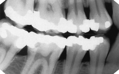
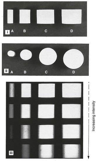
However the final image is captured, it can be described as a two-dimensional picture made up of a variety of black, white and grey superimposed shadows and is thus sometimes referred to as a shadowgraph (see Fig. 1.1).
• The three-dimensional anatomical tissues
• The limitations imposed by a two-dimensional picture and superimposition.
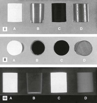
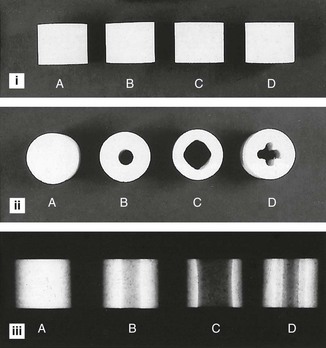
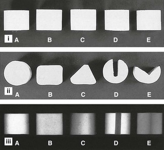
The radiographic shadows
• The white or radiopaque shadows on a film represent the various dense structures within the object which have totally stopped the X-ray beam.
• The black or radiolucent shadows represent areas where the X-ray beam has passed through the object and has not been stopped at all.
• The grey shadows represent areas where the X-ray beam has been stopped to a varying degree.
The final shadow density of any object is thus affected by:
• The specific type of material of which the object is made
• The thickness or density of the material
• The intensity of the X-ray beam used
• The position of the object in relation to the X-ray beam and image receptor
The effect of different materials, different thicknesses/densities, different shapes and different X-ray beam intensities on the radiographic image shadows is shown in Figs 1.2–1.5.
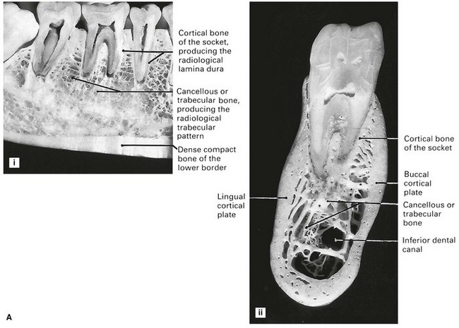
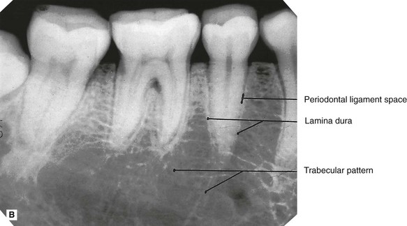
The three-dimensional anatomical tissues
The shape, density and thickness of the patient’s tissues, principally the hard tissues, must also affect the radiographic image. Therefore, when viewing two-dimensional radiographic images, the three-dimensional anatomy responsible for the image must be considered (see Fig. 1.6). A sound anatomical knowledge is obviously a prerequisite for radiological interpretation (see Ch. 16).
Stay updated, free dental videos. Join our Telegram channel
VIDEdental - Online dental courses


