1: Oral and Maxillofacial Radiology

Oral and Maxillofacial Radiology

Interpretation of radiographs is a routine part of the daily practice of oral and maxillofacial surgery. Commonly obtained radiographs at the office include the periapical, occlusal, panoramic, and lateral cephalometric radiographs. Cone beam computed tomography (CBCT) scans are becoming more readily available in many offices. Although this technology is extremely useful, its indications, liabilities, and advantages have to be clearly recognized. As the future unfolds, the advancing technology will improve upon office imaging modalities that will facilitate diagnosis and treatment. Therefore, a knowledge of normal radiographic anatomy and clinical skill in recognizing pathologic conditions become even more essential.

Despite clinicians’ ability to read and interpret many different imaging studies, the oral and maxillofacial radiologist will play an increasingly greater role in the practice of oral and maxillofacial surgeons.

This section includes the radiographic presentation of five important and representative pathologic processes, in addition to a new case demonstrating the use of CBCT. Included in each case is the differential diagnosis of associated conditions, to guide further study.

Figure 1-1 shows the most common location of several radiographically detectable maxillofacial pathologic processes.
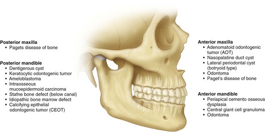
Multilocular Radiolucent Lesion in the Pericoronal Region (Keratocystic Odontogenic Tumor [Odontogenic Keratocyst])

CC

A 20-year-old man is referred for evaluation of a swelling on his right mandible.

MHX/PDHX/Medications/Allergies/SH/FH

Noncontributory. There is no family history of similar presentations.

Nevoid basal cell carcinoma syndrome (NBCCS) is an autosomal dominant inherited condition with features that can include multiple basal cell carcinomas of the skin, multiple KCOTs, intracranial calcifications, and rib and vertebral anomalies. Many other anomalies have been reported with this syndrome (Box 1-1). The prevalence of NBCCS is estimated to be 1 in 57,000 to 1 in 164,000 persons.

Examination

Maxillofacial. The patient has slight lower right facial swelling isolated to the lateral border of the mandible and not involving the area below the inferior border. The mass is hard, nonfluctuant, and nontender to palpation (large cysts may rupture and leak keratin into the surrounding tissue, provoking an intense inflammatory reaction that causes pain and swelling). There are no facial or trigeminal nerve deficits (paresthesia of the inferior alveolar nerve would be more indicative of a malignant process). The intercanthal distance is 33 mm (normal), and there is no evidence of frontal bossing. His occipitofrontal circumference is normal (an intercanthal distance [the distance between the two medial canthi of the palpebral fissures] of greater than 36 mm is indicative of hypertelorism, and an occipitofrontal circumference greater than 55 cm is indicative of frontal bossing; both can be seen with NBCCS).

Neck. There are no palpable masses and no cervical or submandibular lymphadenopathy. Positive lymph nodes would be indicative of an infectious or a neoplastic process. A careful neck examination is paramount in the evaluation of any head and neck pathology.

Intraoral. Occlusion is stable and reproducible. The right mandibular third molar appears to be distoangularly impacted (KCOTs do not typically alter the occlusion). The interincisal opening is within normal limits. There is buccal expansion of the right mandible, extending from the right mandibular first molar area posteriorly toward the ascending ramus. Resorption of bone may include the cortex at the inferior border of the mandible, but this is observed at a slower rate than in intermedullary bone, which is less dense. For this reason, KCOTs characteristically extend anteroposteriorly than buccolingually. This pattern of expansion into less-dense bone explains why maxillary KCOTs show more buccal than palatal expansion and often expand into the maxillary sinus. There is no palpable thrill or audible bruit, both of which are seen with arteriovenous malformations. The oral mucosa is normal in appearance with no signs of acute inflammatory processes.

Thorax-abdomen-extremity. The patient has no findings suggestive of NBCCS (e.g., pectus excavatum, rib abnormalities, palmar or plantar pitting, and skin lesions; see Box 1-1).

Imaging

A panoramic radiograph is the initial screening examination of choice for patients presenting for evaluation of intraosseous mandibular pathology (10% to 20% of KCOTs are incidental radiographic findings). This provides an excellent overview of the bony architecture of the maxilla, mandible, and associated structures. CT scans can be obtained when large lesions are found. CT scans are valuable in that they provide additional information, such as the proximity of adjacent structures (e.g., the mandibular canal), the integrity of cortical plates, and the presence of perforations into adjacent soft tissues. CT scans provide accurate assessment of the size of the lesion and can demonstrate additional anatomic details (or lesions) that do not appear on panoramic radiographs.

A CBCT scan is appropriate for the evaluation of this lesion. Given its higher resolution, lower radiation dose (approximately 20% of the radiation of a conventional [helical] CT ), and lower cost, a CBCT can replace helical CT for evaluation and follow up of such a lesion. The CBCT scan can also be used to create a stereolithic model of the area of interest.

It has been demonstrated that T2-weighted magnetic resonance imaging (MRI) can detect KCOTs in 85% of new cases with a readily recognizable pattern. However, the use of MRI for management of suspected OKCs is not routine.

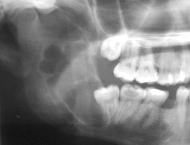
In this patient, the panoramic radiograph reveals a large, multilocular radiolucent lesion with possible displacement of the right mandibular third molar (Figure 1-2). There are also several carious teeth and a retained root tip of the right mandibular second bicuspid (tooth #29). (In a patient with a radiolucent lesion of the mandible presumed to be an odontogenic cystic lesion, a multilocular appearance is associated with a 12-fold increased risk for the diagnosis of KCOT; however, the presence of a unilocular lesion does not exclude the possibility of a KCOT diagnosis.)

Differential Diagnosis

The differential diagnosis of multilocular radiolucent lesions can be divided into lesions of cystic pathogenesis, neoplastic (benign or malignant) lesions, and vascular anomalies (least common). The differential diagnosis of multilocular radiolucent lesions is presented in Box 1-2 and can be further narrowed by the clinical presentation. Special consideration should be given to radiolucent lesions with poorly defined or ragged borders, which have a separate differential.

Box 1-2   Differential Diagnosis of Multilocular Radiolucent Lesions

• Ameloblastoma—The most frequent location is the posterior mandible, and the tumor’s most common radiographic appearance is that of a multilocular radiolucent lesion. This is the most frequently diagnosed odontogenic tumor.

• Keratocystic odontogenic tumor—This lesion cannot be differentiated on clinical and radiographic grounds from an ameloblastoma. KCOTs generally do not cause resorption of adjacent teeth. The orthokeratin variant is usually associated with an impacted tooth.

• Dentigerous cyst—Large dentigerous cysts can have a multilocular appearance on a radiograph, given the existence of bone trabeculae within the radiolucency. However, they are histologically a unilocular lesion. There is a strong association with impacted mandibular third molars. Painless bony expansion and resorption of adjacent teeth are uncommon but can occur.

• Ameloblastic fibroma—The posterior mandible is also the most common site for this lesion. It is predominantly seen in the younger population, and most lesions are diagnosed within the first two decades of life. Large tumors can cause bony expansion. The lesion can manifest as a unilocular or multilocular radiolucent lesion that is often associated with an impacted tooth. Ameloblastic fibro-odontomas are mixed radiopaque-radiolucent lesions.

• Central giant cell tumor—Approximately 70% of these lesions occur in the mandible, most commonly in the anterior region. The tumor’s radiographic appearance can be unilocular or multilocular. These lesions can contain large vascular spaces that can lead to substantial intraoperative bleeding. The aneurysmal bone cyst has been suggested to be a variant of the central giant cell tumor. The majority of these lesions are discovered before age 30.

• Odontogenic myxoma—Although myxomas are seen in all age groups, the majority are discovered in patients 20 to 40 years of age. The posterior mandible is the most common location, and the tumor’s radiographic appearance can be unilocular or multilocular. At times, the radiolucent defect may contain thin, wispy trabeculae of residual bone, given its “cobweb” or “soap bubble” trabecular pattern.

• Aneurysmal bone cyst—Lacking a true epithelial lining, these cysts most commonly occur in the long bones or the vertebral column. They rarely occur in the jaws, but when they do, it is mostly in young adults. They can present as a unilocular or multilocular radiolucent lesion with marked cortical expansion that usually displaces but does not resorb teeth.

• Traumatic bone cyst—This lesion lacks a true epithelial lining and frequently involves the mandibular molar and premolar region in young adults. These cysts can cause expansion and usually show a well-defined unilocular, scalloping radiolucency between the roots without resorption. The lesion always exists above the inferior alveolar canal.

• Calcifying epithelial odontogenic tumor (CEOT)—This is an uncommon tumor. The majority are found in the posterior mandible, mostly in patients age 30 to 50 years. A multilocular radiolucent defect is seen more often than a unilocular radiolucency. Although the tumor may be entirely radiolucent, calcified structures of varying sizes and density are usually seen within the defect. CEOTs can also be associated with an impacted tooth.

• Lateral periodontal cyst (botryoid odontogenic cyst)—Usually found in older individuals (fifth to seventh decades of life), the botryoid variant often shows a multilocular appearance. It is most commonly seen in the premolar canine areas.

• Calcifying odontogenic cyst (COC)—Most commonly found in the incisor canine region, this cyst is usually diagnosed in patients in the mid-30s. Although the unilocular presentation is most common, multilocular lesions have been reported. Radiopaque structures are usually present in approximately one third to one half of the lesions.

• Intraosseous mucoepidermoid carcinoma—This is the most common salivary gland tumor arising centrally within the jaws. Most commonly found in the mandible of middle-aged adults, the tumors can appear radiographically as unilocular or multilocular radiolucent lesions. Association with an impacted tooth has been reported.

• Hyperparathyroidism (brown tumor)—Parathyroid hormone (PTH) is normally produced by the parathyroid gland in response to decreased serum calcium levels. In primary hyperparathyroidism, uncontrolled production of PTH is due to hyperplasia or carcinoma of the parathyroid glands. Secondary hyperparathyroidism develops in conditions of low serum calcium levels (e.g., renal disease), resulting in a feedback increase in PTH. Patients with hyperparathyroidism usually present with a classic triad of signs and symptoms, described as “stones, bones, and abdominal groans.” Patients with primary hyperparathyroidism have a marked tendency to develop renal calculi (“stones”). “Bones” refers to the variety of osseous changes that are seen, including the brown tumor of hyperparathyroidism. These lesions can appear as unilocular or multilocular radiolucent lesions, most commonly affecting the mandible, clavicle, ribs, and pelvis. “Abdominal groans” refers to the tendency of these patients to develop duodenal ulcers and associated pain. When dealing with any giant cell lesions, the clinician must rule out the brown tumor of hyperparathyroidism by evaluating the patient’s serum calcium level (it is elevated in hyperparathyroidism). Patients with brown tumor also have elevated levels of PTH (which is confirmed by radioimmunoassay of the circulating parathyroid levels).

• Cherubism—In this rare developmental inherited condition, painless bilateral expansion of the posterior mandible produces cherublike facies (plump-cheeked little angels depicted in Renaissance paintings). In addition, involvement of the orbital rims and floor produces the classic “eyes upturned toward heaven.” Radiographically, the lesions are usually bilateral multilocular radiolucent lesions. Although rare, unilateral involvement has been reported.

• Intrabony vascular malformations—Arteriovenous malformations are most often detected in patients between 10 and 20 years of age and are more commonly found in the mandible. Mobility of teeth, bleeding from the gingival sulks, an audible bruit, or a palpable thrill should alert the clinician. The radiographic appearance is variable, but the malformation most commonly presents as a multilocular radiolucent lesion. The loculations may be small, giving the honeycomb appearance that produces a “soap bubble” radiographic appearance. Aspiration of all undiagnosed intrabony lesions is warranted to rule out the presence of this lesion, because fatal hemorrhage can occur after an incisional biopsy.

Biopsy

An incisional or excisional biopsy can be performed, depending on the size of the lesion. A smaller cystic lesion can be completely excised, whereas larger lesions require an incisional biopsy to guide final therapy. It is important to aspirate the lesion before incising into it (entering carefully through the cortical bone) to rule out a vascular lesion. The aspiration of bright red blood alerts the surgeon to the presence of a high-flow vascular lesion, such as an arteriovenous malformation, which could result in uncontrollable hemorrhage. In such a case, the procedure should be aborted to allow for further radiographic and angiographic studies to characterize the vasculature of the area. The aspiration of straw-colored (or clear) fluid is characteristic of a cystic lesion, and the absence of any aspirate may be seen with a solid mass (tumors).

Assessment

Expansile multilocular radiolucent mass of the posterior right mandible associated with an impacted right mandibular third molar (25% to 40% of cases are associated with an unerupted tooth).

With this patient under intravenous anesthesia, an incisional biopsy was performed after aspiration of straw-colored fluid that showed the classic histopathology of the KCOT. Histologic features include a thin squamous cell epithelial lining (eight cells or fewer). Because of the lack of rete ridges, the epithelial–connective tissue interface is flat. The epithelial surface is parakeratinized and often corrugated (wavy). The basal cell layer is hyperchromatic and composed of cuboidal cells, which show prominent palisading, giving a “tombstone” effect. The fibrous wall is usually thin and at times shows a mixed inflammatory response. Keratinization of the lumen is not a pathognomonic finding. The fibrous wall may contain epithelial islands that show central keratinization and cyst formation; these are known as daughter-satellite cells.

Treatment

Options that have been used to treat KCOTs include the following:

Resection is advocated only if there have been multiple recurrences after enucleation with an adjunctive procedure (e.g., cryotherapy, Carnoy’s solution, or peripheral ostectomy) or for a large KCOT exhibiting aggressive behavior, such as destruction of adjacent tissues. Several studies demonstrate that enucleation alone (when the diagnosis of KCOT has been established) has a high recurrence rate; therefore, many surgeons advocate enucleation with a local adjunctive procedure, such as cryotherapy, Carnoy’s solution, and/or peripheral ostectomy.

KCOTs do not invade the epineurium; therefore, the inferior alveolar nerve can be separated and preserved. Furthermore, any perforations of the keratinized mucosa should be excised, because they may contain additional epithelial rests, which can lead to recurrences. Aggressive soft tissue excision is not required, because KCOTs do not usually infiltrate adjacent structures. If the cyst is removed in one unit, there is no need for curettage, unless the lining has been shredded or torn.

Some controversy exists regarding the optimal management (extraction versus retention) of teeth involved with an KCOT. It is generally accepted that a KCOT with a scalloped radiographic appearance should have the associated teeth removed, because it is considered impossible to completely remove the thin-walled cystic lining. However, if the KCOT is successfully removed in one unit, the teeth may be spared without compromising recurrence. In most instances there is no need for endodontic therapy, despite surgical denervation. The teeth may not become devitalized due to perfusion of the pulp via accessory canals through the periodontal ligaments.

Some surgeons advocate the application of Carnoy’s solution after enucleation and peripheral ostectomy with application of methylene blue. Carnoy’s solution is composed of 6 ml of absolute alcohol, 3 ml of ferric chloride, and 1 ml of 100% acetic acid. Chloroform is no longer recommended due to its carcinogenic potential. Carnoy’s solution penetrates the bone to a depth of 1.54 mm after a 5-minute application. It is difficult to obtain and needs to be mixed fresh. It does not fixate the inferior alveolar nerve, but some clinicians cover the nerve with sterile petrolatum as a caution.

Synchronous bone grafting is not carried out with this technique. Cryotherapy with liquid nitrogen is an acceptable alternative to the use of Carnoy’s solution. Liquid nitrogen is sprayed within the cavity and penetrates to a depth of about 1.5 mm. Suggested protocols include spraying the cavity for 1 minute and then allowing the bone to thaw. This can be repeated two or three times.

Synchronous grafting with cancellous bone can be accomplished after cryotherapy. Patients should be cautioned since liquid nitrogen does weaken the mandible, and this may result in a pathologic fracture. Sensory nerves within the field may show paresthesia; however, the majority recover within 3 to 6 months.

With both techniques, adjacent soft tissue needs to be protected. An alternate technique is used in cases of buccal or lingual plate perforation and with sinus involvement.

This patient was treated under general anesthesia with enucleation of the lesion followed by the application of methylene blue to guide peripheral ostectomy. The patient was placed on a soft diet to reduce the risk of jaw fracture. The postoperative panoramic radiograph confirmed that the inferior border of the mandible remained intact.

The final pathology report confirmed the diagnosis of a KCOT consistent with the initial incisional biopsy specimen. The patient was placed on a strict recall schedule—every 6 months for the first 5 years and then yearly. The recurrence rate for KCOTs has been reported to range from 5% to 60%. It has been reported that most recurrences are seen within 5 years, although they can develop at any time. Recurrences that arise secondary to residual cyst left in the bone may be apparent within 18 months of surgery.

Complications

The KCOT has been described as having clinical features that include potentially aggressive behavior and a high recurrence rate. Because recurrence is a major concern, clinicians vary in their surgical approach. Resection results in the lowest recurrence rate; however, there is considerable morbidity associated with this radical treatment. The primary mechanisms for recurrence have been postulated to be incomplete removal of all the cystic lining; new primary cyst formation from additional activated rests; or the development of a new KCOT in an adjacent area that is interpreted as a recurrence.

KCOTs have been reported to undergo transformation into ameloblastoma and squamous cell carcinoma, although this occurrence is rare. Other common postprocedural complications include inferior alveolar nerve paresthesia, postoperative infection and, with larger lesions, pathologic mandibular fracture (the highest risk is during the first few weeks after enucleation).

Discussion

Ever since the histologic features of the KCOT were established, many investigators have recognized that two major variants exist, based on microscopic findings: a cyst with a parakeratinized epithelial lining and a cyst with an orthokeratinized epithelial lining.

Crowley and colleagues undertook a comparison of the orthokeratin and parakeratinized variants. In their review, they found that the parakeratinized variant occurred more commonly than the orthokeratinized variant (frequency of 86.2% for the parakeratinized variant compared with 12.2% for the orthokeratinized variant); 1.6% of cysts had both orthokeratin and parakeratin features.

These researchers also found that the parakeratinized variant demonstrated a 42% recurrence rate, compared to only 2.2% for the orthokeratinized variant. In addition, the orthokeratinized variant was more frequently associated with impacted teeth. Given the different clinical behaviors of these two entities, many authors designate them as separate pathologic lesions, with the orthokeratinized variant known as an orthokeratinized odontogenic cyst (OOC). A lesion with both orthokeratin and parakeratin features should be treated as a parakeratinized KCOT (OKC).

As was mentioned earlier, in 2005 WHO designated the OKC a keratocystic odontogenic tumor (KCOT). The lesion was defined as a benign, unicystic or multicystic intraosseous tumor of odontogenic origin with a characteristic lining of parakeratinized, stratified squamous epithelium and the potential for aggressive infiltrative behavior.

Stimulation of residual epithelial cells is a common feature in the development of any cyst. In the case of the KCOT, the epithelial cells implicated are from the rest of Serres or Malassez or from the reduced enamel epithelium. Aberration of a PTCH gene is thought to be a genetic cause for the etiology of the KCOT. Collagenase activity in the cyst’s epithelium, with its resorptive properties, appears to regulate the ability of the lesion to grow expansively in bone.

Identification of individuals who may have NBCCS allows the clinician to arrange for appropriate referrals. NBCCS should be suspected when multiple lesions exist. The diagnosis is confirmed upon finding any two of the major criteria, or one major criterion plus two minor criteria (see Box 1-1). Some abnormalities are pertinent only to the diagnosis and do not require any specific therapy. Other abnormalities may pose further risk to the patient and require the input of other specialists. Spina bifida and central nervous system tumors require referral to a neurosurgeon. In addition, genetic counseling for all patients afflicted with NBCCS is recommended. KCOTs associated with this syndrome are treated in the same manner as an isolated KCOT; however, these lesions have a higher rate of recurrence when associated with NBCCS (which may represent new lesions). KCOTs are often associated with the follicle of a potentially functional tooth and, when possible, marsupialization with orthodontic guidance should be considered.

Bibliography

August, M, Caruso, PA, Faquin, WC. Report of a case: an 18-year-old man with a one-month history of nontender left mandibular swelling. N Engl J Med. 2005; 353:2798–2805.

August, M, Faquin, WC, Troulis, M, et al. Differentiation of odontogenic keratocysts from nonkeratinizing cysts by use of fine-needle aspiration biopsy and cytokeratin-10 staining. J Oral Maxillofac Surg. 2000; 58:935–940.

Barnes L, Eveson JW, Reichart P, et al, eds. Pathology and genetics: head and neck tumours (IARC World Health Organization classification of tumors). Lyon, France: IARC Press, 2005.

Buckley, P, Seldin, E, Dodson, T, et al. Multilocularity as a radiographic marker of the keratocystic odontogenic tumor. J Oral Maxillofac Surg. 2012; 70:320.

Crowley, T, Kaugras, GE, Gunsolley, JC. Odontogenic keratocysts: a clinical and histologic comparison of the parakeratin and orthokeratin variants. J Oral Maxillofac Surg. 1992; 50:22–26.

Evans, DG, Ladusans, EJ, Rimmer, S, et al. Complications of the naevoid basal cell carcinoma syndrome: results of a population based study. J Med Genet. 1993; 30:460–464.

Kolokythas, A. OKC: to decompress or not to decompress—a comparative study between decompression and enucleation vs resection/peripheral ostectomy. J Oral Maxillofac Surg. 2007; 65(4):640–644.

Madras, J. Keratocystic odontogenic tumor: reclassification of the odontogenic keratocyst from cyst to tumor. J Can Dent Assoc. 2008; 74:2.

Marx, RE, Stern, D. Oral and maxillofacial pathology: a rationale for diagnosis and treatment. Chicago: Quintessence; 2003.

Pindborg, JJ, Hansen, J. Studies on odontogenic cyst epithelium: clinical and roentgenographic aspects of odontogenic keratocysts. Acta Pathol Microbial Scand. 1963; 58:283.

Pogrel, MA. Keratocystic odontogenic tumor. In: Bagheri S, ed. Current therapy in oral and maxillofacial surgery. St Louis: Mosby, 2012.

Pogrel, MA, Jordan, RC. Marsupialization as a definitive treatment for the odontogenic keratocyst. J Oral Maxillofac Surg. 2004; 62:651–655.

Pogrel, MA, Schmidt, BL. The odontogenic keratocyst. Oral Maxillofacial Surg Clin N Am. 15(3): xi, 2003.

Shear, M. Primordial cysts. J Dent Assoc S Afr. 1960; 152:1.

Tolstunov, L, Treasure, T. Surgical treatment algorithm for odontogenic keratocyst: combined treatment of odontogenic keratocyst and mandibular defect with marsupialization, enucleation, iliac crest bone graft and dental implants. J Oral Maxillofac Surg. 2008; 66:1025.

Quereshy, F, Barnum, G, Demko, C, et al. Applications of cone beam computed tomography in the practice of oral and maxillofacial surgery. J Oral Maxillofac Surg. 2008; 66:791.

Unilocular Radiolucent Lesion of the Mandible

Examination

General. The patient is a well-developed and well-nourished anxious man (patients are often anxious because they fear a malignant process).

Maxillofacial. There is noticeable right lower facial swelling isolated to the lateral border of the mandible not involving the area below the inferior border. The mass is hard, nonfluctuant, and nontender to palpation, consistent with a noninflammatory process. There are no facial or trigeminal nerve deficits (paresthesia of the right inferior alveolar nerve would be indicative of a malignant process).

Neck. The patient does not have palpable masses or cervical or submandibular lymphadenopathy. Positive lymph nodes would be indicative of an infectious or neoplastic etiology, so a careful neck examination is paramount in the evaluation of any head and neck pathology.

Intraoral. The occlusion is stable and reproducible. There does not appear to be displacement of the dentition in the involved area (dentigerous cysts do not typically alter the occlusion). Interincisal opening is within normal limits. There is significant buccal expansion of the right mandible, extending from the mental foramen posteriorly and ascending into the ramus (large cysts may be associated with a painless expansion of the bone, but most are asymptomatic and do not cause expansion). The patient does not have a palpable thrill or an audible bruit (both are seen with arteriovenous malformations). The oral mucosa is normal in appearance with no signs of any acute inflammatory processes.

Imaging

When evaluating large lesions of the mandible, the panoramic radiograph is an excellent initial study for assessment of the bony and dental anatomy. A CT scan is not essential, but it can better delineate the three-dimensional bony and regional architecture, including involvement of the cortices of the mandible (i.e., cortical perforation is seen with some tumors and locally aggressive cysts) and the lesion’s position in relation to the inferior alveolar canal.

In this patient, a panoramic radiograph (Figure 1-3, A) demonstrates a well-corticated unilocular radiolucent lesion of the right posterior mandible extending from the area of tooth #31 up to the sigmoid notch and coronoid process. The right mandibular third molar (tooth #32) is displaced inferiorly, and the lesion involves the roots of tooth #31, with some resorption and superior displacement of the tooth. After aspiration and incisional biopsy, teeth #31 and teeth #32 were extracted and the cyst was enucleated and curettaged (Figure 1-3, B to E). Six- and 16-week postoperative orthopantograms demonstrate good progressive bony fill of the defect (Figure 1-3, F and G).
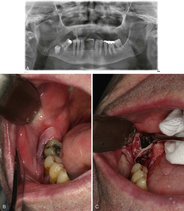
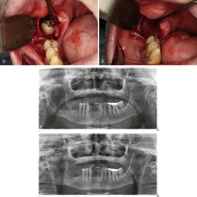
Differential Diagnosis

The differential diagnosis can be divided into lesions of cystic pathogenesis, neoplastic (benign or malignant) lesions, and vascular anomalies (least common). Well-defined borders and the lack of a multilocular appearance are more suggestive of a cystic process, but this distinction is not predictable. The differentials presented in Box 1-3 should be considered; the first three are the most likely.

Stay updated, free dental videos. Join our Telegram channel

Jan 12, 2015 | Posted by in Oral and Maxillofacial Surgery | Comments Off on 1: Oral and Maxillofacial Radiology

VIDEdental - Online dental courses

Get VIDEdental app for watching clinical videos