1
CONE BEAM VOLUMETRIC IMAGING IN CLINICAL PRACTICE
Nothing has captured the dental profession’s imagination in the past few years like the introduction of cone beam volumetric imaging (CBVI), sometimes referred to as cone beam computerized tomography (CBCT) or cone beam volumetric tomography (CBVT). These machines’ image acquisition process differs from that of traditional medical computerized tomography (CT) scanners in that the patient is not usually supine, the image gathered is in a voxel (volume element) format, the x-ray dose absorbed by the patient is substantially lower, appointment availability is much easier, and it is less expensive. In short, although the imaging modality produces significant data volumes like medical CT, it is vastly superior to traditional CT data for specific dental applications.
Box 1-1 Applications of CBVI
3-D virtual model construction
Airway studies related to sleep apnea
Bone structure (dehiscences, fenestrations, and/or periodontal defects in adults)
Impaction related to orthodontics
Implant site assessment including temporary anchorage devices
Inferior alveolar nerve location for third molar extraction
Odontogenic lesion visualization
Orthodontic assessment
Other computer-aided design/computer-assisted manufacture devices
Paranasal sinus evaluation
Space analysis (1:1)
Surgical guide fabrication
Temporomandibular joint visualization
Trauma evaluation
Dentists and dental specialists have seen and appreciated the incredibly precise and profound information produced by CBVI scans, and they are realizing that the data they receive will influence their treatment decisions like no other imaging modality used in the profession in the past 100 years. CBVI makes clinical decision making easier and more precise, patient treatment decisions more accurate, and visualization of the x-ray data more meaningful. Dentistry is moving away from “radiographic interpretation” and into “disease visualization,” and it could not have come at a better time.
PRESENT CONSIDERATIONS FOR CBVI
To date, I have read, or interpreted, almost 3,000 CBVI cases for my dental colleagues. I practice my specialty of oral and maxillofacial radiology from my home in a dedicated radiology office environment via the Internet. I continue to interpret images and create reports for clinicians while I travel, speak, and consult. I operate in the same manner as my medical radiology colleagues and practice my specialty from virtually anywhere in the world because of global Internet access.
Just as there are many different models on the market, I receive data volumes to interpret through many different avenues. Gone are the days when we relied on 2-D grayscale single images attempting to represent 3-D structures, viewed on lightboxes under poor lighting conditions, to help us make our clinical treatment decisions. It is now possible to have 2-D or 3-D color “renderings” of each patient’s anatomy and signs of clinical diseases/disorders.
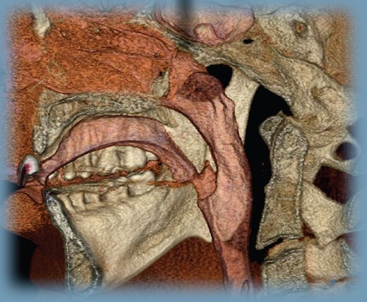
Figure 1-1 shows the CBVI machine’s broad capabilities and power. Whether you are considering purchasing a machine for image acquisition in your practice or are simply accessing this technology by requesting a scan, you should consider the following important questions:
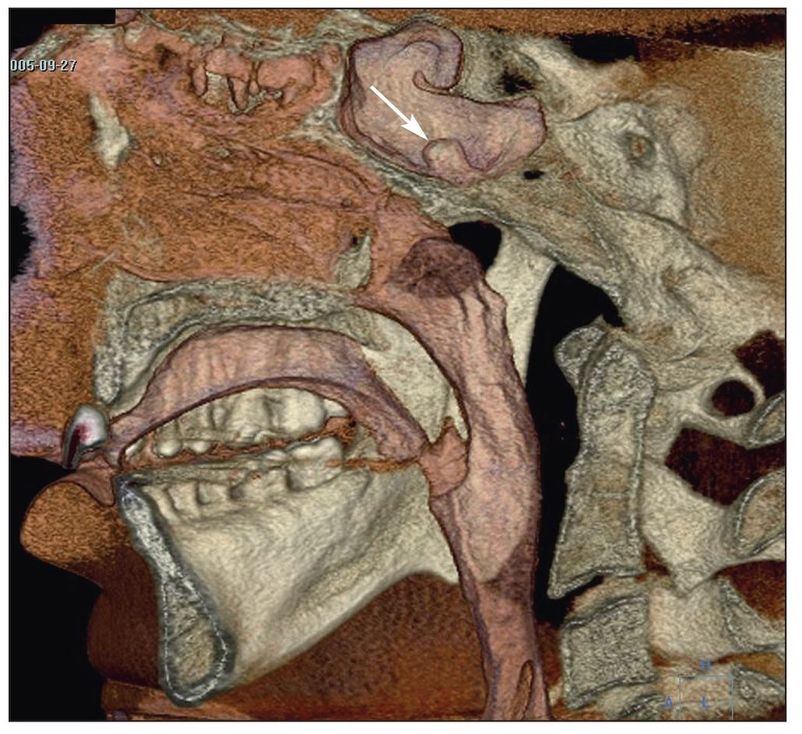
Fig 1-1 Large-volume 3-D color reconstruction of a patient’s airway created with data from an i-CAT machine (Imaging Sciences International) and processed using Accurex software (CyberMed International). Note the cutaway sections of the mandible, the vertebral bodies, and the lesion in the sphenoid sinus, which is an osteoma. The color renderings aid in visualization of the soft tissue lining over the bony lesion (arrow).
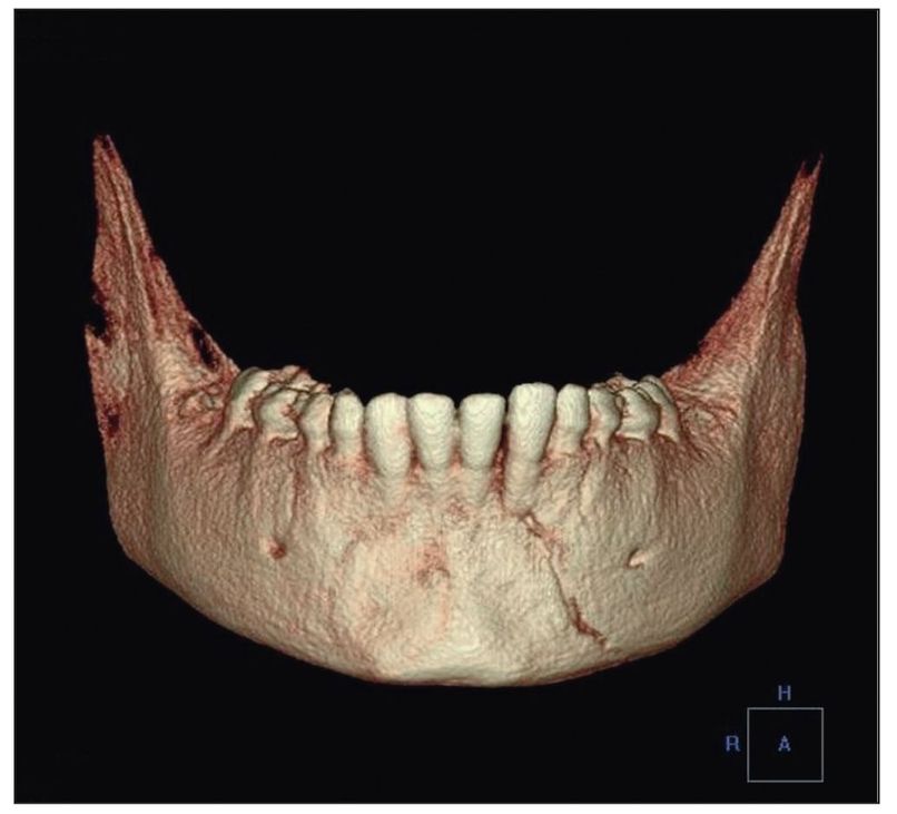
Fig 1-2 Small-volume 3-D color reconstruction of a 9-year-old patient with a fractured mandible, rendered with Accurex. The fracture is easily identified in the anteroposterior view, and the 3-D image can be rotated 360 degrees to see the fracture in any orientation.
- How much data (number of images) do you need?
- How large an area do you wish to evaluate?
- Do you need s/>
Stay updated, free dental videos. Join our Telegram channel
VIDEdental - Online dental courses


