1
Anatomy of the periodontium
Figure 1.1 Longitudinal section through part of a tooth showing healthy periodontal tissues.
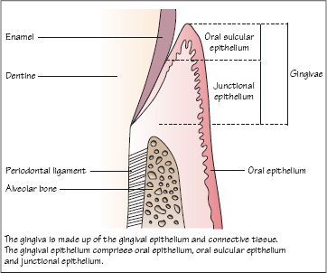
Figure 1.2 Dentogingival fibres, alveolar crest fibres and circular fibres in the gingival connective tissue.
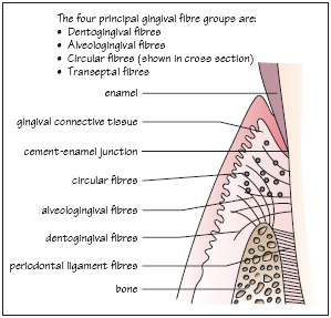
Figure 1.3 Interdental area showing transeptal and circular fibre groups in the gingival connective tissue.
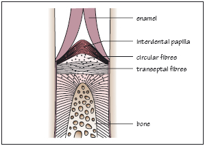
Figure 1.4 The periodontal ligament.
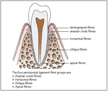
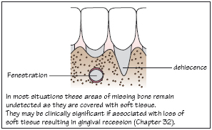
Figure 1.5 Bony fenestration and dehiscence.
The periodontal tissues form the supporting structures of the teeth. The principal components of the periodontium are shown in Fig. 1.1:
The gingivae in health are pink and firm with a knife-edge appearance, scalloped around the teeth. In certain ethnic groups the gingivae may be pigmented. In health, the gingival margin is a few millimetres coronal to the cement–enamel junction. The gingival sulcus (or crevice) is a shallow groove which may be between 0.5 and 3 mm in depth around a fully erupted tooth. The gingival tissues are keratinised and appear paler pink than sites of non-keratinised oral epithelium.
Gingival epithelium
The gingival epithelium comprises (Fig. 1.1):
The gingival sulcus is lined by SE and JE.
Oral epithelium
Stay updated, free dental videos. Join our Telegram channel
VIDEdental - Online dental courses


