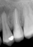
The two maxillary premolar teeth did not respond to thermal and electric tests, while the canine and first molar responded normally.
What did the radiograph reveal (Figure 1.3.2)?
Figure 1.3.2 Radiograph of premolar 24 revealed a periapical radiolucency.

- The 25 had been root canal treated, and the periapical tissues appeared to be healthy.
- The 24 had secondary caries and a periapical radiolucency.
Diagnosis and treatment planning
A diagnosis of chronic periapical periodontitis with suppuration associated with a necrotic root canal was reached for the 24.
What are the histological features of suppuration in a periapical lesion?
Periapical lesions are classified histologically into periapical abscess (i.e. suppuration), granuloma and cyst (Table 1.3.1). Based on the presence of epithelium, abscesses and granulomas can be further classified into epithelialized or non-epithelialized. A periapical abscess is a focus of acute inflammation characterized by an accumulation of polymorphonuclear leukocytes (PMNs) within an already existing chronic granuloma (Figure 1.3.3). Numerous microcavities can be observed in the inflammatory tissue, containing necrotic debris and PMNs. These cavities tend to coalesce, forming larger spaces containing pus. Pus is washed away during histological processing and these cavities appear empty or show some remaining
debris in their lumen. The abscess cavity is surrounded by a severe concentration of inflammatory cells.
Figure 1.3.3 Palatal root of an extracted asymptomatic maxillary molar with chronic periapical periodontitis associated with suppuration, demonstrating histologically the clinical situation in Figure 1.3.1
Stay updated, free dental videos. Join our Telegram channel

VIDEdental - Online dental courses


