PREPARATION FOR ARRANGING TEETH
Figure 1 The mandibular occlusion rim is built up with baseplate wax after the material used for making the interocclusal jaw relation record is removed.
Figures 2 and 3 The occlusion rims have been rebuilt with baseplate wax and are ready for the arrangement of teeth. Note the horizontal overlap.
Figure 4 Note the position of the center line, the canine line and the high lip line. These are the lines which the dentist uses to select teeth for his patient. The center line is transferred from the occlusion rim to the maxillary cast as a permanent reference.
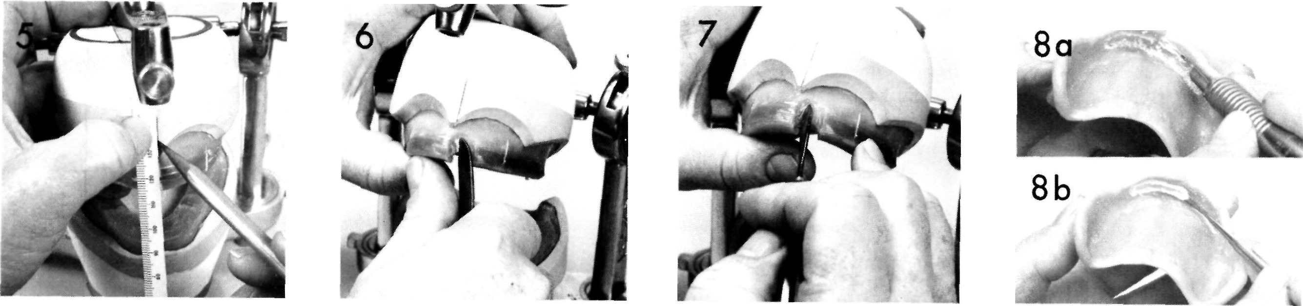
Figure 5 The center line may first be marked with a pencil to insure its proper placement.
Figure 6 A block of wax corresponding to the position of the maxillary left central incisor is removed from the occlusion rim.
Figure 7 The wax surrounding this area is softened with a hot spatula.
Figure 8 Sometimes the baseplate may interfere with proper placement of the teeth. When this occurs a portion of the baseplate is removed. The section is first cut on the tissue surface with a fissure bur (a) and is then lifted out with an instrument (b). The cast opposite the alteration should be lightly coated with Vaseline.
ARRANGING THE MAXILLARY ANTERIOR TEETH
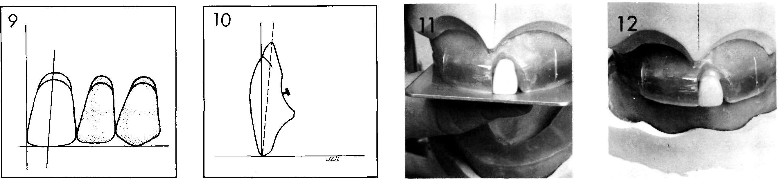
Figure 9 The maxillary central incisor is placed so that the long axis shows a slight distal inclination to the perpendicular. The incisal edge is on the occlusal plane.
Figure 10 The neck of the tooth should be slightly depressed. However, the facial surface of the tooth is nearly perpendicular to the occlusal plane. Avoid over-depressing the neck of this tooth or the tooth will not be in a natural position when viewed in the mouth.
Figure 11 The maxillary left central incisor is placed in the molten wax to conform to the position shown in Figures 9 and 10. The incisal edge is on the occlusal plane. An aluminum plate may be used to orient the teeth to the occlusal plane.
Figure 12 The maxillary left central incisor is in proper position. A small amount of wax has been flowed around the tooth to insure its retention in the wax.
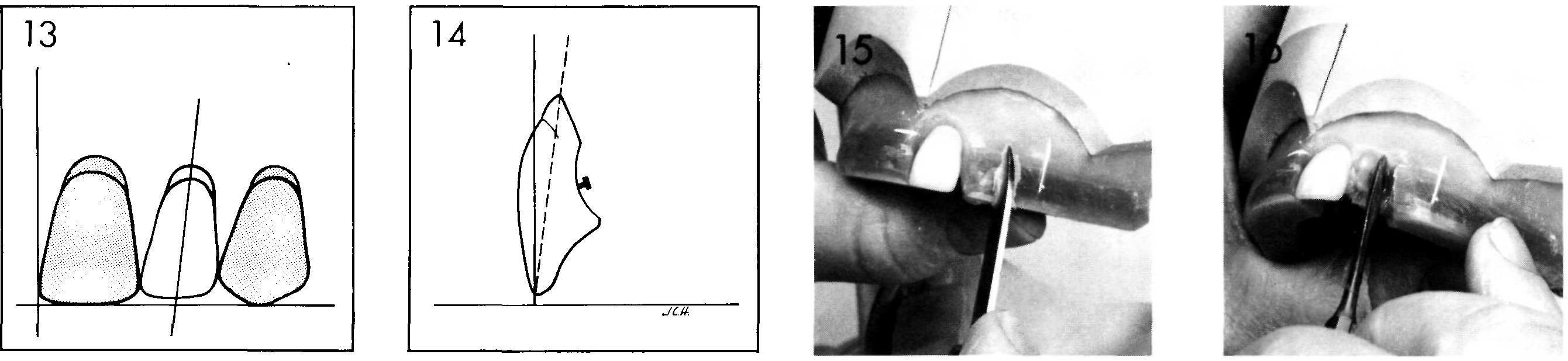
Figure 13 The maxillary lateral incisor has slightly more distal inclination than the central incisor. The incisal edge is usually ½ to 1 mm. above the occlusal plane.
Figure 14 The neck of the maxillary lateral incisor is depressed more than the central incisor, although the facial surface will be nearly in line with the central incisor.
Figure 15 A block of wax is removed in the left lateral incisor area.
Figure 16 The wax is softened with a hot wax spatula.
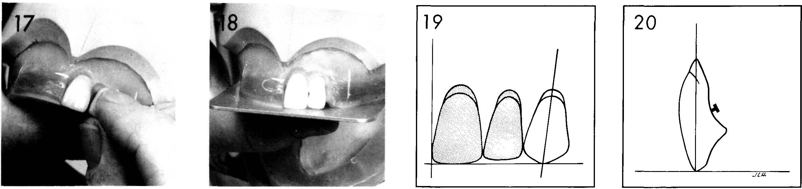
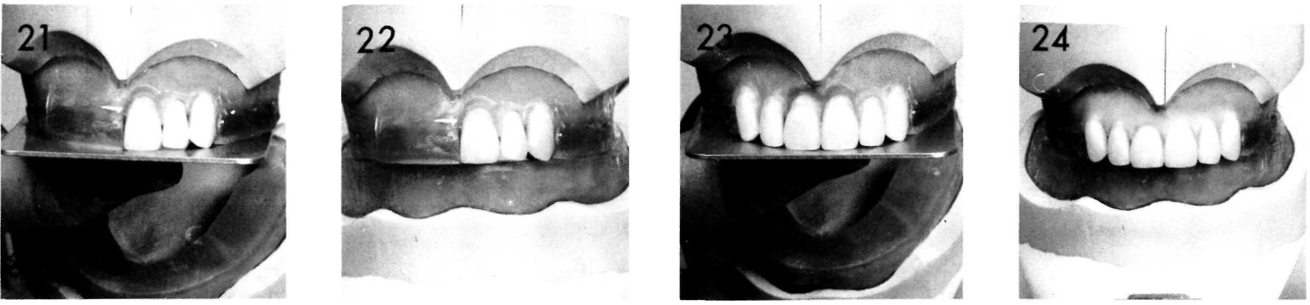
Figure 17 The lateral incisor is placed in position in the softened wax.
Figure 18 The maxillary left lateral incisor is placed to conform to the position shown in Figures 13 and 14. Note that the incisal edge is slightly above the occlusal plane and that the neck is slightly depressed.
Figure 19 The maxillary canine is placed so that the long axis is almost vertical. It may have a slight distal inclination but should not lean mesially. The incisal tip is on the occlusal plane.
Figure 20 The neck of the maxillary canine is prominent.
Figures 21 and 22 The maxillary left canine is placed in position using the same technique previously used for the central and lateral incisors. The canine tooth is an important tooth in any tooth arrangement because it forms the corner of the dental arch. The maxillary canine has two planes on the facial surface; the mesial plane should follow the contour of the anterior teeth while the distal plane will be in line with the posterior teeth. The neck of the tooth is prominent as it supports the corner of the patient’s mouth.
Figure 23 The maxillary right anterior teeth are set in a similar fashion. Note the relation of the incisal edges to the occlusal plane. Note also the prominence of the gingival third of the maxillary central incisors and canines. The gingival third of anterior teeth is primarily responsible for lip support. Depressing the gingival third causes the upper lip to have a flaccid appearance. (The proper angulation of the maxillary anterior teeth may be judged by imagining that the artificial tooth has grown a root. By judging the amount of resorption—using the position of the incisive papilla as a guide—this imaginary root should assume a natural position in the dental arch. Utilization of this image will result in having the maxillary incisor teeth in a natural and correct angulation.)
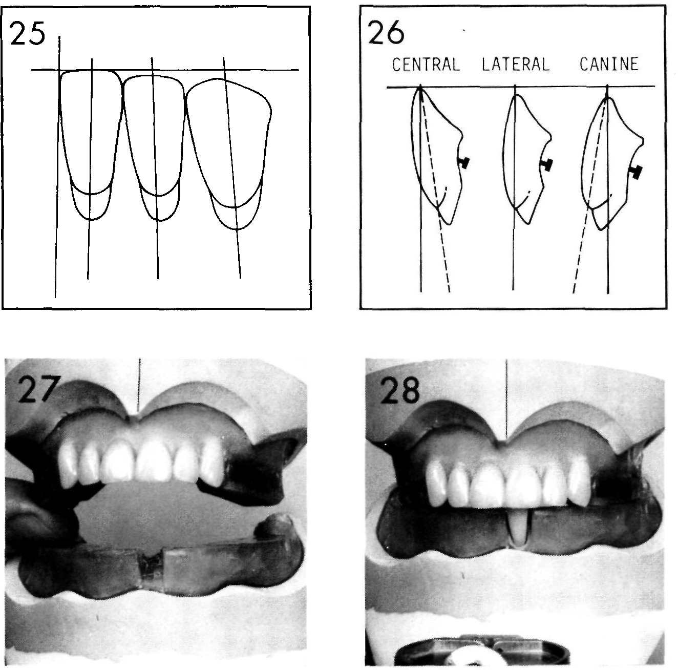
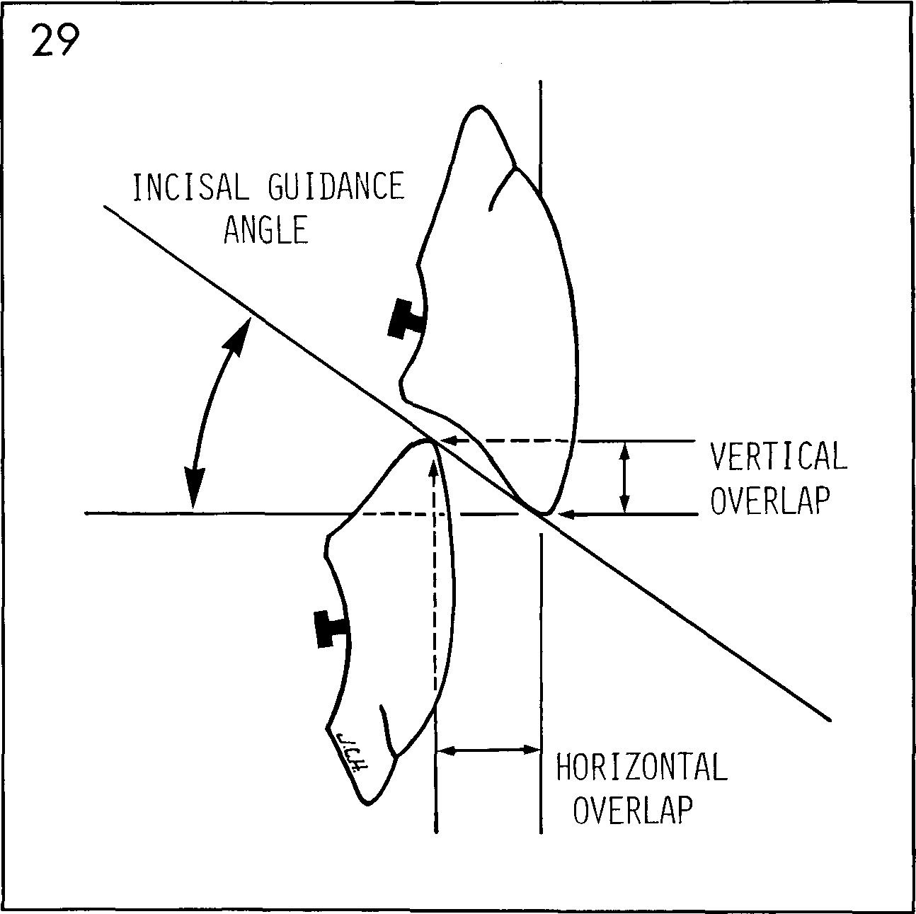
Figure 24 The arrangement of the maxillary anterior teeth has been completed and the wax smoothed around the teeth.
ARRANGING THE MANDIBULAR ANTERIOR TEETH
Figures 25 and 26 The mandibular central incisors are set with the long axis perpendicular to the occlusal plane with the neck depressed. The mandibular lateral incisors are set with a slight distal inclination and with the facial surface at a right angle to the occlusal plane. The mandibular canines have more distal inclination than the lateral incisors and the neck is set prominently.
Figures 27 and 28 A block of wax is removed from the mandibular occlusion rim, the wax softened and the mandibular central incisor placed in position.
Figure 29 The incisal guidance angle is the angle formed by a line drawn through the incisal edges of the maxillary and mandibular incisors and the horizontal plane. (The incisal guide table is the mechanical equivalent of the incisal guidance angle.) The person arranging artificial teeth determines the incisal guidance angle by the level at which he sets the mandibular and maxillary incisors. In denture construction, the mandibular incisors should never touch the maxillary incisors in centric occlusion and the incisal guide angle should be kept as low as possible to enhance free movement of the teeth in protrusive and lateral excursions. These objectives can usually be realized and still achieve good esthetics.
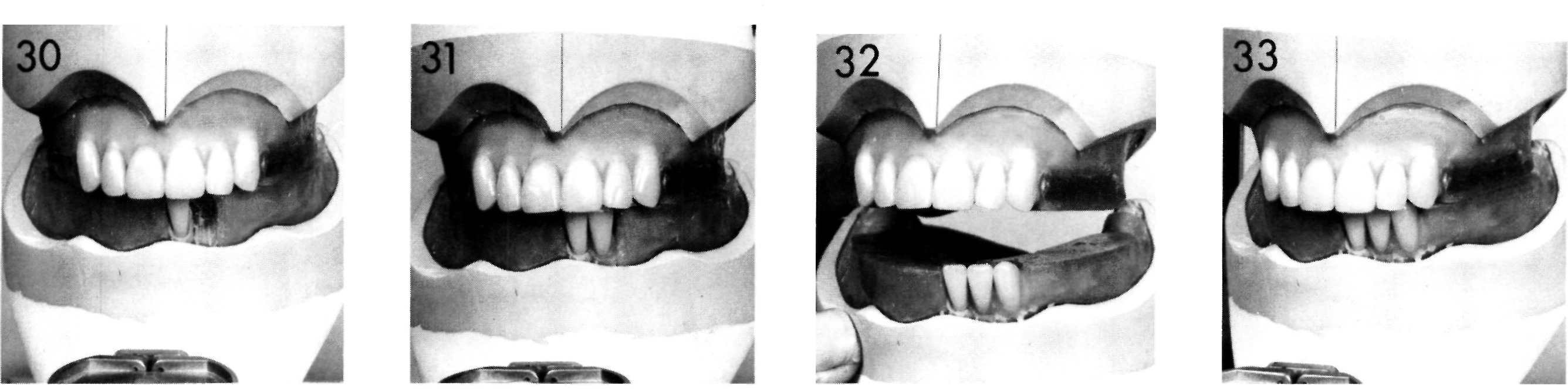
Figures 30 and 31 The same procedure is used to set the mandibular left lateral incisor in proper position.
Figures 32 and 33 The mandibular left canine is set in position as indicated by the facial surface of the occlusion rim and the inclinations shown in Figures 25 and 26.
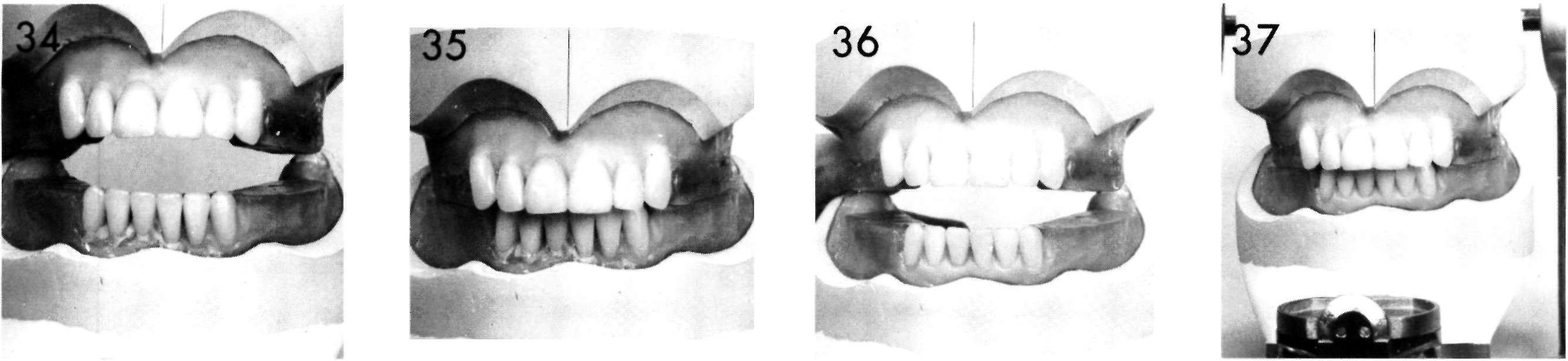
Figure 34 The mandibular right anterior teeth are arranged in a similar manner.
Figure 35 Note the vertical and horizontal overlap.
Figures 36 and 37 Wax has been added and roughly contoured around the anterior teeth. Excess wax should be removed from the surface of the teeth.
Figure 38 The anterior teeth should be examined from the occlusal aspect to insure that the facial surfaces of the teeth follow the original contours of the occlusion rim.
Figure 39 Artificial teeth should not be arranged in a stereotyped manner. Some variations, without losing natural inclinations, will make artificial teeth appear more natural. These variations may be laps, rotations, differences in the level of incisal edges, or esthetic spacing. These deviations should be placed in the teeth by the dentist at the time of the try-in, or by the technician upon prescription by the dentist.
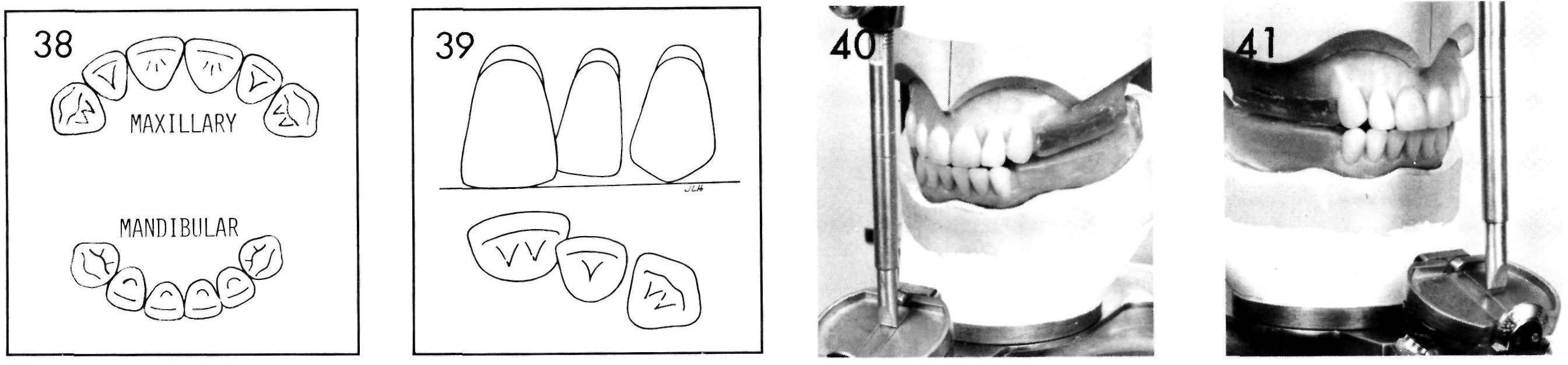
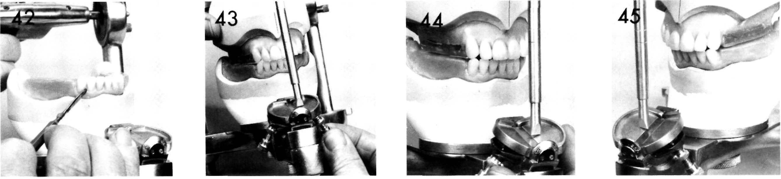
Figure 40 The arrangement of the anterior teeth is not complete until they have been checked in protrusive and lateral excursions. Here they are being checked in protrusive. Note that the teeth touch and that the incisal pin is contacting the incisal guide table. To insure proper contact, each mandibular anterior tooth may be checked for excursive movements while it is being set.
Figure 41 The right lateral movement is checked. Note that the pin is above the incisal guide table.
Figure 42 The mandibular right canine and lateral incisor are lowered slightly to provide better contact and lower the incisal guide plane.
Figure 43 The lateral wing of the incisal guide table is raised until it contacts the incisal pin. The extent to which the lateral wings may be raised is determined to some extent by the type of posterior teeth to be used; the higher the cusps, the steeper the lateral guides may be set. However, free articulation is improved by keeping the wings as low as possible.
Stay updated, free dental videos. Join our Telegram channel
VIDEdental - Online dental courses


