Osteology of the Skull
• To name the bones of the neurocranium and viscerocranium
• To identify the various bones and sutures as seen from anterior, lateral, posterior, inferior, and interior views of the skull
• To name the openings, foramina, and canals as seen from the aforementioned views
• To describe the boundaries of the three cranial fossae and what lies within them
• To describe the pterygoid processes of the sphenoid bone and their components
• To describe in detail the various parts and landmarks of the maxillae
• To describe in detail the various parts and landmarks of the mandible
• To describe where growth takes place in the maxillae and mandible to allow for increase in arch length
The following eight bones make up the neurocranium, the bones surrounding the brain:
The 14 bones that make up the viscerocranium, the bones of the face, include the following:
VIEWS OF THE SKULL
Anterior View
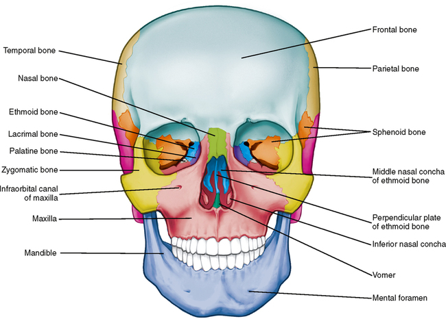
In the anterior or frontal view of an adult skull (Fig. 26-1), the area from the eyes up to the top of the skull is made up of the frontal bone. The area below the eyes down to the occlusal plane between the upper and lower teeth comprises the paired zygomatic or cheekbones and the paired maxillae. The nasal bones form the bridge of the nose, and the lower jaw is formed by the single mandible. The inner or medial corner of the eye cavity (orbit) contains a small lacrimal bone. Within the nasal cavity the vertical nasal septum is composed of the vomer and the perpendicular plate of the ethmoid bone. The inferior nasal conchae are found in the lower, lateral portions of the nasal cavity. More details of the bones of the nasal cavity are discussed in Chapter 27.

If you look back into the orbit, in the medial wall you can see the orbital portion of the ethmoid bone and the lesser and greater wings of the sphenoid, forming the superior orbital fissure in the posterior part of the orbit. The roof of the orbit is formed primarily by the orbital plate of the frontal bone, the lateral wall of the orbit is primarily zygomatic bone, and the floor of the orbit is primarily the maxilla and some of the zygomatic bone. In Fig. 26-1, note that the rim of the orbit is formed from equal parts of the frontal, zygomatic, and maxillary bones. At the lateral edges of the skull, some parts of the parietal and temporal bones are visible, as well as the greater wing of the sphenoid.
Lateral View
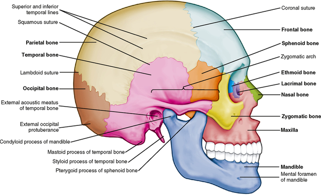
From a side or lateral view, parts of the following bones can be seen: the frontal; zygomatic; maxilla; mandible; nasal; lacrimal; a small bit of the ethmoid bone in the medial wall of the orbit; the greater wing of the sphenoid; the temporal with its squamous, mastoid, and zygomatic portions; the parietal; and the occipital. Fig. 26-2 shows jagged suture lines separating one bone from another. It may be difficult to see some of them, but it is more important to be able to visualize the relationship of one bone to another rather than to differentiate every single suture.
Inferior View
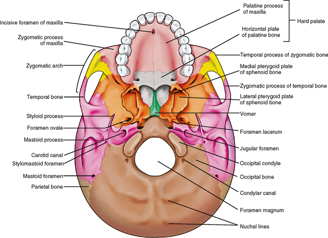
The most difficult view of the skull for the beginning student of anatomy is the inferior view. There are numerous points or landmarks of study, and it is difficult to see the suture lines between the bones in many instances. Nevertheless, in the anterior region illustrated in Fig. 26-3, the hard palate, which is formed by the palatal processes of the maxillae and the palatal processes of the palatine bones, is visible. Just behind and above the palate, a small portion of the vomer bone can be seen, forming the lower part of the nasal septum. Just behind that and running the full width of the skull is the sphenoid bone, which is discussed in greater detail later in this chapter. It is difficult to see the suture line between the sphenoid and the occipital bones because it disappears when a person is about 18 years of age. The area marked by the dashed line in Fig. 26-3 is known as the sphenoccipital synchondrosis. This is a major area of endochondral bone formation, which is an important factor in the development of facial profiles and types of malocclusions. From this view, portions of the zygomatic bones, the temporal bones, and just a tiny portion of the posterior part of the parietal bones can also be seen.
Interior View
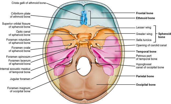
One other view of the skull should be considered: a view of the inside of the skull with the top of the skull removed (Fig. 26-4). Much of the front of the skull is formed by the frontal bone; however, a small area near the midline in the front is part of the ethmoid bone with its crista galli and cribriform plate. Immediately behind these bones is the sphenoid bone with its greater and lesser wings, and behind and lateral to it are the temporal and parietal bones. The occipital bone lies posterior to this.
LANDMARKS OF THE SKULL
Anterior View
In Fig. 26-5, you can again see that the rim of the orbit is formed by the frontal, zygomatic, and maxillary bones. The supraorbital notch or foramen is seen in the upper rim of the orbit in the frontal bone. The nerve and blood supply to the forehead come through this opening. Toward either side at the top, you can see a part of the coronal suture, also called the frontoparietal suture. A few sutures, such as the coronal, have special names, but most of them are named by the two bones they join. In Fig. 26-5, you can also see the nasal or piriform aperture. Below the orbit, in the maxillae, are the infraorbital foramina, which transmit the infraorbital vessels and nerves to the upper lip, lower eyelid, and side of the nose; you can also see the intermaxillary suture. The canine eminence is the ridge of bone over the maxillary canine. The depressions in the maxillae, just above the canines, are the canine fossae. The alveolar processes are the areas in the maxillae and the mandible that form the sockets for the teeth. You can also clearly see the mental foramen in the mandible. The mental nerves and vessels to the chin, lower lip, and facial anterior gingiva of the mandi/>
Stay updated, free dental videos. Join our Telegram channel

VIDEdental - Online dental courses


