LARYNX
Overview and Topographic Anatomy
Overview and Topographic Anatomy
GENERAL INFORMATION
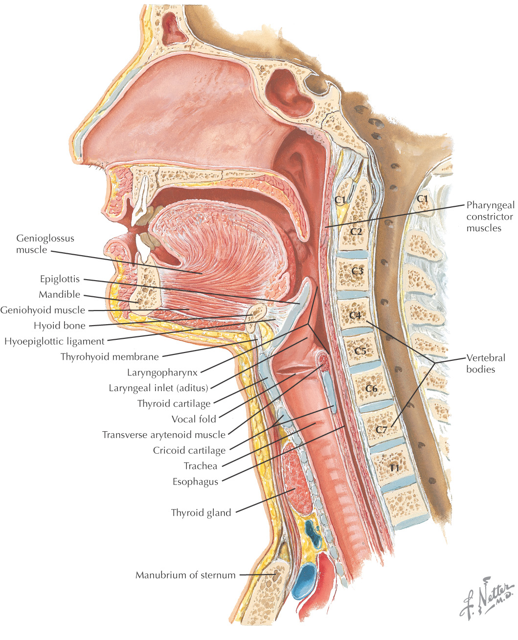
Larynx: connection between the pharynx and the trachea
Prevents foreign bodies from entering the airways
Designed for the production of sound (phonation)
Shorter in women and children
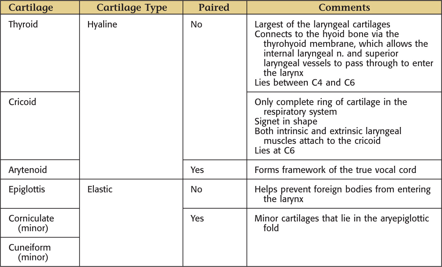
Formed by 9 cartilages: 3 paired and 3 unpaired
Located in the midline opposite the 3rd to the 6th cervical vertebrae
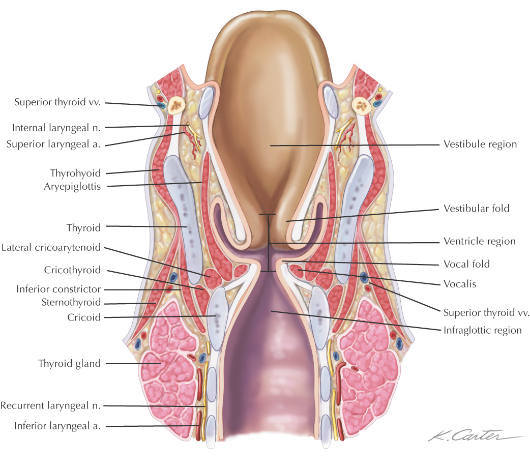
Regions of the larynx:
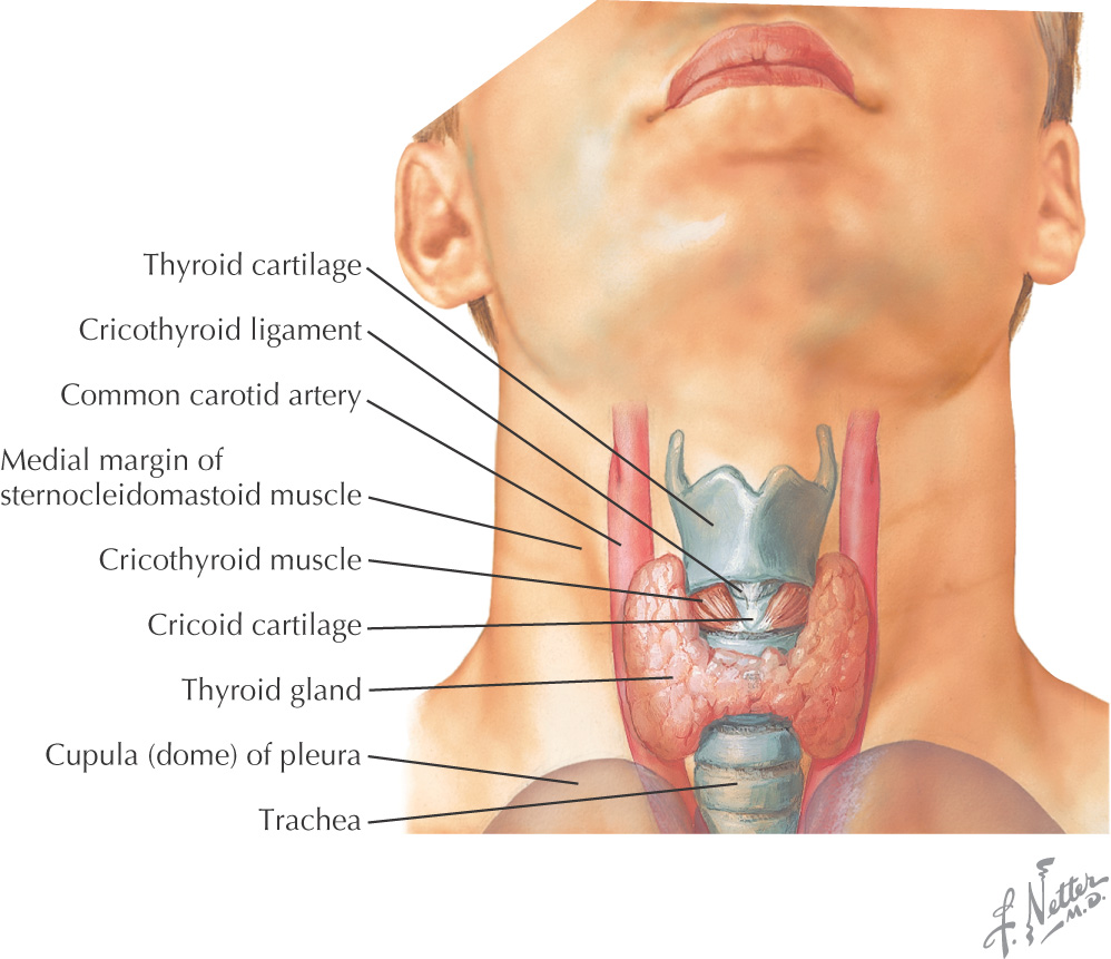
Relations of the Larynx
• Anterolateral—infrahyoid muscles, platysma
• Lateral—lobes of the thyroid gland, carotid sheath
• Posterior—it forms the anterior wall of the laryngopharynx
• Superior—base of tongue and vallecula
Cartilages
GENERAL INFORMATION
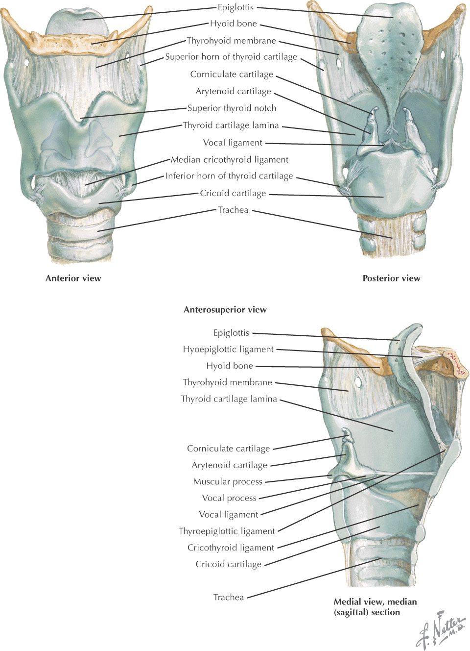
THYROID CARTILAGE
|
Anatomic Feature |
Comments |
|
2 lateral laminae |
2 plates that meet at an acute angle in the anterior midline |
|
Laryngeal prominence |
Also known as the Adam’s apple |
|
Thyroid notch |
Superior portion of the laryngeal prominence, which forms a V shape |
|
Superior tubercle |
Superior border of the oblique line |
|
Oblique line |
Attachment for sternothyroid, thyrohyoid, and inferior constrictor mm. (extrinsic muscles of the larynx) |
|
Inferior tubercle |
Inferior border of the oblique line |
|
Superior horn |
Provides lateralmost attachment for the thyrohyoid membrane |
|
Inferior horn |
|
Stay updated, free dental videos. Join our Telegram channel
VIDEdental - Online dental courses


