Introduction
The purpose of this study was to assess the reliability of the Frankfort horizontal (FH), sella-nasion horizontal, and optic planes in terms of their variabilities in relation to a true horizontal line in orthognathic surgery patients.
Methods
Thirty-six consecutive presurgical orthognathic patients (13 male, 23 female; age range, 16-35 years; 30 white, 6 African Caribbean) had lateral cephalometric radiographs taken in natural head position, with a plumb line orientating the true vertical line, and the true horizontal line perpendicular to the true vertical. The inclinations of the anatomic reference planes were compared with the true horizontal.
Results
The FH plane was found to be on average closest to the true horizontal, with a mean of −1.6° (SD, 3.4°), whereas the sella-nasion horizontal and the optic plane had means of 2.1° (SD, 5.1°) and 3.2° (SD, 4.7°), respectively. The FH showed the least variability of the 3 anatomic planes. The ranges of variability were high for all anatomic planes: −8° to 8° for the FH, −8° to 15° for the sella-nasion horizontal, and −6° to 13° for the optic plane. No significant differences were found in relation to patients’ sex, skeletal patterns, or ethnic backgrounds.
Conclusions
The clinically significant variability in the inclinations of anatomic reference planes in relation to the true horizontal plane makes their use unreliable in orthognathic patients.
Highlights
- •
Use of horizontal anatomic reference planes is unreliable.
- •
Their unreliability is magnified in orthognathic surgery patients.
- •
Patients should be evaluated in physiologic natural head position.
- •
True vertical and true horizontal lines may be used for esthetic analysis.
- •
The importance of such analysis in orthognathic patients is emphasized.
Orthognathic surgery can lead to significant esthetic and functional changes in the dentofacial complex. Therefore, accurate diagnosis and treatment planning are imperative. Since the simultaneous introduction of lateral cephalometric radiographs by Broadbent in 1931, diagnosis and treatment planning in orthodontics has relied heavily on the use of reference planes. Numerous cephalometric planes have been proposed in the literature; however, the 2 most frequently documented are the intracranial sella-nasion plane (SN; drawn from sella, the point representing the geometric center of the pituitary fossa in the midsagittal plane, to nasion, the intersection of the internasal and frontonasal sutures, in the midsagittal plane) and the Frankfort horizontal plane (FH; drawn from porion, the most superior point of the outline of the external auditory meatus, to orbitale, the lowest point on the inferior orbital rim). These cephalometric planes have their shortcomings, including individual variability and difficulties in landmark identification. Therefore, researchers have proposed the use of other planes to find the most reliable and least variable. Sassouni recommended constructing a plane relying on multiple landmarks for greater accuracy and proposed using the “optic plane” as a substitute for the FH plane. The optic plane is constructed as follows: a “supraorbital plane” is drawn tangent to the superior contour of the anterior clinoid process and the roof of the orbit; an “infraorbital plane” is drawn tangent to the lower border of sella turcica and the floor of the orbit; the optic plane bisects the angle formed by the supraorbital and infraorbital planes. Sassouni thought that the optic plane was a preferable alternative to the FH plane because it was based on more easily identifiable skeletal structures. Burstone et al suggested the construction of a horizontal line through sella, 7° down from the SN line, which will be referred to in this article as the SN-horizontal (SN-h) plane.
Reference planes can be either intracranial or extracranial; intracranial planes may be significantly affected by landmark identification and location, whereas extracranial planes may provide less variability. Taking lateral cephalometric radiographs in natural head position (NHP) has been recommended because it provides a physiologically natural position of the head, such as with a person viewing a distant object at eye level. Recording the NHP as such not only enhances reproducibility but also allows cephalometric planes to be compared with a true vertical line (TrV), drawn parallel to a plumb line hanging from a ceiling, and a true horizontal line (TrH), drawn perpendicular to the TrV.
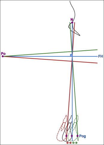
It has been suggested that using TrH as an extracranial reference plane may be more reliable than the commonly used intracranial planes, and this is particularly important in orthognathic patients. Therefore, it is crucial that when intracranial planes are used in orthognathic patients, the plane must have an inclination close to the TrH. The importance of the variability of intracranial reference planes is demonstrated in Figure 1 . Even a few degrees of difference in the inclination of a horizontal plane, such as the FH plane from the TrH, may lead to a considerable effect on the results of skeletal or soft-tissue analyses. For example, in a subject with average lower and middle anterior facial heights, a 2° difference in the inclination of the FH plane from the TrH can cause a difference of 4 mm in the sagittal position of pogonion in relation to a vertical line perpendicular to the FH plane.

When selecting an ideal reference plane, one should consider the ease and accuracy of identifying the structures and landmarks on the lateral cephalometric radiographs. Reliability is enhanced by locating landmarks that are in clear contrast from the adjacent structures and those that are stable and not affected by growth. In orthognathic patients, most craniofacial growth is completed before surgery, and thus the landmarks selected will undergo minimal changes. The reference planes should have good reliability, good intraindividual reproducibility, and low interindividual variability, and they should closely resemble the natural horizontal balance of the head or have an average orientation close to the TrH or TrV.
The FH plane is frequently used as part of soft-tissue and skeletal cephalometric analyses as a basis for planning jaw positions. Therefore, any significant individual variation may lead to erroneous findings. A 0° meridian reference line dropped vertically from soft-tissue nasion is routinely used in relation to the FH plane. This relies on the assumption that the upper facial morphology is normal, particularly the sagittal position of soft-tissue nasion. As such, the sagittal positions of the middle and lower facial structures are determined in relation to the 0° meridian, which is itself drawn perpendicular to the FH plane. Although this concept is easy to follow, its accuracy is hindered by several limitations. In surgical patients with great individual anatomic variations, the inclination of the FH plane is likely to be variable; consequently, the 0° meridian may also be incorrect. Furthermore, the morphology of the nasal radix and glabellar region is extremely variable, ranging from a deep concavity to a flat nasal radix; this ultimately affects the sagittal position of soft-tissue nasion. As a result of these limitations of the 0° meridian, it is recommended that patients should be evaluated in NHP and a TrV constructed with a plumb line hanging from the ceiling. A line drawn perpendicular to the TrV effectively replaces the FH plane. This is particularly important for orthognathic patients. Nevertheless, anatomic reference planes continue to be used, and their reliability should therefore be evaluated.
Significant individual variability in the inclination of reference planes may lead to incorrect diagnoses and treatment planning. The potential individual variability in the inclination of the FH plane in orthognathic surgery patients has been anecdotally noted. However, the hypothesis that the FH, SN-h, and optic planes have significant individual variability in orthognathic patients has not been scientifically evaluated, and there are no previous data on the reliability of anatomic reference planes in relation to the TrH plane in orthognathic surgery patients. The principal objective of this investigation was to compare the inclinations of 3 intracranial reference planes of the craniofacial complex (FH, SN-h, and optic) with the TrH line in presurgical orthognathic patients.
Material and methods
Ethical approval was granted for the study by the National Research Ethics Service of the United Kingdom, reference number 13/LO/0519.
The sample size was determined by carrying out a power calculation before inviting participants. Anticipating a standard deviation of 0.2 in the percentage deviations from the gold standard, we found that a total of 36 subjects would allow the 95% confidence intervals estimating the percentage deviation of the inclination of the reference lines from the gold standard to have a precision within 0.07.
The inclusion criteria were (1) adult patients (≥16 years) competent to provide consent, (2) undergoing orthognathic treatment, (3) with no cleft lip and palate or other congenital deformities, and (4) with no previous surgical procedures on the maxilla or the mandible.
A total of 44 consecutive presurgical patients between the ages of 16 and 35 who were to undergo combined orthodontic and orthognathic surgical treatment in 1 center consented to take part in the study. Eight subjects were excluded because of unusable cephalometric radiographs (plumb line absent from the radiograph), previous surgery to the maxilla or mandible, and a previously operated cleft palate. Thirty-six participants (13 male, 23 female) met the inclusion criteria, forming the participants.
Each patient had 1 lateral cephalometric radiograph at the presurgical diagnostic records appointment. To obtain NHP, the subjects were instructed to stand upright and look straight ahead into the image of their eyes in a small mirror placed at the level of their eyes on the wall opposite the cephalostat. All radiographs were taken by the same operator (A.M.Z.). A plumb line was constructed by attaching a piece of string to a weight suspended from the front of the cephalostat, indicating a TrV line on the radiograph. The cephalometric tracings were undertaken by hand on a light box, with clear acetate tracing paper secured by tape, and a sharp pencil. The TrH was obtained by drawing a line perpendicular to the TrV line. The FH plane was drawn from porion to orbitale. The SN plane was drawn from sella to nasion, and the SN-h was drawn as a horizontal line through sella, 7° down from the sella-nasion line. The optic plane was drawn as the bisection of the angle formed by the supraorbital and infraorbital planes.
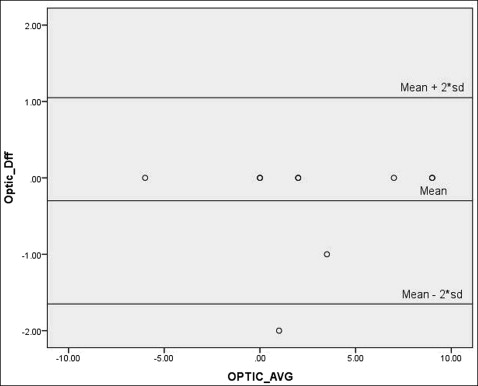
The inclinations of the FH, SN-h, and optic planes were compared with the TrH line. Angles formed below the TrH were designated as negative, and angles formed above the line as positive. Intraexaminer reliability was checked by retracing 10 randomly selected radiographs after 3 weeks and analyzing with the Bland-Altman test.
Results
A Bland-Altman test was carried out to evaluate the intraexaminer reproducibility of the repeated measurements, which demonstrated good reproducibility ( Fig 2 ).

The Pearson correlation coefficients between the 2 measurements for the cephalometric planes were 0.975 for the FH plane, 0.987 for the SN-h plane, and 0.99 for the optic plane. These demonstrated good correlations between the measurements, indicating good intraexaminer reliability.
Descriptive data analysis was carried out to determine means, standard deviations, ranges (minimum, maximum), and coefficients of variation for the different planes. The coefficient of variation is the standard deviation divided by the mean, expressed as a percentage, and is a measure of relative variability. It helps to place the standard deviation in perspective by relating it to the size of the mean and was calculated because, for some reference planes, the mean value was small compared with the standard deviation.
Table I illustrates the interindividual variability of the craniofacial reference lines in relation to the TrH. The reference plane found to be the least variable and thereby closest in mean inclination to the TrH was the FH, with a mean of −1.6° (SD, 3.4°). This was closely followed by SN-h with a mean of 2.1° but a larger SD (5.1°). The optic plane was found to be the farthest in mean inclination from TrH with mean and SD values of 3.2° and 4.7°, respectively. Nevertheless, the range of variability for all reference planes was clinically significant.
| Reference plane | Mean (°) | SD (°) | Range of variability (°) | CV (%) | P value | |
|---|---|---|---|---|---|---|
| Minimum | Maximum | |||||
| FH | −1.6 | 3.4 | −8 | 8 | 45.9 | – |
| SN-h | 2.1 | 5.1 | −8 | 15 | 178.7 | 0.002 ∗ |
| Optic | 3.2 | 4.7 | −6 | 13 | 67.7 | <0.0001 ∗ |
∗ Significantly different from FH (comparison angle), with overall significance of P <0.0001.
The 3 main planes in question were compared between sexes using the independent samples t test ( Table II ). The results demonstrated no significant difference in variability between them for all 3 planes.
| Reference plane | Male (n = 13) | Female (n = 23) | Male vs female | |||
|---|---|---|---|---|---|---|
| Mean (°) | SD (°) | Mean (°) | SD (°) | t test | P value | |
| FH | −1.5 | 2.1 | −1.7 | 3.9 | 1.3 | 0.2 |
| SN-h | 3.6 | 6.2 | 1.2 | 4.2 | 1.4 | 0.2 |
| Optic | 5.1 | 4.6 | 2.1 | 4.5 | 1.9 | 0.07 |
An independent samples t test was carried out to determine the variability of the reference planes between patients with Class II and Class III skeletal patterns ( Table III ). No participant had a skeletal Class I pattern; as a result, a t test rather than an F test was used to compare the variability between skeletal Class II and Class III. No significant differences were noted between the 2 categories.


