The aim of this article was to report a clinical case of orthodontic treatment in a patient with Class II malocclusion and ankylosis of a maxillary first molar. Surgical luxation was performed, followed immediately by traction with an orthodontic arch with straps. The results obtained were satisfactory, and occlusal equilibrium was improved.
Dentoalveolar ankylosis is an anomaly of eruption that involves anatomic fusion of the alveolar bone with the cementum or dentin. The periodontal ligament disappears, and the cementum and dentin can be resorbed and replaced by bone, resulting in fusion.
Ankylosis can occur during any eruptive period or after the occlusion is established. If there is dentoalveolar ankylosis, vertical growth and development of the alveolar bone are affected, diminishing the height and not allowing vertical movement of the affected tooth, which will remain below the occlusal plane, giving the impression of being submerged.
Ankylosis of a tooth can cause various complications, such as loss of arch length, inclination of the adjacent teeth, risk of caries and periodontal disease of the neighboring teeth because of the difficulty of cleaning, food impactation, reduction in the vertical height of the teeth next to the infraoccluded tooth with extrusion of the antagonist teeth and consequent alteration in the occlusal plane, lateral open bite (lateral open occlusal relationship), tongue habits, and deviation of the midline to the side of the infraoccluded tooth.
According to Lim et al,
Five treatment approaches have been suggested for impacted teeth: no treatment, orthodontic treatment, prosthetic buildup, segmental osteotomy, and extraction. No treatment might be the option when the infraocclusion is mild and the tooth can be periodically observed. Orthodontic treatment combined with luxation might be an acceptable approach in some cases, although there are risk factors including fracture, recurrence of ankylosis, and the need for endodontic treatment. Prosthetic buildup is possible if infraocclusion is less than 5 mm. Segmental osteotomy is a surgical procedure in which alveolar bone including the affected tooth is sectioned and repositioned. Surgical removal might be appropriate for a nonrestorable tooth with severe infraocclusion and tipping of the adjacent teeth. This approach, however, often results in an exaggerated bony defect.
There have been few case reports regarding treatment of submerged permanent molars. Several articles described the successful orthodontic treatment combined with surgical luxation in 3 adolescent patients with ankylosed permanent posterior teeth. Also, there have been 2 examples in adults.
The aim of this article was to describe a clinical case of ankylosis of a molar in an adolescent patient, in which surgical luxation followed by traction with a fixed orthodontic appliance was performed.
Diagnosis and etiology
This female patient was 14 years 5 months of age and sought orthodontic treatment with the chief complaint of “crooked teeth.” She was in a good state of general health.
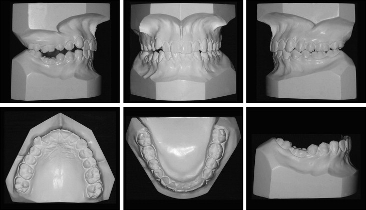
The patient had an Angle Class II subdivision right molar relationship, with a moderate overbite, a maxillary midline deviation to the right, lack of occlusal contact between the mandibular right first molar and the maxillary right first molar with an open occlusal relationship (open bite) in this region, and a mild arch-length discrepancy in the mandibular arch ( Figs 1 and 2 ).

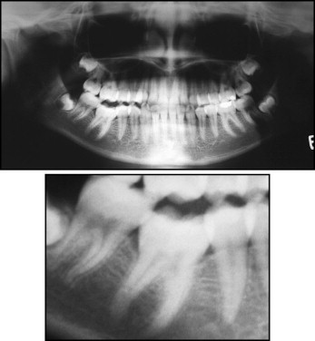
The patient had a straight profile with proportional facial thirds and no asymmetries. Radiographically, she had all of her teeth, with the third molars still forming. The infraocclusion of the mandibular right first molar produced a bone defect in this region ( Fig 3 ).

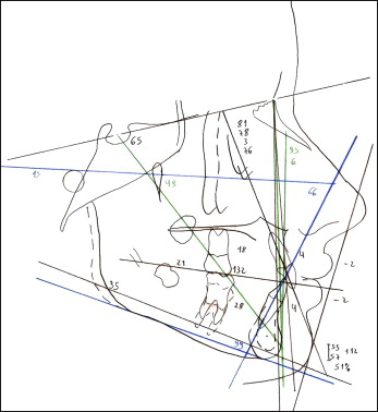
Cephalometrically, the patient had a Class I skeletal relationship, a trend toward horizontal facial growth, a straight profile (LS-S, 0 mm; LI-S, 0.5 mm), retroclined maxillary incisors and well-positioned mandibular incisors ( Fig 4 ).

A percussion test was performed on the mandibular right molar, by using the handle of a clinical mirror for this purpose. A sharp sound was produced, different from the muffled sound of the neighboring teeth, and ankylosis was confirmed.
Treatment objectives
The treatment objectives were (1) tooth alignment and leveling, (2) correction of the Class II dental relationship, (3) occlusal repositioning of the ankylosed mandibular molar, (4) obtainment of space in the maxillary arch to align the teeth, (5) reduction of the vertical overbite, and (6) correction of the bone defect in the ankylosed molar region.
Treatment alternatives
The treatment alternatives were (1) orthodontic treatment, and surgical luxation and respositioning of the ankylosed molar; (2) orthodontic treatment to improve the relationship of the other teeth, followed by restoration of the ankylosed tooth; and (3) extraction of the ankylosed tooth and placement of orthodontic appliances, followed by an osseointegrated implant in the place of the extracted tooth.
Treatment progress
After reviewing all of the information, we decided to attempt traction of the ankylosed mandibular molar with fixed orthodontic appliances after surgical luxation. Initially, orthodontic brackets with 0.022 × 0.028-in slots were placed on all teeth. A sequence of 0.014-in, 0.016-in, and 0.018-in steel archwires accomplished the initial alignment and leveling. At this stage, Class II mechanics were used on the right side to correct the Class II relationship. Then a 0.020-in mandibular archwire was placed with a compressed open-coil spring positioned between the mandibular right second molar and the mandibular right second premolar. To increase the anchorage of the anterior teeth, the teeth were ligated together.
After space was opened for the mandibular right first molar ( Fig 5 ), an orthodontic ring was made for this tooth, and a 0.020-in archwire with L-shaped loops was prepared. At this stage, the patient was referred to the maxillofacial surgeon for the luxation ( Fig 6 ).


