Treatment for Class II Malocclusion
Class II malocclusion can be corrected with a variety of treatment options, depending on how patient presents clinically and radiographically. The age and growth of the patient greatly contributes to the choice of appliance and treatment. This section briefly covers some common treatments used for class II correction and the influencing factors that can compromise a treatment. Several factors affect the specialist’s choice of treatment, such as social environment, compliance, growth and facial profile; these are the determining elements in planning for the most effective treatment outcome.
Principles of Treatment for Class II Malocclusion
Patients can present with a skeletal class II due to a maxillary excess, a mandibular deficiency, or both. If the skeletal class II is caused by maxillary excess, patients present with a backward mandibular growth rotation. This results in an increased anterior facial height. Patients with a deficient mandible typically present with a normal nasolabial angle, a smaller chin owing to a retrusive mandible, protrusion of the maxillary teeth and everted lips. The retruded position of the mandible in relation to the maxilla causes incompetent lips. In severe cases, the lower lip rests palatally to the upper incisors, resulting in minimal lip support.
Some patients may present with a class II malocclusion and a convex profile that is caused by a dentoalveolar anomaly in the absence of an underlying skeletal discrepancy. The clinical and radiographical analysis are the key factors in differentiating the underlying problems. Generally, skeletal class II can be treated by growth modification with orthopaedic appliances in growing patients, and fixed appliance therapy with intermaxillary elastics and camouflage (extraction) treatment or orthognathic surgery in severe cases.
Functional Appliances and Headgear
If the diagnosis is skeletal class II from a retrognathic mandible, a combination of fixed appliances and orthopaedic devices, such as a twin block, can be used in children during active growth to posture the mandible forward. Depending on the patient, the functional appliance can be incorporated as part of phase I or phase II treatment for correction of the anteroposterior discrepancies in the jaws, while fixed appliances correct poor alignment of the teeth. The functional appliances can be used before, after or simultaneously with fixed appliances. Orthopaedic devices that are activated with facial musculature activity are called myofunctional appliances. These appliances can be fabricated in several designs to posture the mandible forward and mitigate the skeletal class II effect during an active growth phase.
Patient compliance and motivation are critically important for optimal results, particularly for removable appliances. The most suitable candidates for a functional appliance are growing patients, particularly undergoing rapid growth. The temporomandibular joint allows displacement of the condyles from the glenoid fossae. The primary goal of functional appliances is positioning the mandible downwards and forwards by increasing the endochondral growth. This is achieved via distraction to the condyles as its postured out of the glenoid fossae. Full‐time or part‐time (a minimum of 14–16 hours) wear of the removable appliances is highly recommended, for a duration of at least 12 months. Initially, the appliance can be uncomfortable for the patient but it is critical for practitioners to stress the importance of wearing the appliance at every review appointment, usually every six to eight weeks. Since the appliance is fitted during mixed dentition, regular adjustments may be indicated to prevent obstruction to the eruption of permanent dentition. Nightly wear of the appliance must be stressed since most growth takes place at night. Overcorrection is preferably made by orthodontists to minimise the risk of relapse. Once an active phase of treatment is completed, nightly wear of the appliance will provide adequate retention for a few months.
Functional appliances can be fixed or removable. Some can be designed with anterior capping to open the bite in cases of deep bite. Conversely, some can be fabricated with posterior capping to prevent open bites in cases with increased overjet. To fabricate these appliances, laboratory technicians require immaculate impressions with an edge‐to‐edge wax bite registration or digital scans of the teeth to ensure that the correct bite is captured. The bite registrations are used during fabrication to design the appliance in a manner that postures the mandible forward. Any distortions in the impressions will directly impact the fit of the appliance and this is eliminated with the use of digital scanners.
The bite registration for functional appliances differs from the bite registration taken as a record. As shown in Figure 7.1, small pieces of wax are softened in warm water and wrapped around the bite stick for the patient to bite on. The patient must bite into the indentations with the upper and lower incisors biting edge to edge (Figure 7.2). Care must be taken that the patient does not posture the mandible too far forward. Every specialist would have their own unique design to the chosen functional appliance. There is a variety of appliances, some of which are described below.

Figure 7.1 A wax bite for functional appliances.

Figure 7.2 Biting edge to edge.
Twin Block
The twin block was introduced by William Clark in 1977. This appliance is commonly removable, although a modified fixed design is also available. It has two components: an upper and a lower block (Figure 7.3). The interlocking of the blocks in the mouth allows the forward posturing of the mandible. Full‐time wear of this appliance provides a rapid optimal outcome if the patient is compliant and motivated. Twin blocks can also be designed to be worn with a headgear. Expansion of the upper arch may be needed if the patient presents with a narrow upper arch, so the upper block may be fabricated with an expansion screw in the middle to expand the upper arch and separate the palatine suture as instructed by the orthodontist.

Figure 7.3 A twin block.
The Bionator
The bionator was introduced by Wilhelm Balters in 1950s. This removable orthopaedic appliance contains a single acrylic element that postures the lower incisors edge‐to‐edge with the upper incisors. A heavy wire loop is incorporated across the upper anterior segment. This functional appliance is designed to keep the cheeks away from the posterior teeth to allow expansion to a degree and restrict soft tissue pressure on the buccal segment. The bionator can be designed to be worn with a headgear by incorporating a face bow into the appliance (Figure 7.4).

Figure 7.4 A bionator with a face bow.
The headgear inhibits maxillary growth as the mandible grows. This type of treatment is most effective if the patient presents with an anteroposterior maxillary excess. The headgear not only restrains the maxillary dentoalveolar structures but also acts as a great source of anchorage to prevent unwanted movement of anchor teeth (Figure 7.5). If the headgear is designed as a source of anchorage it is termed extraoral anchorage. If the headgear distalises the upper permanent molars, it is termed extraoral traction. The factors that differentiates the two is the duration of wear and the force applied to either maintain the dentoalveolar structures or initiate movement. To achieve the most effective treatment outcomes with the headgear, the direction of the force may require modification throughout the treatment.

Figure 7.5 Headgear components: A) The fabric strap that sits on the head, around the neck or a combination of the two. B) Safety strap to allow the appliance to disengage easily. C) A face bow with whisks that is usually attached to the intraoral appliance to allow attachment to the fabric and safety strap.
The headgear can function in three force systems, as it affects the centre of resistance of the maxillary molar in various ways (Figure 7.6):
- Cervical: a low pull as the strap rests on the neck and attaches to the bow. The force is generated below the centre of resistance of upper molars and the force created is down and back. This results in distalisation and extrusion of the upper posterior segment, allowing the mandible to rotate posteriorly backwards and downwards to open the bite. This is effective in treating class II division 2, as the molars are extruded to open the deep bite.
- Occipital: a high pull allowing the force vector to pass through or close to the centre of resistance of the upper molars. The extrusion of the molars is eliminated or minimised. This is useful in treating class II division 1, as the molars are distalised and intruded.
- A combination of cervical and occipital: a straight pull; as the vector force also passes through or close to the centre of resistance of the upper molars, it reduces or prevents extrusive forces. It applies intrusive forces to the molars.

Figure 7.6 Vector and direction of force. Cervical (left): a low pull, as the strap rests on the neck and attaches to the bow; the force is generated below the centre of resistance of the upper molars. Occipital (centre): a high pull, allowing the force vector to pass through or close to the centre of resistance of the upper molars. A combination of cervical and occipital (right): a straight pull, as the vector force also passes through or close to the centre of resistance of the upper molars.
The selection of the magnitude, direction, duration and timing of extraoral forces is determined by the specialist.
Case Study
The patient in Figure 7.7 presented with an Angle class II division 1 malocclusion characterised by proclined maxillary incisor teeth and a retrusive mandible on a brachyfacial skeletal base. The treatment objective was an interceptive orthopaedic development. A removable functional appliance (twin block) was worn full time to address the skeletal nature of the malocclusion. The maxillary component of the appliance had a labial bow incorporated into the design to retrocline the upper incisors by wearing a headgear. Upon completion of treatment, a removable orthopaedic night‐time retainer was issued for retention (Figure 7.8). The patient was kept on a recall until the permanent dentition was established to commence phase II.
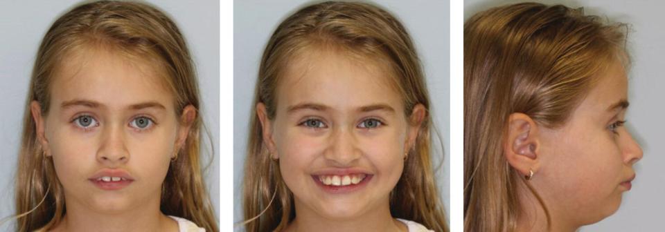


Figure 7.7 Angle class II division 1 malocclusion; initial records
(courtesy of Dr Shimanto K. Purkayastha).
Stay updated, free dental videos. Join our Telegram channel
VIDEdental - Online dental courses


