TOOTH DEVELOPMENT
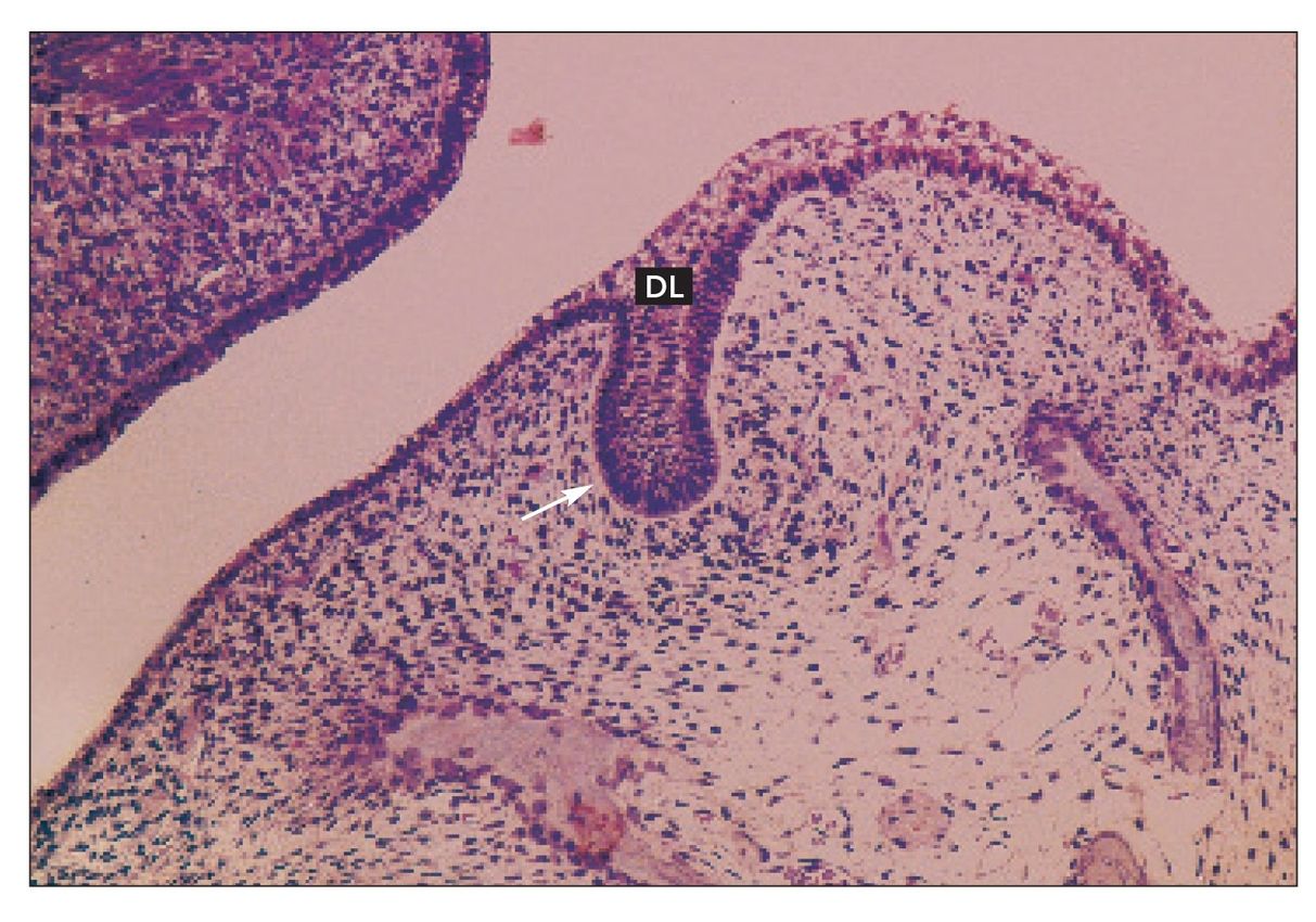
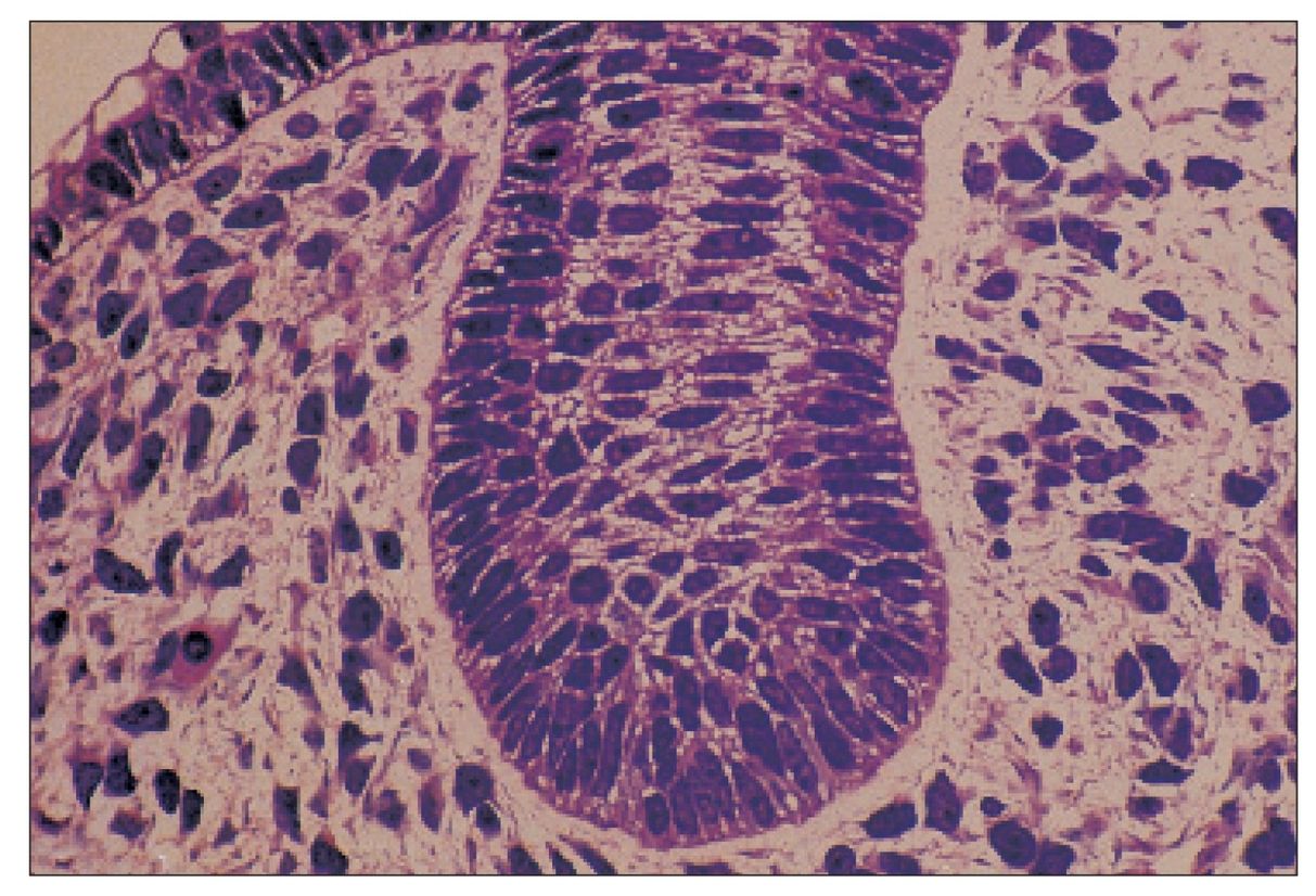
Tooth bud
Tooth bud developing in the mandible of Macaca mulatta. The bud (arrow) has proliferated from the dental lamina (DL) into the underlying ectomesenchyme (H and Lee stain; ×160).
Cap stage tooth
The dental organ (DO) sits on the dental papilla (DP) and is surrounded by the thickening dental sac (arrows) (H and Lee stain; ×160).
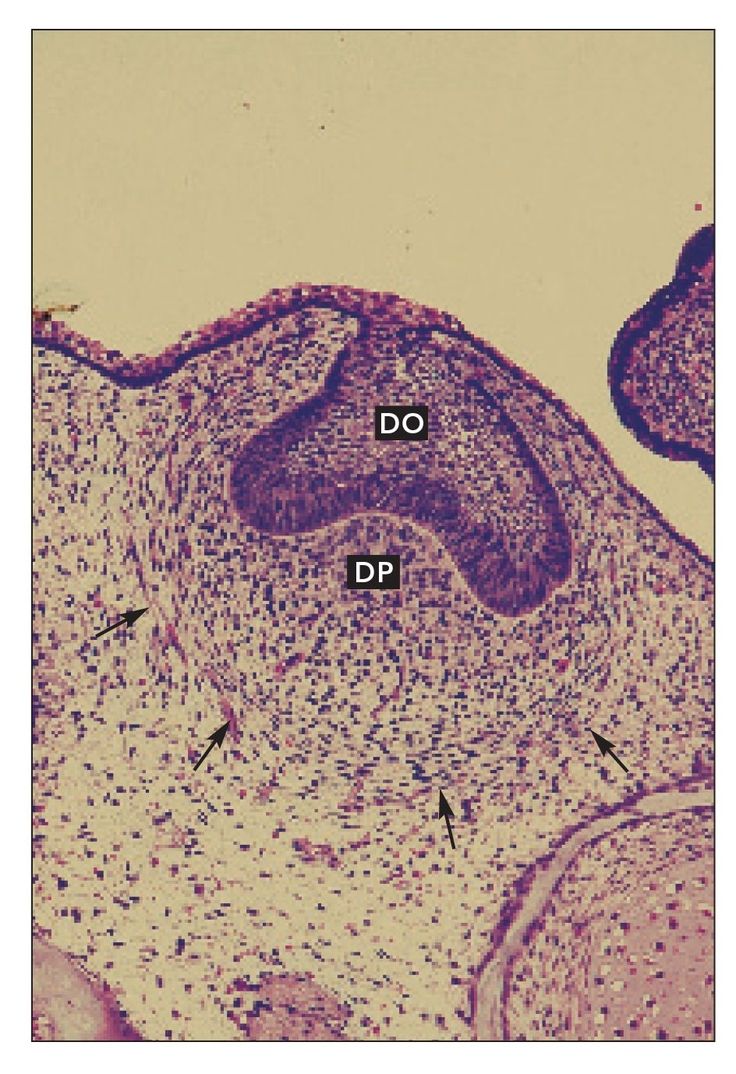
FIG 1-4
Cap stage tooth
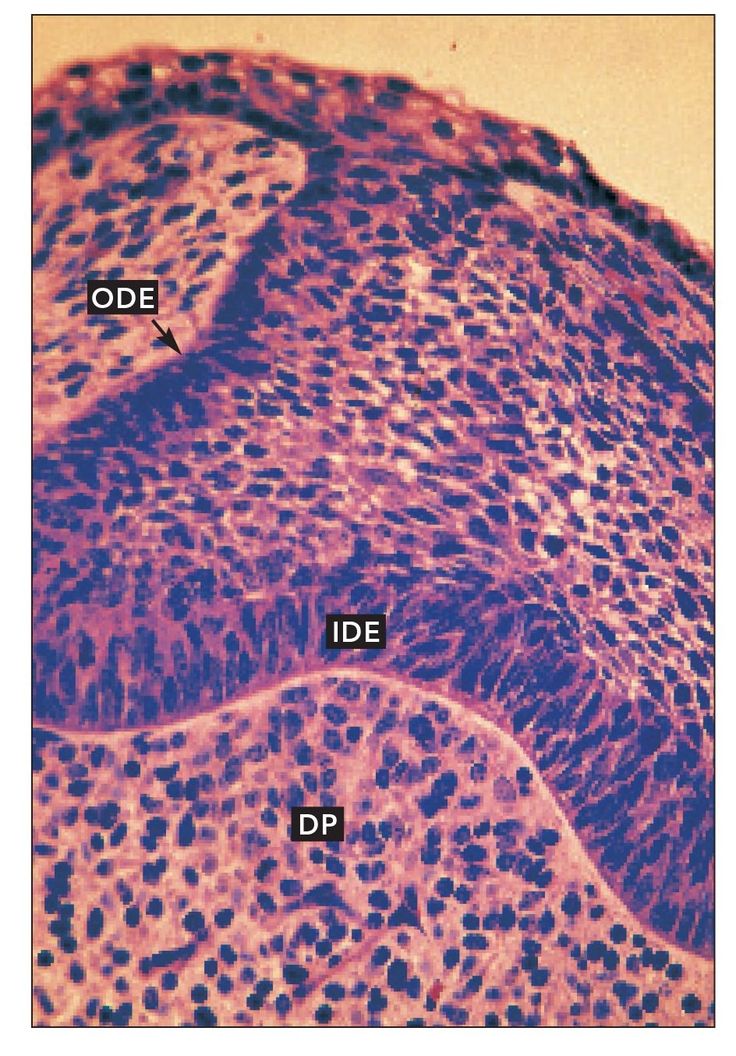
Higher magnification of the cap stage developing tooth shown in Fig 1-3. The outer dental epithelium (ODE) and inner dental epithelium (IDE) are clearly visible. The inner dental epithelium rests on the dental papilla (DP), which remains undifferentiated (H and Lee stain; ×400).
Bell stage tooth
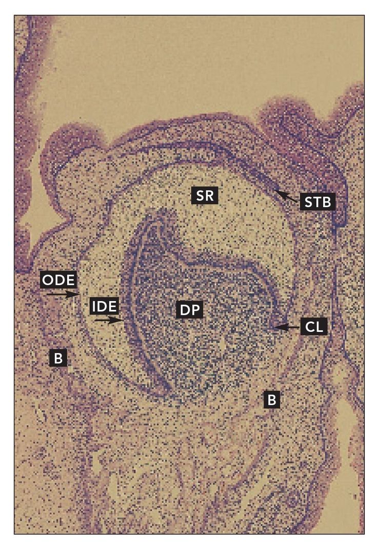
Early bell stage developing tooth in Macaca mulatta. Stellate reticulum (SR) has differentiated between the outer dental epithelium (ODE) and inner dental epithelium (IDE). The cervical loop (CL) is well defined. A secondary tooth bud (STB) extends from the remaining dental lamina. The dental papilla (DP) is very cellular and clearly separate from the developing bone (B) and connective tissue in the dental sac (H and Lee stain; ×40).
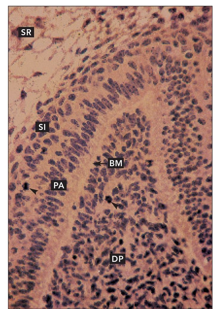
FIG 1-6
Bell stage tooth
Higher magnification of the prospective cusp tip of the early bell stage tooth shown in Fig 1-5. Preameloblasts (PA) are tall and columnar. They are separated from the dental papilla (DP) by a basement membrane (BM). The cells of the dental papilla are still morphologically homogenous. Stratum intermedium (SI) has differentiated between the preameloblasts and the stellate reticulum. Mitoses are visible in the dental papilla and stratum intermedium (arrowheads) (×160).
Stay updated, free dental videos. Join our Telegram channel

VIDEdental - Online dental courses


