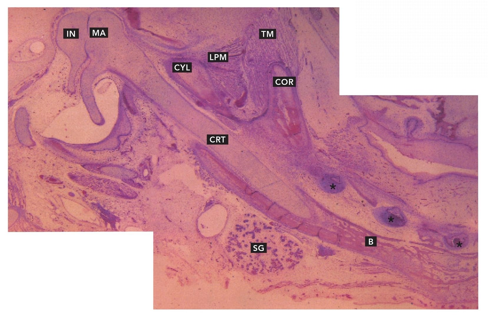
FIG 11-1
Developing (fetal) temporomandibular joint
Sagittal section through the developing temporomandibular joint and mandible of a human fetus. The body of the mandible (B) is developing lateral to and around Meckel’s cartilage (CRT). The coronoid process (COR) with attached temporalis muscle (TM) and condyle (CYL) with attached lateral pterygoid muscle (LPM) complete the mandible. Developing teeth (asterisks) are also visible. The distal end of Meckel’s cartilage has given rise to the incus (IN) and malleus (MA) within the developing ear, but the malleus is still confluent with the cartilage. Developing submandibular gland (SG) lies inferior to the mandible (H and E stain; ×16).
FIG 11-2
Developing condyle
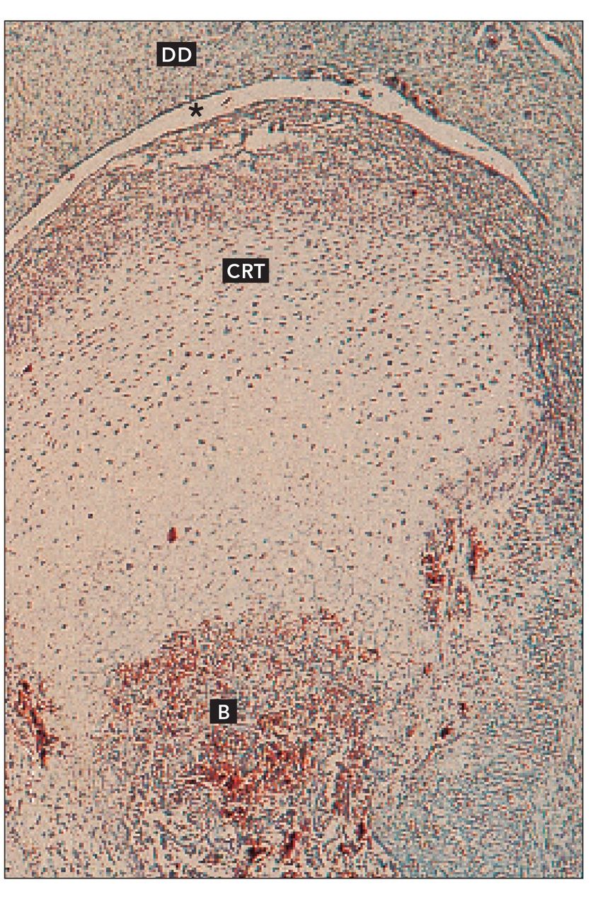
Frontal section of developing mandibular condyle. Cartilage (CRT) is developing atop mandibular bone (B). Developing disc (DD) and the inferior joint space (asterisk) are also visible (H and E stain; ×64).
FIG 11-3
Young condyle
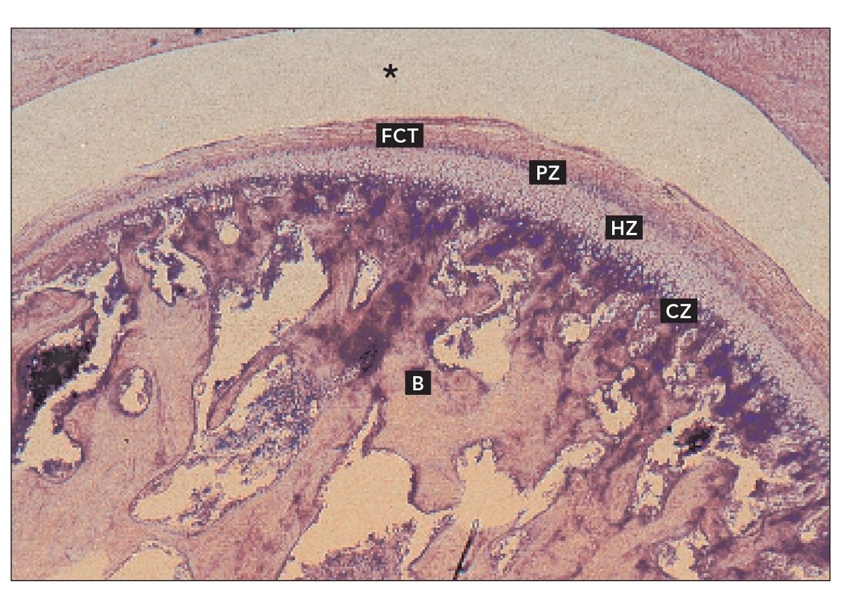
Articulating surface of a young condyle. Trabecular bone of the condyle (B) supports the functional layers of the articular surface. The surface is covered by fibrous connective tissue (FCT). Beneath the articular surface is a layer of cells forming the proliferative zone (PZ), which provides cartilage cells. The cartilage cells form several layers. Cells in the deeper layers hypertrophy in a zone of hypertrophic cartilage (HZ). The hypertrophic cells become calcified and form the basis for new bone. The process is similar to endochondral ossification. The inferior joint space (asterisk) is visible above the articular surface (H and E stain; ×40).
FIG 11-4
Mature temporomandibular joint
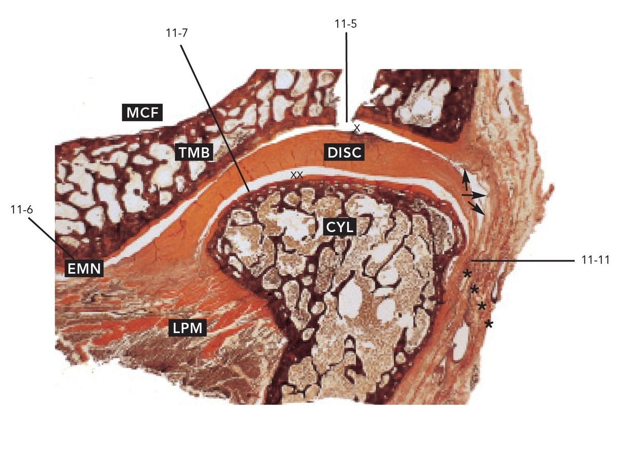
Sagittal section of the mature temporomandibular joint. The two bones of the joint are the mandibular condyle (CYL) and temporal bone (TMB). The middle cranial fossa (MCF) lies above the joint. The disc divides the joint cavity into upper (X) and lower (XX) joint spaces. The disc attaches anteriorly to the connective tissue of the joint capsule and temporal bone anterior to the articular eminence (EMN). The lateral pterygoid muscle may partially insert into the connective tissue of the a/>
Only gold members can continue reading.
Log In or
Register to continue