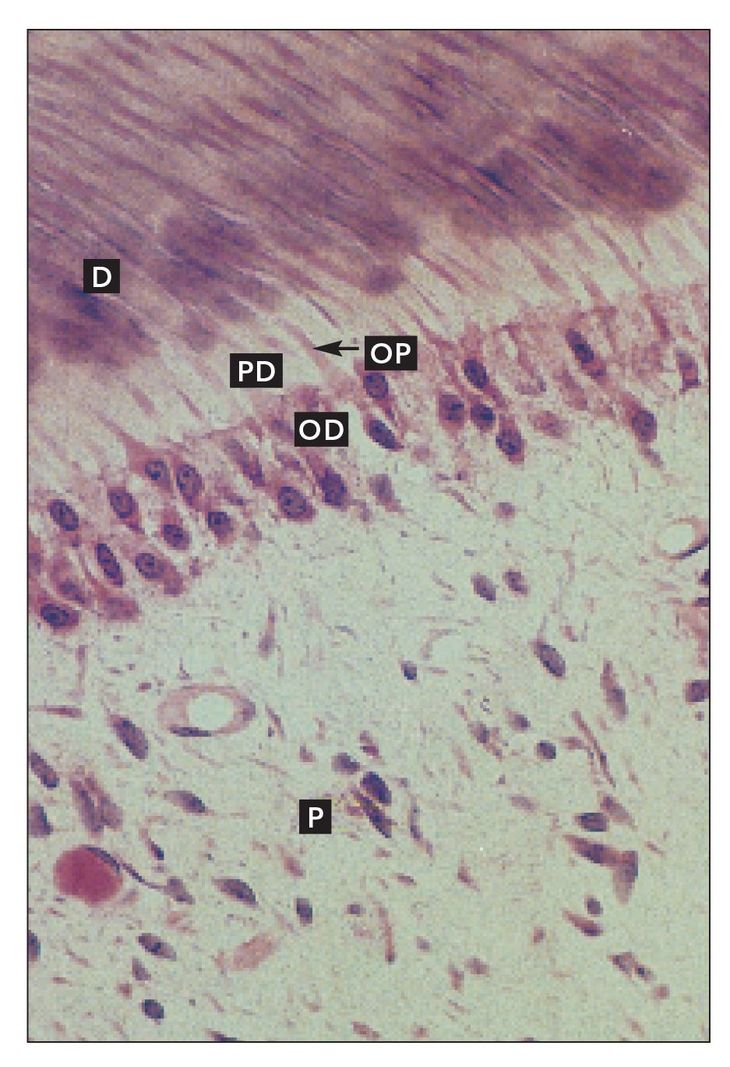
FIG 3-1
Coronal pulp
Mature odontoblasts (OD) in coronal pulp (P). Odontoblastic processes (OP) extend into the predentin (PD) and mineralizing dentin (D) (H and Lee stain; ×640).
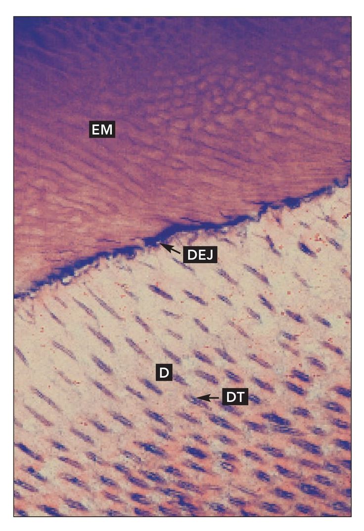
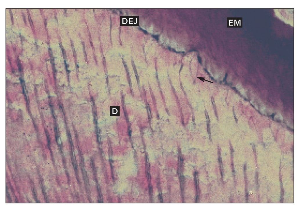
FIG 3-2
Dentinoenamel junction
The dentinoenamel junction (DEJ) in a developing tooth. Enamel matrix (EM) is present. Dentinal tubules (DT) are spaced more widely at the DEJ than further into the dentin (D) (H and Lee stain; ×640).
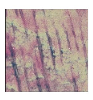
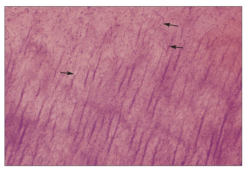
FIG 3-3
Dentinal tubules
Branching dentinal tubules (arrow) in developing dentin (D) near the dentinoenamel junction (DEJ). Other branches are faintly visible in the deeper dentin (H and Lee stain; ×640).
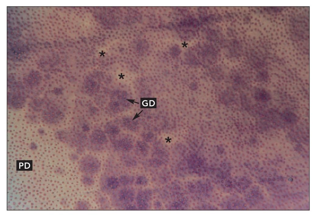
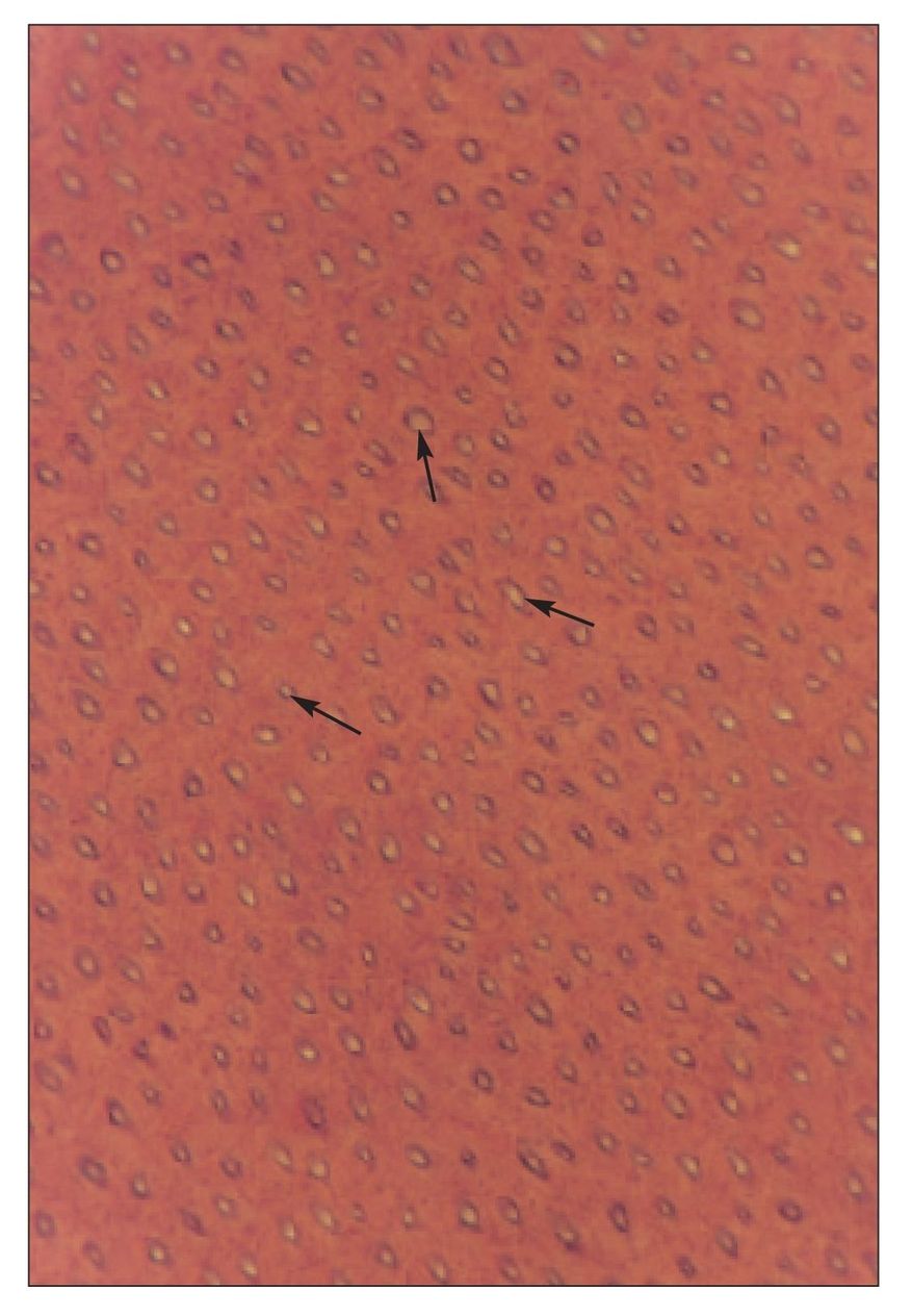
FIG 3-5
Dentinal tubules
Cross section of dentinal tubules near predentin (PD) shows the globular nature of calcification in circumpulpal dentin. Variably shaped calcification centers, referred to as globular dentin (GD), leave relatively uncalcified areas around the asterisks (H and Lee stain; ×400).
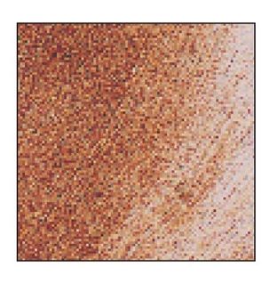
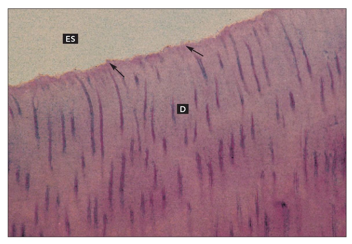
FIG 3-6
Dentinoenamel junction
Dentinoenamel junction (arrows) in a mature tooth. Enamel is represented by the enamel space (ES). Dentinal tubules are visible in the dentin (D) (H and Lee stain; ×640).
FIG 3-8
Dentin
Decalcified tissue section of dentin (D). Secondary dentin (SDN) can be distinguished from primary dentin (D) at the point at which the dentinal tubules change direction (arrows). Predentin (PD) and odontoblasts (OD) are also visible (H and Lee stain; ×400)./>
Only gold members can continue reading.
Log In or
Register to continue