Introduction
The aim of this study was to evaluate the morphologic symmetry of the maxillary and mandibular teeth between the left and right quadrants in 3 dimensions using advanced engineering software.
Methods
The total sample comprised 120 dental casts of 60 patients with dental and skeletal Class I, Class II, and Class III malocclusions. They were divided into 3 groups of 40 dental casts (20 maxillary, 20 mandibular) belonging to 20 patients. The dental casts were digitized with an intraoral 3-dimensional scanner (TRIOS; 3Shape, Copenhagen, Denmark). Segmentation and superimposition procedures were carried out using Rapidform software (Inus Technology, Seoul, Korea). Teeth in the left and right quadrants (except for the second molars) in both jaws were superimposed using 3-point registration followed by surface-based registration; 3-Matic software (Materialise, Leuven, Belgium) was used for deviation analysis.
Results
The maximum mean deviations observed in the positive and negative directions were 0.14 ± 0.10 mm in the maxilla (for the Class I group) and 0.16 ± 0.09 mm for the Class III group. The differences of the maximum deviation amounts among the malocclusion groups were 0.47 ± 0.08 mm in negative direction in the maxillary teeth and 0.79 ± 0.17 mm in the mandibular arch.
Conclusions
In the 3 malocclusion groups investigated, morphologic deviations were low and clinically insignificant. Symmetry of tooth morphology did not differ among Class I, Class II, and Class III malocclusions.
Highlights
- •
An evaluation of dental symmetry was carried out in various malocclusions.
- •
A total of 120 maxillary and mandibular dental casts were digitized.
- •
Mean positive and negative deviations between the left and right teeth were analyzed.
- •
Dental symmetry does not differ among malocclusions.
Facial appearance, specifically the lower third of the face and particularly the mouth, has been deemed to play a vital part in social interactions, expressions of emotions, and physical attractiveness. Subsequent to the many studies of concerned with evaluation of facial symmetry, the big picture concerning the oral environment has been scrutinized into so-called microelements from macroelements to mini-elements over the years, and 1 such component is morphologic tooth symmetry. Montero et al, in a study of 548 subjects, observed that tooth symmetry was graded to be the main element for a beautiful smile in a third of the population.
Several techniques have been used in assessing tooth size and shape, such as linear measurements in various directions either manually or with computer software. However, these methods have inherent incapabilities of depicting variations in tooth shape, form, and surface topography and are limited to providing mainly tooth size. Considering the complex 3-dimensional (3D) morphology of teeth, merely measuring in designated landmarks is insufficient to depict the true picture. On the other hand, the data acquired with surface laser scanners in x, y, and z coordinates and analyzed with reverse engineering technology enables us to observe morphologic differences precisely based on biologically meaningful structures in acquiring 3D superimpositions.
Although few studies have evaluated tooth symmetry even with linear measurements, there is yet no study that assessed both maxillary and mandibular teeth in 3 dimensions. We hypothesized that symmetry between the left and right posterior and anterior teeth in both jaws is different among Class I, Class II, and Class III malocclusions. The aim of this study was to evaluate the morphologic symmetry of maxillary and mandibular teeth in the left and right quadrants in 3 dimensions using advanced engineering software in the context of Class I, Class II, and Class III malocclusions.
Material and methods
Ethical approval for the study was granted by the ethics committee of the School of Medicine, Ege University, İzmir, Turkey.
This study was conducted on 120 dental casts of 60 patients (mean age, 13.9 years; range, 12-15 years) having Class I, Class II, and Class III malocclusions. The mean ages of the patients in malocclusion groups were 13.7 ± 1.5, 14.2 ± 1.9, and 13.5 ± 1.1 years, respectively. Forty maxillary and mandibular dental casts belonging to 20 patients were included in each malocclusion group. The dental malocclusions were determined as Class I, Class II, and Class III according to the maxillomandibular molar relationship. ANB angle and Wits appraisal were used for assessing the skeletal anomalies. Subjects having an ANB angle of 2° ± 2° were identified as skeletal Class I, whereas ANB angles greater 4° and less than 0° were determined as skeletal Class II and Class III, respectively. Subjects exhibiting a Wits appraisal of −1 ± 2 mm were categorized as Class I, whereas discrepancies more than 1 mm and less than −3 mm were identified as Class II and Class III, respectively. Only those showing consistency between the ANB angle and the Wits appraisal were included in the study to overcome the effects of occlusal plane inclination and vertical growth pattern on the measurements. Additional selection criteria of the casts were (1) a fully erupted permanent dentition except for the second molars in both jaws; (2) no tooth agenesis or extractions; (3) no restorations, abrasions, or tooth anomalies; (4) no clinically visible discrepancies on the gingival margins of collateral teeth; (5) maximum of 3 mm of crowding; and (6) no evident facial and dentoalveoler asymmetry. Records of patients with genetic disorders such as syndromes were not included in the study.
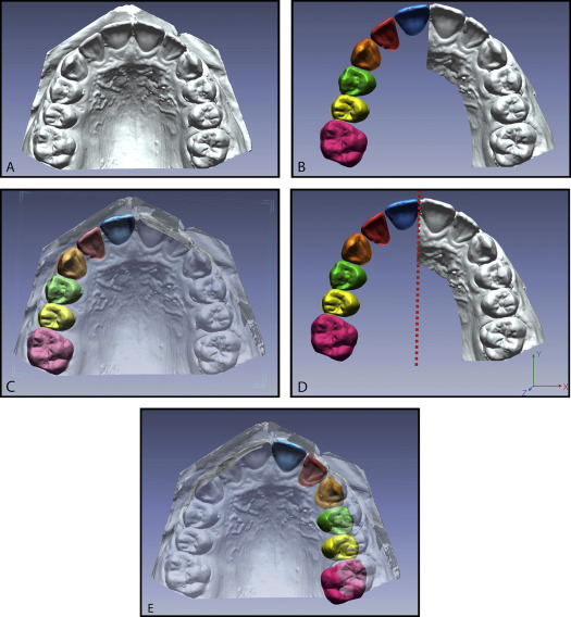
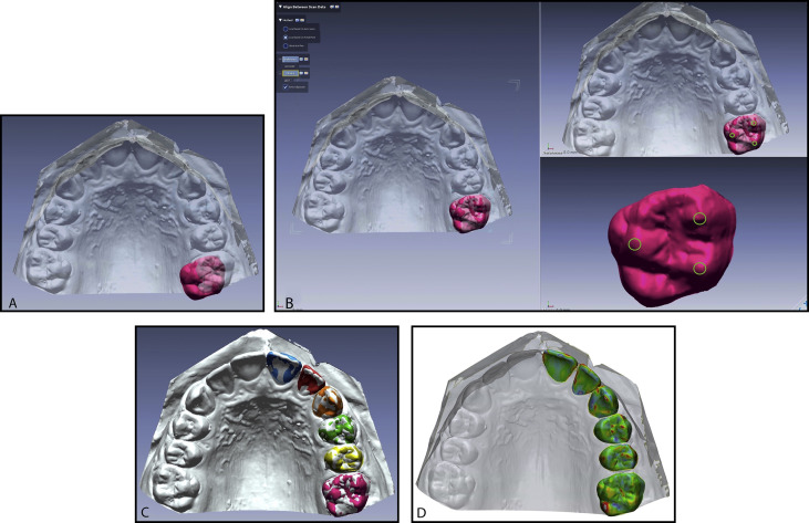
Dental casts were digitized using an intraoral 3D scanner (TRIOS; 3Shape, Copenhagen, Denmark). Segmentation and superimposition procedures on casts converted to digital data were carried out using engineering software (Rapidform; Inus Technology, Seoul, Korea). Before the 3D deviation analysis, the teeth were superimposed twice using point-based and surface-based registrations. First, the crowns of anterior and posterior teeth in the right quadrant were segmented at the gingival margin ( Fig 1 , A-C ). Then mirror images of these teeth were acquired relative to the median palatine suture to have the buccal surfaces of the teeth facing the same direction to make the initial superimposition accurately ( Fig 1 , D and E ). The initial superimposition was conducted on the same 3 points that were designated on the tooth surface in both the left and right quadrants ( Fig 2 , A and B ). These points were the buccal and palatinal cusp tips for the posterior teeth; the deepest points in the gingival contour, and the mesial and distal contact points were used for the anterior teeth. After these low-sensitivity superimpositions, the images were converged for the second superimpositions. In the next step, the images were aligned onto each other using best-fit method ( Fig 2 , D ). This 2-step alignment process provided greater alignment accuracy; also, the initial superimpositions could shorten the time needed for the best-fit alignment. After aligning the images, 3-Matic software (Materialise, Leuven, Belgium) was used for deviation analysis. Maximum positive and negative deviations and mean positive and negative deviations were evaluated between the 2 images of 100% mesh points. Furthermore, percentages of the meshes having deviations between −0.5 and 0.5 mm were calculated. In cases of clinically invisible but still present gingival margin discrepancies, discordant regions were excluded from the analysis. Assessments where right teeth were positioned anterior to the left contralaterals were rated as positive values, and negative values implied vice versa.


Statistical analysis
The data were analyzed with SPSS software (version 20.0; IBM, Armonk, NY). A significance level of 0.05 was used. Data were tested for normality with the Shapiro-Wilk test. Descriptive statistics were reported for each parameter as mean and standard deviation or standard error. One-way analysis of variance was used for the evaluation of positive and negative maximum and mean deviation amounts in the Class I, Class II, and Class III malocclusion groups for each maxillary and mandibular tooth. The Bonferroni post hoc was applied for multiple comparison tests among the groups. To assess method errors, segmentation, superimposition, and deviation analyses were carried out on 15 randomly selected teeth by the same investigator (G.S.D.) after an interval of 1 month, and intraobserver reliability was evaluated using intraclass correlation coefficient.
Results
Measurements repeated after a month showed intraobserver reliability of 0.912 to 0.976 (intraclass correlation coefficient).
Maximum deviation amounts observed in the maxillary and mandibular dentitions in the positive direction were 0.72 ± 0.46, 1.00 ± 0.51, and 1.14 ± 0.77 mm for the Class I, Class II, and Class III malocclusion groups, respectively. The corresponding values in the negative direction were 0.70 ± 0.35, 0.97 ± 0.35, and 1.10 ± 0.54 mm ( Table I ). The maximum mean deviations observed in the positive and negative directions in the maxilla belonged to the Class I group with 0.14 ± 0.10 mm; in the mandible, a value of 0.16 ± 0.09 mm was found in the Class III group ( Table II ). Measurements in the different tooth groups showed mean deviations below the aforementioned threshold level in the positive and negative directions. Table II depicts the mean deviations in the positive and negative directions pertaining to the maxillary and mandibular dentitions.
| Tooth pairs ∗ | Maximum positive deviation | Maximum negative deviation | ||||||
|---|---|---|---|---|---|---|---|---|
| Mean (SD) | Mean (SD) | Mean difference (SE) | P | Mean (SD) | Mean (SD) | Mean difference (SE) | P | |
| Maxilla | ||||||||
| 11 – 21 | 0.62 (0.21) a | 0.60 (0.49) b | 0.014 (0.13) | 0.998 | 0.69 (0.16) a | 0.97 (0.40) b | −0.29 (0.09) | 0.048 |
| 0.62 (0.21) a | 0.68 (0.40) c | −0.06 (0.10) | 0.995 | 0.69 (0.16) a | 0.73 (0.48) c | 0.04 (0.11) | 0.928 | |
| 0.60 (0.49) b | 0.68 (0.40) c | 0.07 (0.13) | 0.993 | 0.97 (0.40) b | 0.73 (0.48) c | 0.24 (0.13) | 0.108 | |
| 12 – 22 | 0.49 (0.16) a | 0.52 (0.24) b | −0.031 (0.07) | 0.956 | 0.62 (0.21) a | 0.74 (0.28) b | −0.12 (0.09) | 0.699 |
| 0.49 (0.16) a | 0.76 (0.40) c | −0.30 (0.11) | 0.033 | 0.62 (0.21) a | 0.78 (0.47) c | −0.16 (0.11) | 0.477 | |
| 0.52 (0.24) b | 0.76 (0.40) c | −0.24 (0.10) | 0.094 | 0.74 (0.28) b | 0.78 (0.47) c | −0.04 (0.13) | 0.944 | |
| 13 – 23 | 0.47 (0.21) a | 0.57 (0.30) b | −0.10 (0.09) | 0.625 | 0.58 (0.40) a | 0.50 (0.28) b | 0.09 (0.12) | 0.720 |
| 0.47 (0.21) a | 0.73 (0.43) c | −0.26 (0.10) | 0.101 | 0.58 (0.40) a | 0.96 (0.40) c | −0.38 (0.12) | 0.060 | |
| 0.57 (0.30) b | 0.73 (0.43) c | −0.16 (0.14) | 0.537 | 0.50 (0.28) b | 0.96 (0.40) c | −0.47 (0.08) | 0.001 | |
| 14 – 24 | 0.51 (0.23) a | 0.50 (0.23) b | 0.01 (0.07) | 0.989 | 0.58 (0.21) a | 0.53 (0.19) b | 0.04 (0.09) | 0.863 |
| 0.51 (0.23) a | 0.60 (0.26) c | −0.09 (0.07) | 0.443 | 0.58 (0.21) a | 0.86 (0.42) c | −0.28 (0.12) | 0.040 | |
| 0.50 (0.23) b | 0.60 (0.26) c | −0.10 (0.07) | 0.404 | 0.53 (0.19) b | 0.86 (0.42) c | −0.33 (0.11) | 0.012 | |
| 15 – 25 | 0.39 (0.19) a | 0.60 (0.24) b | −0.22 (0.08) | 0.015 | 0.65 (0.25) a | 0.58 (0.24) b | 0.07 (0.06) | 0.722 |
| 0.39 (0.19) a | 0.48 (0.25) c | −0.10 (0.08) | 0.425 | 0.65 (0.25) a | 0.69 (0.43) c | −0.04 (0.11) | 0.917 | |
| 0.60 (0.24) b | 0.48 (0.25) c | 0.12 (0.08) | 0.250 | 0.58 (0.24) b | 0.69 (0.43) c | −0.11 (0.11) | 0.521 | |
| 16 – 26 | 0.64 (0.27) a | 0.78 (0.45) b | −0.14 (0.11) | 0.560 | 0.70 (0.35) a | 0.97 (0.35) b | −0.26 (0.10) | 0.070 |
| 0.64 (0.27) a | 1.11 (0.51) c | −0.48 (0.13) | 0.009 | 0.70 (0.35) a | 0.84 (0.43) c | −0.13 (0.15) | 0.806 | |
| 0.78 (0.45) b | 1.11 (0.51) c | −0.34 (0.16) | 0.146 | 0.97 (0.35) b | 0.84 (0.43) c | 0.13 (0.16) | 0.798 | |
| Mandible | ||||||||
| 31 – 41 | 0.52 (0.25) a | 0.53 (0.26) b | −0.01 (0.08) | 0.998 | 0.59 (0.27) a | 0.95 (0.41) b | −0.36 (0.10) | 0.003 |
| 0.52 (0.25) a | 0.59 (0.28) c | −0.07 (0.08) | 0.714 | 0.59 (0.27) a | 0.51 (0.26) c | 0.08 (0.10) | 0.735 | |
| 0.53 (0.26) b | 0.59 (0.28) c | −0.07 (0.08) | 0.746 | 0.95 (0.41) b | 0.51 (0.26) c | 0.44 (0.10) | 0.000 | |
| 32 – 42 | 0.54 (0.27) a | 0.53 (0.22) b | 0.01 (0.07) | 0.988 | 0.59 (0.19) a | 0.96 (0.53) b | −0.37 (0.12) | 0.024 |
| 0.54 (0.27) a | 0.46 (0.27) c | 0.08 (0.07) | 0.644 | 0.59 (0.19) a | 0.69 (0.25) c | −0.10 (0.08) | 0.412 | |
| 0.53 (0.22) b | 0.46 (0.27) c | 0.07 (0.07) | 0.735 | 0.96 (0.53) b | 0.69 (0.25) c | 0.26 (0.13) | 0.157 | |
| 33 – 43 | 0.60 (0.21) a | 1.00 (0.51) b | −0.40 (0.17) | 0.031 | 0.63 (0.22) a | 0.89 (0.41) b | −0.26 (0.10) | 0.098 |
| 0.60 (0.21) a | 0.54 (0.23) c | 0.06 (0.08) | 0.777 | 0.63 (0.22) a | 0.76 (0.44) c | −0.13 (0.10) | 0.558 | |
| 1.00 (0.51) b | 0.54 (0.23) c | 0.46 (0.14) | 0.001 | 0.89 (0.41) b | 0.76 (0.44) c | 0.13 (0.17) | 0.746 | |
| 34 – 44 | 0.48 (0.21) a | 0.80 (0.44) b | −0.32 (0.12) | 0.034 | 0.53 (0.29) a | 0.69 (0.35) b | −0.16 (0.11) | 0.379 |
| 0.48 (0.21) a | 0.52 (0.35) c | −0.04 (0.06) | 0.929 | 0.53 (0.29) a | 0.52 (0.24) c | 0.01 (0.11) | 0.998 | |
| 0.80 (0.44) b | 0.52 (0.35) c | −0.28 (0.11) | 0.068 | 0.69 (0.35) b | 0.52 (0.24) c | 0.16 (0.11) | 0.338 | |
| 35 – 45 | 0.72 (0.46) a | 0.27 (0.31) b | 0.45 (0.16) | 0.018 | 0.66 (0.28) a | 0.32 (0.40) b | 0.34 (0.12) | 0.022 |
| 0.72 (0.46) a | 0.19 (0.29) c | 0.54 (0.16) | 0.004 | 0.66 (0.28) a | 1.10 (0.54) c | −0.45 (0.15) | 0.020 | |
| 0.27 (0.31) b | 0.19 (0.29) c | 0.08 (0.16) | 0.864 | 0.32 (0.40) b | 1.10 (0.54) c | −0.79 (0.17) | 0.000 | |
| 36 – 46 | 0.45 (0.26) a | 0.28 (0.41) b | 0.17 (0.07) | 0.234 | 0.61 (0.33) a | 0.28 (0.29) b | 0.33 (0.10) | 0.020 |
| 0.45 (0.26) a | 1.14 (0.77) c | −0.69 (0.23) | 0.024 | 0.61 (0.33) a | 0.99 (0.37) c | −0.38 (0.10) | 0.007 | |
| 0.28 (0.41) b | 1.14 (0.77) c | −0.86 (0.21) | 0.000 | 0.28 (0.29) b | 0.99 (0.37) c | −0.70 (0.10) | 0.000 | |
∗ Tooth numbering according to the Fédération Dentaire Internationale system.
| Tooth pairs ∗ | Mean positive deviation | Mean negative deviation | ||||||
|---|---|---|---|---|---|---|---|---|
| Mean (SD) | Mean (SD) | Mean difference (SE) | P | Mean (SD) | Mean (SD) | Mean difference (SE) | P | |
| Maxilla | ||||||||
| 11 – 21 | 0.14 (0.10) a | 0.07 (0.03) b | 0.07 (0.04) | 0.267 | 0.12 (0.08) a | 0.06 (0.04) b | 0.06 (0.03) | 0.025 |
| 0.14 (0.10) a | 0.07 (0.04) c | 0.07 (0.04) | 0.325 | 0.12 (0.08) a | 0.10 (0.05) c | 0.02 (0.03) | 0.615 | |
| 0.07 (0.03) b | 0.07 (0.04) c | −0.004 (0.01) | 0.980 | 0.06 (0.04) b | 0.10 (0.05) c | −0.04 (0.03) | 0.198 | |
| 12 – 22 | 0.07 (0.03) a | 0.06 (0.02) b | 0.01 (0.01) | 0.910 | 0.08 (0.03) a | 0.06 (0.02) b | 0.02 (0.01) | 0.193 |
| 0.07 (0.03) a | 0.10 (0.04) c | −0.03 (0.02) | 0.068 | 0.08 (0.03) a | 0.06 (0.01) c | 0.02 (0.009) | 0.179 | |
| 0.06 (0.02) b | 0.10 (0.04) c | −0.04 (0.02) | 0.025 | 0.06 (0.02) b | 0.06 (0.01) c | 0.000 (0.008) | 0.998 | |
| 13 – 23 | 0.10 (0.05) a | 0.05 (0.01) b | 0.05 (0.03) | 0.333 | 0.06 (0.02) a | 0.05 (0.02) b | 0.008 (0.008) | 0.644 |
| 0.10 (0.05) a | 0.05 (0.01) c | 0.04 (0.03) | 0.429 | 0.06 (0.02) a | 0.06 (0.02) c | −0.001 (0.008) | 0.998 | |
| 0.05 (0.01) b | 0.05 (0.01) c | −0.004 (0.008) | 0.909 | 0.05 (0.02) b | 0.06 (0.02) c | −0.008 (0.008) | 0.583 | |
| 14 – 24 | 0.06 (0.03) a | 0.06 (0.02) b | 0.001 (0.009) | 0.999 | 0.07 (0.03) a | 0.06 (0.01) b | 0.01 (0.008) | 0.596 |
| 0.06 (0.03) a | 0.05 ( 0.03) c | 0.01 (0.01) | 0.769 | 0.07 (0.03) a | 0.05 (0.02) c | 0.01 (0.009) | 0.481 | |
| 0.06 (0.02) b | 0.05 ( 0.03) c | 0.009 (0.008) | 0.641 | 0.06 (0.01) b | 0.05 (0.02) c | 0.002 (0.009) | 0.981 | |
| 15 – 25 | 0.10 (0.05) a | 0.06 (0.03) b | 0.04 (0.02) | 0.147 | 0.07 (0.03) a | 0.06 (0.02) b | 0.02 (0.01) | 0.305 |
| 0.10 (0.05) a | 0.10 (0.04) c | 0.003 (0.03) | 0.981 | 0.07 (0.03) a | 0.04 (0.01) c | 0.03 (0.01) | 0.028 | |
| 0.06 (0.03) b | 0.10 (0.04) c | −0.03 (0.02) | 0.208 | 0.06 (0.02) b | 0.04 (0.01) c | 0.01 (0.01) | 0.478 | |
| 16 – 26 | 0.10 (0.08) a | 0.11 (0.07) b | −0.006 (0.03) | 0.978 | 0.07 (0.03) a | 0.10 (0.05) b | −0.03 (0.02) | 0.114 |
| 0.10 (0.08) a | 0.10 (0.06) c | 0.002 (0.03) | 0.996 | 0.07 (0.03) a | 0.07 (0.05) c | −0.008 (0.02) | 0.951 | |
| 0.11 (0.07) b | 0.10 (0.06) c | 0.008 (0.03) | 0.957 | 0.10 (0.05) b | 0.07 (0.05) c | 0.03 (0.02) | 0.429 | |
| Mandible | ||||||||
| 31 – 41 | 0.06 (0.02) a | 0.10 (0.04) b | −0.04 (0.02) | 0.034 | 0.07 (0.01) a | 0.11 (0.06) b | −0.03 (0.01) | 0.157 |
| 0.06 (0.02) a | 0.11 (0.05) c | −0.05 (0.02) | 0.006 | 0.07 (0.01) a | 0.10 (0.05) c | −0.03 (0.01) | 0.245 | |
| 0.10 (0.04) b | 0.11 (0.05) c | −0.01 (0.02) | 0.729 | 0.11 (0.06) b | 0.10 (0.05) c | 0.006 (0.01) | 0.925 | |
| 32 – 42 | 0.05 (0.02) a | 0.06 (0.03) b | −0.007 (0.01) | 0.843 | 0.06 (0.02) a | 0.05 (0.02) b | 0.01 (0.007) | 0.172 |
| 0.05 (0.02) a | 0.08 (0.04) c | −0.02 (0.01) | 0.128 | 0.06 (0.02) a | 0.08 (0.04) c | −0.02 (0.01) | 0.328 | |
| 0.06 (0.03) b | 0.08 (0.04) c | −0.02 (0.01) | 0.338 | 0.05 (0.02) b | 0.08 (0.04) c | −0.03 (0.01) | 0.035 | |
| 33 – 43 | 0.07 (0.03) a | 0.06 (0.04) b | 0.01 (0.01) | 0.459 | 0.07 (0.03) a | 0.05 (0.02) b | 0.02 (0.01) | 0.269 |
| 0.07 (0.03) a | 0.06 (0.03) c | 0.01 (0.01) | 0.498 | 0.07 (0.03) a | 0.08 (0.04) c | −0.008 (0.02) | 0.792 | |
| 0.06 (0.04) b | 0.06 (0.03) c | −0.001 (0.01) | 0.998 | 0.05 (0.02) b | 0.08 (0.04) c | −0.03 (0.01) | 0.064 | |
| 34 – 44 | 0.06 (0.02) a | 0.11 (0.05) b | −0.05 (0.01) | 0.001 | 0.06 (0.03) a | 0.09 (0.04) b | −0.03 (0.02) | 0.103 |
| 0.06 (0.02) a | 0.07 (0.03) c | −0.007 (0.01) | 0.836 | 0.06 (0.03) a | 0.07 (0.03) c | −0.006 (0.01) | 0.918 | |
| 0.11 (0.05) b | 0.07 (0.03) c | 0.04 (0.02) | 0.005 | 0.09 (0.04) b | 0.07 (0.03) c | 0.02 (0.01) | 0.219 | |
| 35 – 45 | 0.06 (0.03) a | 0.06 (0.03) b | 0.005 (0.01) | 0.973 | 0.07 (0.03) a | 0.05 (0.03) b | 0.02 (0.01) | 0.257 |
| 0.06 (0.03) a | 0.16 (0.09) c | −0.09 (0.03) | 0.008 | 0.07 (0.03) a | 0.06 (0.03) c | 0.009 (0.01) | 0.747 | |
| 0.06 (0.03) b | 0.16 (0.09) c | −0.1 (0.02) | 0.005 | 0.05 (0.03) b | 0.06 (0.03) c | −0.01 (0.01) | 0.557 | |
| 36 – 46 | 0.07 (0.03) a | 0.05 (0.03) b | 0.02 (0.01) | 0.225 | 0.06 (0.03) a | 0.05 (0.02) b | 0.01 (0.01) | 0.865 |
| 0.07 (0.03) a | 0.12(0.09) c | −0.05 (0.01) | 0.025 | 0.06 (0.03) a | 0.13 (0.09) c | −0.05 (0.02) | 0.026 | |
| 0.05 (0.03) b | 0.12(0.09) c | −0.07 (0.01) | 0.000 | 0.05 (0.02) b | 0.13 (0.09) c | −0.06 (0.02) | 0.007 | |
Stay updated, free dental videos. Join our Telegram channel

VIDEdental - Online dental courses


