Introduction
Two-dimensional maxillary superimposition techniques have been routinely used in clinical practice, but a 3-dimensional plane has yet to be introduced and validated. The purposes of this study were to propose a new plane for regional superimposition of the maxillary complex and then to validate it through clinical data.
Methods
Pretreatment and posttreatment palatal expansion records were used. The magnitudes of the transverse expansion at the levels of the first premolars and the first molars were assessed using the proposed superimposition plane and then were compared with the gold standard plaster model measurements. Descriptive statistics and agreement testing were performed to compare the methods.
Results
When comparing the superimposition and plaster measurement methods, the mean errors for intermolar and interpremolar distances were 0.57 and 0.59 mm, respectively. Both the intraclass correlation coefficient and the Bland-Altman plot demonstrated high agreement between the 2 methods (intraclass correlation coefficient greater than 0.9).
Conclusions
The proposed maxillary superimposition plane yields clinically suitable results when compared with the gold standard technique, with a mean error of less than 0.6 mm for typical intra-arch measurements. This new landmark-derived maxillary plane for superimposition is a promising tool for evaluating maxillary dentoalveolar changes after treatment.
Highlights
- •
We validated a new 3D maxillary superimposition plane using a landmark-derived plane method.
- •
Measurements on plaster models were compared with those from the 3D superimposition technique.
- •
The results had excellent agreement with those from the plaster models (gold standard).
Over the past decade, the use of cone-beam computed tomography (CBCT) has increased significantly in dentistry and orthodontics. The advanced imaging capabilities of CBCT have enabled 3-dimensional (3D) cephalometric analysis, upper airway and temporomandibular joint assessment, and evaluation of dental anomalies, to name only a few. Moreover, the superimpositions from these 3D cephalometric studies can be used to quantify growth and therefore evaluate treatment effects over time.
Currently, there are 2 well-published methods for 3D cephalometric superimposition: the voxel-based superimposition method and the landmark-derived plane superimposition method. Both methods use parts of the cranial base, a structure known to have completed growth before the adolescent growth spurt, therefore making it a stable reference structure for superimposition. The voxel-based method provides a color-coded 3D head reconstruction superimposition demonstrating bone changes over time. This method attempts to match the cranial bases voxel by voxel between 2 CBCT images taken at 2 times from the same subject and then computes the difference between all other points. Although a preliminary study showed it as a promising method, more research exploring the accuracy of this type of superimposition is needed to further validate it. The second method comprises a landmark-derived plane and is similar to the traditional 2-dimensional (2D) cephalometric superimposition methods that are familiar to orthodontists. The landmark-derived plane method makes use of several identifiable landmarks, such as foramina located in the cranial base, to align the 2 planes based on a 3D Cartesian coordinate system. The main drawbacks of this technique are operator-dependent errors in identifying the landmarks and the need for precise, time-stable landmarks. Three-dimensional regional superimposition techniques, including the maxillary and mandibular regions, have only been sparsely described in the literature. Current superimposition techniques use the cranial base to describe the changes in maxillary and mandibular hard and soft tissues with treatment. In contrast, maxillary superimposition assesses the changes in the dentition before and after treatment specifically and more precisely in that region. Traditionally, in 2D cephalometry, the line of best fit on the palatal plane has been found to be a time-stable landmark for superimposition. The main drawback of this 2D superimposition analysis is the inability to precisely quantify movement, which 3D superimposition analysis should be able to address. In 3 dimensions, the zygomatic arch superimposition has been suggested as a stable reference point on which the nasomaxillary complex could be superimposed ; however, this has not yet been validated.
The maxilla comprises 2 fused bones that grow dynamically in all planes of space during different periods of adolescence. Contrary to the cranial base, which is a more or less stable structure before the adolescent growth spurt, the maxilla grows throughout this period, therefore clouding cephalometric measurements (it is difficult to differentiate movement resulting from treatment vs movement from the patient’s natural skeletal growth).
The purposes of this study were to propose and validate a maxillary superimposition plane using the transformation and optimization method described by Lagravère et al. Thirty patients who had maxillary expansion were evaluated using the proposed superposition technique, and pretreatment and posttreatment plaster models were used as the gold standard to evaluate this new technique in a clinical setting.
Material and methods
The records of 30 patients who were treated with rapid palatal expansion in a clinical research trial at the University of Alberta, Edmonton, Alberta, Canada, orthodontic clinic were selected randomly and analyzed. Ethics approval was obtained through the University of Alberta research ethics board. The inclusion criteria were dental age, treatment received, and complete and readable records. Only patients with a dental age of 12 (ie, all permanent dentition present except the second and third molars) were included, since it was assumed that most clinically significant maxillary transverse growth was completed by this dental age. Sex, rapid maxillary expander type, and chronologic age were not considered, since methods rather than treatment effects were compared.
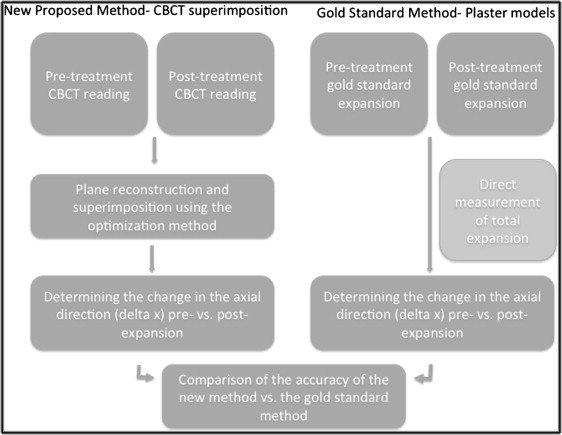
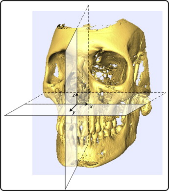
The methodology of this research is summarized in Figure 1 . The gold standard technique (plaster model measurements) of measuring the transverse dimension change from preexpansion and postexpansion was compared with the proposed 3D plane superimposition technique using pretreatment and posttreatment CBCT images to look at changes in the x-axis (transverse direction) ( Fig 2 ). All measurements were made by the same experienced and calibrated orthodontist (G.L.). Both data collection methods are explained below.


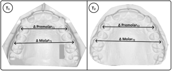
Plaster models were used for the measurement of the total dental expansion in the transverse dimension, referred to as the x-axis ( Fig 2 ). Total delta X (ie, the total change in the x dimension) between the premaxillary and postmaxillary expansion models at the levels of the first premolars (from tip to tip of each buccal cusp) and the first molars (from tip to tip of each mesiobuccal cusp) ( Fig 3 ) were measured. A digital caliper (Mitutoyo, Aurora, Ill), a commonly accepted tool to measure a linear distance on plaster models, was used with an accuracy of 0.01 mm. Each measurement was repeated twice and checked for gross error (more than a 0.2-mm difference). Once both series of measurements were found to be exempt of gross measuring errors, the averages of both measures were taken as the final delta X from the plaster model.

CBCT images from the premaxillary and postmaxillary expansion treatments were used to test the maxillary plane superimposition technique. Data from large field-of-view CBCT images taken at 0.25-mm voxel size were obtained using a Newtom 3G volumetric scanner (Aperio, Verona, Italy) taken at 110 kV, 6.19 mAs, and 8-mm aluminum filtration. The time between the CBCT images was less than a year for each patient to minimize any measurement error introduced by the patient’s normal transversal growth. It was assumed that most clinically significant normal maxillary transverse growth was completed by this dental age. A relatively short treatment time interval helps to guarantee that the landmarks used in the superimposition are stable. Furthermore, the plaster models and CBCT images were taken on the same day for each patient before and after expansion.
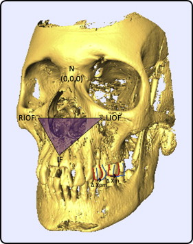
To create a plane for superimposition, 4 accurate and reliable landmarks were used. A detailed explanation on how this superimposition and Cartesian coordinate transformation technique was performed can be found in the studies of Lagravère et al. Briefly, the landmarks selected to create the maxillary superimposition plane were nasion, bilateral infraorbital foramina, and the incisive foramen. All landmarks were analyzed and located using Avizo software (version 6.0; Visualization Sciences Group, Burlington, Mass) with both the volume-rendering and the multiplanar-rendering views for an increased accuracy. All 4 landmarks were validated by the authors (work in process). The landmarks were shown to have intraclass correlation coefficients (ICCs) for the intrarater and interrater reliability calculations greater than 0.924, 0.992, and 0.973 in the x, y, and z planes, respectively. The superimposition method was performed using a series of coordinate transformations performed with MATLAB software (R2008a; MathWorks, Natick, Mass). Using nasion as the center of the coordinates and 3 other stable landmarks (left and right infraorbital foramina and incisive foramen) to form virtual planes, the selected dental landmarks were computed and then compared.
As was done with the plaster models, the expansions of the first premolars and the molars were examined with the superimposition plane as demonstrated in Figure 4 . The differences in the x-axis premaxillary and postmaxillary expansions of the maxillary premolars and molars were computed. The expansion (delta X) is the overall expansion of both sides (right and left).

Statistical analysis
The records of 30 patients were selected for this study without doing any power analysis because of the lack of preliminary studies in the field at the time of the data collection. SPSS software (version 16.0; SPSS, Chicago, Ill) was used for all statistics. Descriptive statistics were used to describe the errors between the plaster and the CBCT superimposition expansion measurements. Bland-Altman plots and ICCs were used to examine the levels of agreement between the CBCT superimposition technique and the plaster model measurements, respectively.
Results
The descriptive statistics are summarized in Table I . The mean errors between the 2 methods were 0.59 ± 0.67 mm and 0.57 ± 0.42 mm at the levels of the premolars and the molars, respectively. Regarding the level of agreement, the ICC ( Table II ) was higher than 0.9 at the level of each teeth, showing almost perfect agreement between the 2 methods. Bland-Altman plots ( Figs 5 and 6 ), another way to graphically demonstrate agreement between 2 measurement methods, also showed strong agreement between the CBCT and the plaster model measurements.


