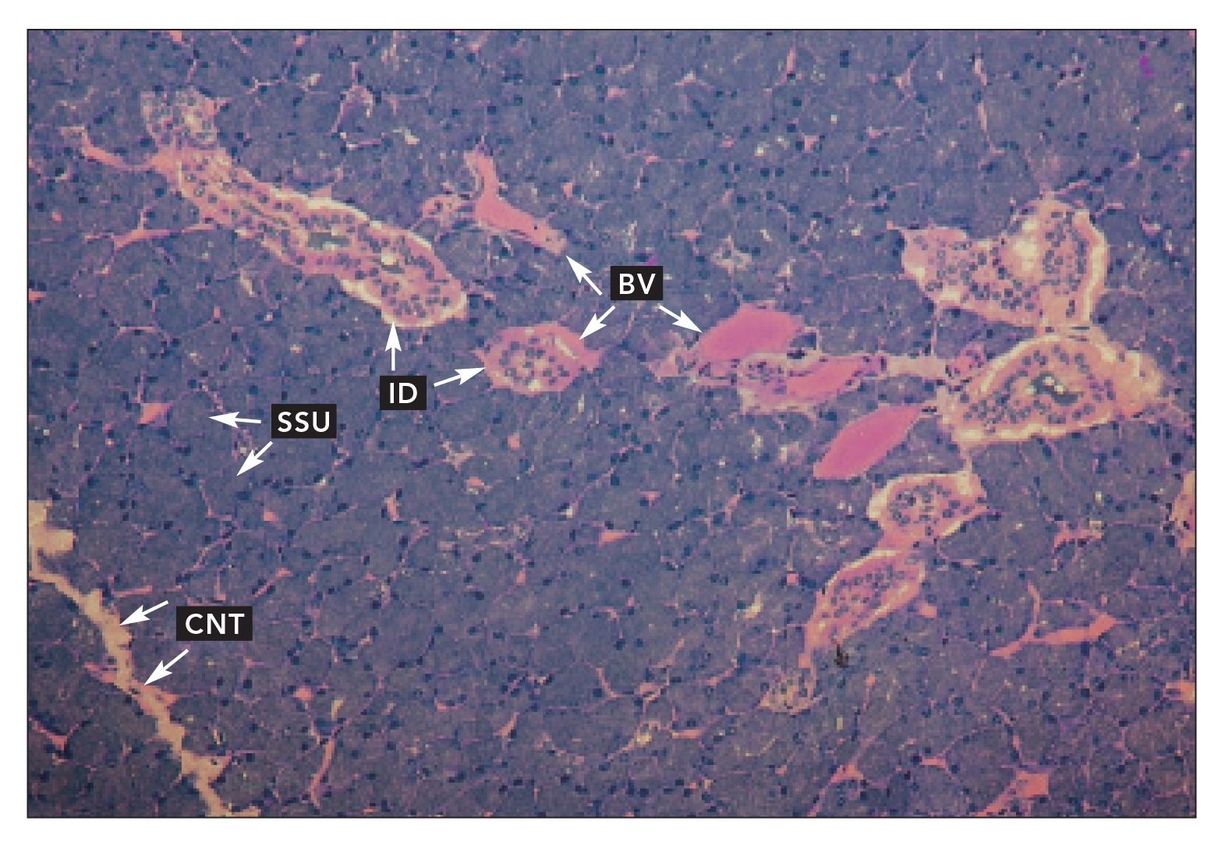
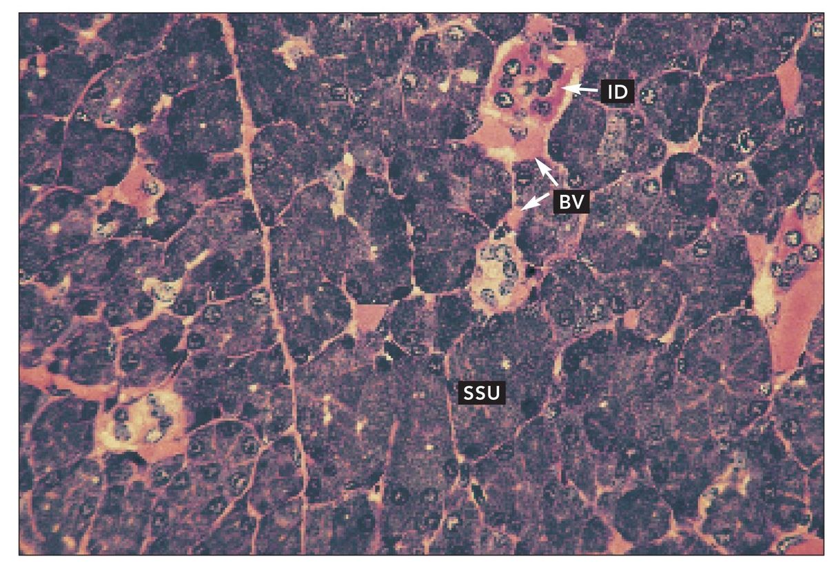
FIG 10-1
Parotid salivary gland
Thin section of parotid salivary gland. Serous secretory units (SSU) secrete into intercalated ducts (ID). Blood vessels (BV) are numerous. The gland is divided into lobes and lobules by connective tissue septa (CNT) (H and Lee stain; ×160).
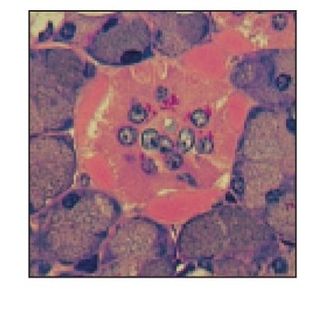
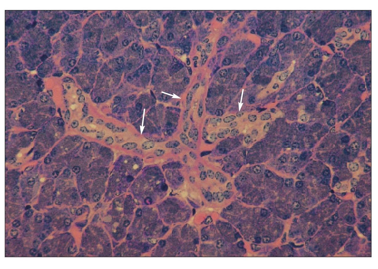
FIG 10-2
Parotid salivary gland
Higher magnification of parotid salivary gland. Intercalated ducts (ID) and accompanying blood vessels (BV) are numerous among the serous secretory units (SSU) (H and Lee stain; ×400).
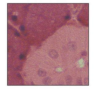
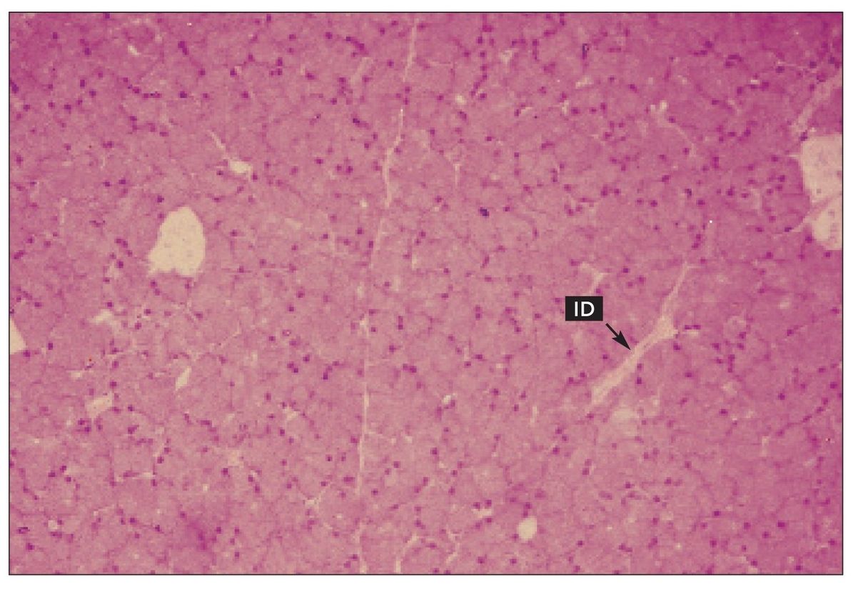
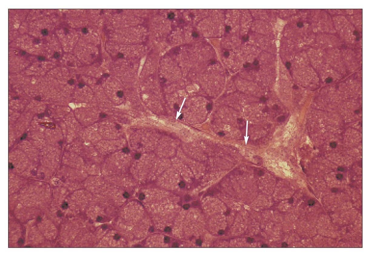
FIG 10-3
Intercalated ducts
Long, branched intercalated ducts (arrows) in the parotid salivary gland (H and Lee stain; ×400).
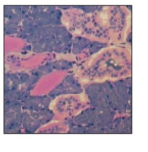
FIG 10-4
Intercalated duct
Low-power view of an intercalated duct (ID) in a paraffin-embedded specimen of parotid gland (H and E stain; ×160).