Salivary Diagnostics and the Oral Microbiome

References
[31]
[32]
[2]
[33]
[34]
[35]
[16]
[36]
[37]
[38]
[3]
Number of subjects
5
71; 98
7
3
120
12
3
10
5
1
242
Locations analyzed
Multiple sites
Saliva; Sp-ging
Two sites
Two sites
Saliva
Saliva
Multiple sites
Un-saliva
Un-saliva
Multiple sites
Multiple sites
Specific sites
Buccal
Saliva tongue
Saliva O-P
Buccal
St-saliva
Buccal
H-palate
H-palate
Sp-ging
H-Palate
Sb-ging
St-saliva
Teeth
Saliva
Teeth tongue
Sp-ging
Tongue
Sb-ging
Tonsils
Teeth
Un-saliva
Sp-ging
Tongue
Throat
Tongue
Tonsils
Notes
4 time points
12 worldwide locations
3 time points


Hpalate hard palate, OP oropharyngeal, Sbging subgingival, Spging supragingival, StSaliva stimulated saliva, Unsaliva unstimulated saliva
A313900_1_En_5_Fig1_HTML.gif

Fig. 5.1

Shared oral microbiome. Average phylum- and genus-level classification of common bacteria colonizing the oral cavity (Data compiled from studies highlighted in Table 5.1)
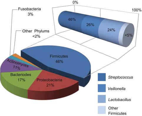
Pooled sample studies are working toward identifying a global oral microbiome profile for healthy individuals. One study found 72 % of the genus level or above, which accounted for 99.8 % of all the organisms present in the mouth, was the same among unrelated healthy volunteers [16]. Another group found that 15 genera were found to overlap between 10 unrelated healthy individuals [40]. Of these genera, 13 were found to be in common between both studies: Streptococcus, Corynebacterium, Neisseria, Rothia, Veillonella, Actinomyces, Granulicatella, Fusobacterium, Haemophilus, Prevotella, Campylobacter, Capnocytophaga, and Cardiobacterium [16, 40]. This overlap creates an outline of the typical organisms and their relative amounts present within healthy individuals, defining the so-called oral “core” microbiome.

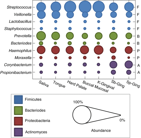
Thorough evaluation of oral micro-niches has recently become possible thanks to the culture-independent methods of high-throughput sequencing. Multiple studies have been published outlining the normal microorganisms found in various locations in the mouth. Analysis in one study revealed that at each oral site tested, there was an average of 200–300 different “species-level” phylotypes [16]. Interestingly, comparison between these unrelated healthy volunteers showed a significant overlap in the typical organisms present in each location. When samples were compared across various sampling sites in the mouth, saliva showed the closest relationship to the tongue first, then palate, mucosa and gingiva, and finally plaque [3, 15, 16, 32, 38, 47] (Fig. 5.2). Firmicutes were the dominant species in saliva followed by Bacteroidetes, Proteobacteria, and Actinobacteria [16, 32, 34, 36]. Together, these four phyla made up >96 % of the total bacterial composition. Salivary samples were also found to be relatively stable in composition over time among healthy individuals [2, 36, 48, 49]. Samples taken at three separate time points over a period of 29 days showed consistent results in their overall salivary microbiome profile [36]. Overall, these studies revealed that each oral location tested resulted in a relatively unique profile or relative number of microorganisms and these healthy communities are stably maintained over time.

A313900_1_En_5_Fig2_HTML.gif

Fig. 5.2

Comparison between bacterial abundance from typical sites in the oral cavity. Relative prevalence of selected oral bacteria shown for a healthy population. KGingival keratinized gingiva, Sbging subgingival plaque, SpGing supragingival plaque, F Firmicutes, P Proteobacteria, B Bacteroides, A Actinomyces (Based on data from [3, 15])
This persistent maintenance of the “normal” flora appears to be a key feature not only in the oral cavity but also in the gut, skin, and vaginal locations [24, 11, 15, 16, 31, 48, 50]. Strikingly, the oral environment was found to have the most consistent “core” microbiome maintained between unrelated people in comparison to other microbial sites such as the gut [2, 36, 40]. The other microbiome sites in the body showed a diverse population of bacteria, but there was considerable variation between unrelated individuals [2, 40]. Defining this “core” microbiome is one of the primary steps that allow for future comparison studies to define profiles that shift away minimally or aggressively from the normal profile.

Shaping the Normal Oral Microbiome

Oral bacteria have preferences for specific sites in the mouth including the teeth, tongue, hard and soft pallet, buccal mucosa, tonsils, gingival tissue, and saliva. Several factors influence the formation and maintenance of the oral ecosystem including the host (host tissues, fluids and signals, diet, genetics), the local environment (pH, temperature, oxygen, amount of nutrients), and the microorganisms themselves (adherence, coaggregation, inter-/intraspecies interactions, virulence mechanisms). The particular community of microorganisms is normally maintained in a state of symbiosis with the host. Yet this diversity can be shifted with changes in the local host condition along with the influence from beneficial and/or antagonistic microbial interactions. In fact, many oral pathogens are opportunist. These pathogens only show virulence when presented with a susceptible host or when a normal host undergoes a number of changes within their oral physiology. Multiple studies have shown that microbial species show distinct properties when part of a multispecies community versus growing in isolation [5159]. Specifically, work by Foster and Kolenbrander [54] showed that the adherence of certain oral species to an artificial tooth surface was dependent on specific microbial binding partners. These multispecies communities generally respond to changes as a group. The microbial population is continuously undergoing a process of turnover. New microorganisms colonize and persist at the expense of others and based on complex interactions will then influence subsequent population compositions [57].
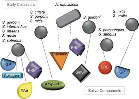
The foundation of these oral communities starts with the deposit of the salivary pellicle on surfaces. The pellicle is primarily composed of glycoproteins derived from saliva that are recognized by bacterial adhesions, allowing selective binding of mainly Gram-positive cocci to the pellicle surface. In addition to providing a binding surface for bacteria, saliva also provides nutrients for bacterial growth from both endogenous (glycoproteins) and exogenous (carbohydrates and peptides) sources. The saliva also shapes the composition by introducing antimicrobial elements and clearance of bacterial species (see later). Bacteria that have attached to the pellicle generate bacterial products as they grow and multiply that influences the subsequent colonizers. A number of organisms are dependent on coaggregation in order to form part of the microbial community [57]. Once the initial microbial population is established, there is a shift observed in composition as more organisms attach and also based on growth rate differences. The majority of these bacterial communities must be constantly rebuilt in response to the mouth’s natural cleansing activities such as saliva production, abrasion, and swallowing.

The Role of Saliva in Forming the Oral Microbial Environment

Saliva is a complex mixture of salivary gland secretions, gingival crevicular fluid, microorganisms, microbial by-products, epithelial cells, and other chemical components. Saliva is secreted by three primary glands, the sublingual, the parotid, and the submandibular, as well as by many minor glands [60]. The amount and type of components present in saliva are known to affect many aspects of oral health and influence bacterial growth [19, 54, 57, 6166]. The bicarbonates, phosphates, and urea within saliva act to modulate the pH and buffer the oral cavity. Salivary proteins contribute to oral microbial metabolism, aggregation, and attachment as well as bacterial cleansing. Immunoglobulins, enzymes, and proteins in saliva manage bacterial growth through antimicrobial action. Finally, saliva is primarily composed of water and therefore provides the necessary moisture for bacterial survival.
The normal pH of saliva is slightly acidic from 6 to 7; however, the range varies based on salivary flow, with values near 7.8 during high flow, and the pH can approach 5.3 at low flows [60]. Bacteria are able to survive in a wide range of pH conditions, but the healthy microbiome composition is primarily supported by a neutral pH range [19]. Circulating bicarbonate and phosphate systems work together to buffer saliva and maintain pH levels [60]. Bacteria also help to buffer saliva by breaking down urea to ammonia and CO2, resulting in an increase in pH [67, 68]. The buffering action of saliva is greatly dependent on high flow rate, allowing regulation of the pH in regions such as the oral plaque environment on and around the teeth [69]. The amount of saliva produced in the mouth displays regional variation, with different areas of the mouth producing various volumes. This variation in saliva volume has been directly linked to the regional clearance rate of acid produced by oral bacteria, generating pH micro-niches within the oral cavity [60]. This variation explains some of the regional differences in bacterial composition seen in the oral cavity (Fig. 5.2). A shift toward acidic pH is known to support the growth of acid-producing and acid-tolerant organisms while killing off acid-susceptible bacteria.
There is a large variability in individual flow rates of saliva [70]. Salivary flow is commonly triggered by mechanical chewing and the taste or smell of food. Other factors that influence salivary flow rates are pain, certain medications, and various local and systemic diseases [7176]. The salivary glands are linked to the sympathetic and parasympathetic nervous system [77]. There are also a number of neurotransmitters and hormones that can influence the rate of salivary flow. Insufficient salivary flow has been clearly associated with a significantly increased risk of oral disease [64, 72, 74, 78, 79] coinciding with a shift in the oral flora [55, 61, 72, 80, 81] (see later). Nutritional changes and deficiencies have also been shown to affect salivary function [19, 8284] (see later). Salivary flow rates correlate with clearance of fermentable carbohydrates and buffering around high-dental-plaque zones.

Colonization of oral surfaces by bacteria depends on binding to the salivary pellicle (Fig. 5.3). The acquired salivary pellicle is a proteinaceous layer that covers all exposed surfaces in the oral cavity. This complex layer is composed of approximately 90–130 different proteins derived from saliva and crevicular fluid serum [60]. These proteins have been grouped based on various roles including binding calcium and/or phosphate, interaction with other proteins, antimicrobial activity, inflammatory response, immune defense, lubrication, and/or buffering and remineralization. Pellicle formation depends in part on the presence of intact mucins and proline-rich proteins (PRPs). Mucins are glycoproteins mainly secreted by sublingual, submandibular, and palatal glands [8789]. PRPs constitute the majority of the human parotid saliva [90]. Mucins and PRPs help initiate colonization by healthy oral flora and are incorporated into the bacterial biofilm structure [87, 88, 91, 92]. Healthy bacteria and the mucin pellicle component provide a protective layer that shields against acids and excessive wear of oral surfaces [88, 93]. If excess colonization takes place, mucins are able to aggregate oral bacteria and clear them from oral surface [93]. Mucins are also critical in hydration and lubrication in the oral cavity and thought to play a role in binding toxins [9395].

A313900_1_En_5_Fig3_HTML.gif

Fig. 5.3

Oral bacteria mediate initial attachment to salivary pellicle. Early colonizers bind to various salivary molecules to initiate attachment to oral surfaces. These connections are mediated by surface adhesions (solid lines) or fibrillar appendages (dashed lines). These early colonizers provide the main binding capability of the biofilm. Few late colonizers are known to interact with salivary molecules, but one example is Fusobacterium nucleatum interacting with statherin [85]. Saliva component abbreviations shown GP glycoprotein, PSA parotid salivary agglutinin, PRP proline-rich protein, SmSP submandibular salivary protein, SCP salivary component in pellicle, Streptococcus genus, except for Actinomyces naeslundii (Based on compiled information in [41, 86])
The antimicrobial activity of saliva is primarily maintained by production of immunoglobulins and enzymes. Salivary glands secrete immunologic agents to protect the teeth and mucosal surfaces. IgA, IgG, and IgM are common immunological salivary components. Secretory IgA is the dominant immunoglobulin on all mucosal surfaces, and it works to neutralize viruses and to aggregate bacteria and functions as an antibody to bacterial antigens [96, 97]. Mucin had been shown to work with secretory IgA and bind bacterial pathogens with greater affinity than either molecule working alone [98]. IgM is thought to work in a similar way to IgA but is more susceptible to proteolytic degradation [99]. IgG is primarily added to saliva via crevicular fluid and has been found to inhibit colonization of the oral pathogen Streptococcus mutans [100]. Saliva-based enzymes (lactoferrin, lysozyme, peroxidase, amylase) protect the teeth from microbial insults [19, 60]. Nutritional immunity is enacted when lactoferrin binds salivary ferric iron, a necessary nutrient for some oral bacterial species [101, 102]. Lysozyme destroys bacterial cell walls and also promotes clearance through bacterial aggregation [19, 103]. Peroxidase catalyzes the production of bactericidal by-products such as thiocyanate [104]. Amylase is a well-known digestive enzyme secreted by the parotid gland [105]. It functions to modulate the adhesion of certain oral species (e.g., Streptococcus gordonii and Streptococcus mitis) while forming part of the salivary pellicle and as a component in free saliva [19]. Finally, amylase has been specifically found to inhibit the growth of bacterial pathogens Legionella pneumophila, Neisseria gonorrhoeae, and Neisseria meningitidis [106109].
Different salivary glands produce saliva with various levels of key salivary components [87, 110]. Differences are seen in the growth of oral bacteria on saliva from different glands [111]. This alone demonstrates that variations in saliva components are able to influence the microbiome of a healthy adult. At the outset, studies have looked at the direct effect on saliva composition and flow rates based on medication, radiation treatment, diet, or disease/trauma directly linked to the salivary gland functionality [73, 76, 79, 101, 112, 113]. A classic example is seen with the autoimmune disease Sjögren’s syndrome. Patients with Sjögren’s have chronic inflammation that affects their salivary glands leading to reduced salivary flow [76, 113]. Radiation has also been shown to decrease not only the salivary flow rate but also buffering capacity and pH [79, 114]. Together, altering the buffering capacity, salivary flow, and/or protein levels can all lead to major changes in the oral environment.

Mechanisms of Oral Bacterial Colonization

Bacterial Adherence and Coaggregation

Coaggregation is a process whereby two or more species of bacteria adhere to each other via specific adhesin-receptor interactions. Coaggregation is highly selective and dependent on interaction of multiple binding receptors in order to build the structured and complex multispecies community that resides in the oral cavity. Coaggregation allows for colonization of microbial species that lack pellicle receptor sites. This colonization is based on cell-cell interactions that were found to be mediated by existing surface molecules from both viable and dead cells attached to the salivary pellicle or floating in free saliva [54, 57, 58]. These interactions are dependent on multiple factors, including physical proximity, nutrient sources, bacterial factors, and host factors. The spatial location of adhesins and their receptors has been identified using antibodies and microscopy [115]. Confocal microscopy revealed that bacterial cells with complimentarily binder/receptors were found to be adjacent to each other within the biofilm structures [86, 116]. Further studies with electron microscopy demonstrated that there appears to be a competition for binding sites between common partners when using the same binding mechanisms. A classic example of this is seen when coccoid cells (streptococci) bind to long rods such as Fusobacterium nucleatum or Corynebacterium matruchotii forming a “corncob structure” [117].

If different mechanisms are used in coaggregation, then some organisms may serve as a bridge to connect other species. Indeed, Prevotella loescheii was found to link Streptococcus oralis to Actinomyces israelii [118120]. The presence of simple sugars in the local environment is also able to change the coaggregation dynamic of several interspecies pairs [63, 119, 121124]. Dextrans are produced by streptococci, encouraging coaggregation and forming part of the intracellular matrix in the biofilms [121]. The enhanced binding of multiple bacterial partners also has the potential to prevent colonization of pathogenic species as the binding sites are blocked [124]. Collectively, the ability of multiple species to coaggregate with each other plays a key role in the spatial architecture and volume of oral biofilms (Fig. 5.4).

A313900_1_En_5_Fig4_HTML.gif

Fig. 5.4

Spatial model of oral bacterial colonization. These bacteria present some common relationships seen in the oral cavity. The salivary component recognition and coaggregation between bacterial binding partners is shown. Bacteria used as part of biomarker studies in cancer diagnosis are indicated in black (see Table 5.4), and biomarkers for oral health diseases are shown in gray (see Table 5.2). The diamond pattern denotes bacterial shifts associated with oral health diseases and selected systemic disease states (see Tables 5.2 and 5.3). Fusobacterium nucleatum, in particular, was found to be associated with several different diseases. The saliva component abbreviations shown are proline-rich proteins (PRPs), bacterial cell fragments (BCFs), and salivary agglutinin (SAs). The bacterial species shown are Aggregatibacter actinomycetemcomitans (A.a.), Actinomyces israelii (A.i.), Actinomyces naeslundii (A. n.), Actinomyces oris (A.o.), Capnocytophaga gingivalis (C.g.), Capnocytophaga ochracea (C. o.), Capnocytophaga sputigena (C. s.), Eikenella corrodens (E. c), Eubacterium spp. (E. sp.), Fusobacterium nucleatum (F.n.), Haemophilus parainfluenzae (H.p.), Lactobacillus spp. (L.sp.), Porphyromonas gingivalis (P.g.), Prevotella denticola (P. d.), Prevotella intermedia (P.i), Prevotella loescheii (P.l.), Prevotella melaninogenica (P.m.), Propionibacterium acnes (P.a.), Selenomonas flueggei (S.f.), Streptococcus gordonii (S.g.), Streptococcus mitis (S.m.), Streptococcus mutans (S.mu.), Streptococcus oralis (S.o.), Streptococcus sanguinis (S.s.), Treponema denticola (T.d.), Veillonella spp (V.sp.) (This image was adapted from [86] with data added from [166168] and Tables 5.2, 5.3, and 5.4)

Bacterial Metabolic Products, Food Webs, and Nutrient Sharing

Bacterial metabolic products and food sharing also have the ability to shape the biofilm, encouraging the growth of some species while deterring others. For example, lactic acid is produced by a number of microorganisms in the mouth as a result of carbohydrate fermentation. Some of the most notable acid producers are from Streptococcus and Actinomyces. This lactic acid can then be used by Veillonella allowing for the production of menadione that enriches for growth of Porphyromonas and Prevotella. Fusobacterium creates fatty acids that are used by Treponema. Porphyromonas gingivalis then works together with Treponema to generate products used by Mogibacterium timidum [169].
Cooperative nutritional behaviors have also been studied for bacteria growing in saliva as its sole nutrient source [54, 57]. Some species are able to be grown individually with saliva as the sole food source, but many common oral bacteria are not. Veillonella parvula, Aggregatibacter actinomycetemcomitans, and Fusobacterium nucleatum are unable to grow in saliva by themselves. However, pairwise and all together, these three species are able to thrive in saliva. Actinomyces oris paired with Porphyromonas gingivalis, V. parvula paired with A. oris, and V. parvula paired with P. gingivalis all show increased growth in saliva; yet, together the three-species group refuses to grow. Streptococcus oralis, P. gingivalis, and V. parvula grow together, while S. oralis paired with P. gingivalis does not. This dependency on the presence of certain species before others can survive leads to a defined spatial architecture within the oral biofilms. This spatial dependence is thought to influence the survival of certain microorganisms within these dynamic oral biofilms, and coaggregation used in conjunction with saliva represents a key mechanism of group bacterial clearance [88, 170] and facilitates symbiotic relationships [58].
In contrast to coaggregation and food webs, oral bacteria may also have antagonistic relationships. Thus, production of certain by-products by established microbiota may act to exclude or inhibit growth of other incompatible microbes. A classic example is the production of acid by S. mutans, which eliminates competitors that are less tolerant of low pH [171174]. Streptococcal species may also compete by production of hydrogen peroxide or bacteriocins in an effort to establish certain bacterial species as dominant in a particular host.

The Dynamic Oral Microbiome: Shifting Bacterial Compositions in Response to Change

Changes in salivary flow can greatly influence the microbial composition present in the oral cavity [54, 55, 60, 72, 80, 114]; pH buffering, bacterial nutrient access, and components of the host response all shape the oral environment [19, 60]. While bacterial populations are able to remain relatively stable throughout the day with multiple shifts in saliva production levels and access to fermentable substrates, prolonged difference in the salivary composition or flow rate can result in a dramatic shift in the oral population. Bacterial mechanisms to tolerate external stress have been well documented [172, 175177]. Cell-cell communication and biofilm formation oftentimes allow these microbial communities to quickly adapt and survive short-term extreme changes in the environment (e.g., lack or food, host antimicrobial factors). Yet, these complex communities inevitably demonstrate changes in species composition after persistent changes occur in the host environment. On the microbial scale, even minute changes that are not readily perceivable by the host, if consistent, will impact the microbial composition. These shifts in host-microbial homeostasis demonstrate that, instead of considering our microbiome as something peripheral, we need to recognize our flora as an integral part of our body systems.
Next, we will discuss how the oral microbiome shifts away from the oral core microbiome in response to oral-specific diseases and systemic diseases/conditions.

Diseases of the Oral Cavity

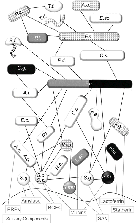
Some of the most well-documented relationships between health, disease, and the oral flora can be observed with the common dental ailments of tooth decay (caries) and periodontitis, including gingivitis. These diseases represent two of the most common chronic diseases seen worldwide [178180]. Several decades of research by dental scientists have clearly illustrated that dental caries and periodontitis are caused by ecological changes in the homeostatic balance between host and bacterial flora (Table 5.2). While initial work assumed that the presence or absence of a single species would be linked to cause and effect for these diseases, it was quickly discovered that these diseases develop along a more complex story line. These oral diseases involve regulation by many sources such as multispecies bacterial connections, host-bacterial interactions, and behavioral or environmental shifts. This body of work begins to demonstrate that changes in the microbial balance can both lead to and result from active oral disease profiles in humans. Oral biofilms have been studied extensively in oral diseases. There is a general progression seen in the buildup of the oral composition as early/primary colonizers attach to the salivary pellicle, additional bacteria bind to the primary colonizers, and finally late colonizers join the group only after a complex community is present. These later colonizers include a number of key oral disease-linked bacteria.

Table 5.2

Bacteria connected to oral health conditions
Oral health condition
Caries
Periodontitis/gingivitis
Healthy
Bacteria
Actinomyces spp.
Aggregatibacter actinomycetemcomitans
Actinomyces spp.
Lactobacillus spp.
Campylobacter rectus
Capnocytophaga sputigena
Propionibacterium acidifaciens
Fusobacterium nucleatum
Cardiobacterium hominis
Streptococcus mutans/sobrinus
Porphyromonas gingivalis
Haemophilus parainfluenzae
Veillonella spp.
Prevotella intermedia/nigrescens
Rothia dentocariosa/mucilaginosa
Treponema denticola/forsythensis
Streptococcus sanguinis
References
[46, 134145]
[27, 146151]
[2, 3, 16, 3138]
Bold indicates organism was used as disease biomarker

Caries

Dental caries lesions are localized demineralization of tooth enamel as a result of acid production by dental plaque biofilms. Caries progression has been well documented from superficial white spot lesion to deep-dentin cavitations [56]. This decay is due to chronic acid production from bacterial fermentation of carbohydrates in the vicinity of the tooth enamel [56]. Previous work looking for the causative agent of dental caries showed that mutans streptococci (Streptococcus mutans and Streptococcus sobrinus) appeared to be the primary acid producers associated with enamel caries [56, 134136, 171, 181]. These bacteria were consistently shown to create the extreme acidic environment necessary for caries conditions, a pH <5.5 [182184]. More recent research has shown that Lactobacillus spp. are also prevalent at active caries site and appear to create the necessary cariogenic conditions in the absence of the S. mutans or S. sobrinus [137, 138, 185]. These species are all aciduric, meaning that they thrive in low pH environments, while other members of the oral flora do not survive long exposures to acid conditions. The loss of competing species during low pH events creates a feedback loop that ultimately yields more acid production, due to the enrichment of aciduric species in the population, leading to the progressive destruction of enamel and dentin, if left untreated. Thus, caries patients display an increasing accumulation of these cariogenic bacteria. Modern techniques using sequencing technology have allowed for further characterization of not just a few select species but analysis of the entire microbiome at the disease sites. More detailed microbiome sequencing reveals an apparent distinct overall microbial shift as the tooth deterioration progresses from mild white spot lesion to caries dentin involvement and finally to severe disease with deep-dentin lesions [21, 46]. The attachment of specific oral flora to the salivary pellicle was directly tied to the particular binding partners that were present or absent during multispecies biofilm formation [51, 116]. Organisms that would normally be able to bind to the pellicle may be outcompeted for binding sites by other common oral species, while others are completely dependent on “helper” bacteria to mediate any active binding to the pellicle surface. These multispecies interactions appear to significantly influence the makeup of the biofilm composition as well as the circulating bacteria in saliva. These interactions must be constantly reestablished following removal from common oral practices such as swallowing, mechanical abrasion from mastication, and toothbrushing.
Lapses in oral hygiene and increased access to fermentable carbohydrates correlate with increased acid production from certain oral species leading to a localized drop in pH at sites with high levels of dental plaque [83, 186, 187]. This drop in pH then starts an environmental feedback cycle shifting some of the oral flora in the mouth. The decrease in pH starts to kill off susceptible oral bacterial species and selects for acid-tolerant organisms while simultaneously dissolving mineral from the tooth enamel. As the caries disease progresses, the environment has been shown to enrich growth of acid-producing and acid-tolerant bacteria including S. mutans, S. sobrinus, Propionibacterium acidifaciens, Lactobacillus spp., Veillonella spp., and Actinomyces spp. [138, 188]. Allowed to persist, this localized loss of mineral from of tooth enamel can lead to significant structural deterioration and pain.
While these acid-tolerant species increase in numbers, it should be emphasized that the remaining oral species do not completely disappear, but instead are less prevalent at the disease site. It was mentioned earlier that the oral environment normally undergoes a number of environmental shifts throughout the day; nonetheless, the overall composition of the oral flora remains constant in healthy individuals [177, 189]. Thus, bacterial and host biomarker profiles can then be used to infer the risk of developing dental disease as well as monitor dental treatment effectiveness. For example, caries risk has been assessed by the amount of lactobacilli, mutans streptococci, and Actinomyces spp. in the patient’s saliva [46, 135, 137, 139, 140, 185]. Young children with high levels of S. mutans and S. sobrinus were more than five times as likely to develop dental caries than individuals with lower levels [135, 136, 141, 190]. Sweden has used salivary levels of lactobacilli (>100,000/ml saliva) and mutans streptococci (>1 million/ml saliva) to determine caries risk for more than 30 years [142144]. The progression of dental caries was also studied by looking at the microbial composition in white spot lesions, dentin lesions, and deep-dentin lesions [46]. They saw a continuous shift in the bacterial composition as the caries sites progress through the stages of disease. Together, these works indicate that while the presence of key bacterial species is necessary for caries development, a microbial community is crucial to supporting the persistence of those pathogenic species in the oral cavity. This change appears to be driven by changes in the local environmental conditions, such as a lower pH and changes in the host response. Therefore, monitoring known caries pathogens as well as the rest of the microbial community should provide a global picture of the oral disease state and can act as a tool to predict caries risk and treatment outcomes.

Periodontology

Periodontal diseases range from basic inflammation of the gum tissue to major damage of oral connective tissue and bone. Gingivitis is mild to moderate inflammation of the gum tissue, and diseased tissue can normally be reversed to a healthy state with a proper oral hygiene regimen [191, 192]. In contrast, periodontitis is diagnosed when inflammation begins to cause permanent damage to the connective tissue surrounding a tooth or the teeth. As the body tries to respond to the presence of bacterial biofilms on the tooth, the host’s immune system actually starts to damage oral tissue. Left untreated, this host-pathogen interaction can lead to loss of gum tissue, loss of attachment of connective tissue to the tooth and bone, and can potentially lead to the loss of teeth [193]. Extensive research has linked the pathology of gingivitis and periodontitis to certain microbial species such as Porphyromonas gingivalis, Treponema denticola, Tannerella forsythia, Aggregatibacter actinomycetemcomitans, Prevotella intermedia/nigrescens, and Campylobacter rectus [146, 147, 194, 195]. These etiological agents are dependent on other members of the oral flora to allow for colonization, and many times there is also an increase in salivary viral load (see later). These bacteria are late colonizers of the biofilm and are not often allowed access to gum tissue when excellent oral hygiene standards are followed.
Reminiscent of dental caries, periodontitis progression is underpinned by a global shift in microbial species [13, 23, 196]. Extensive work has looked at outlining the composition of the periodontal microbiota in order to help develop more effective treatments and diagnostic testing [12, 13, 193]. The predicted periodontitis microbiome shift was analyzed using the microarray technology HOMIM to discover the major differences between healthy control patients and those patients that responded well to treatment versus those that suffered from refractory periodontitis [23]. More species of bacteria in total were detected in diseased patients in comparison to healthy subjects, where 28 % of the 300 species were not detected in healthy individuals. The putative periodontal pathogens were found in much higher numbers than those with healthy periodontium. Those patients with treatable periodontitis were also distinguishable from those with refractory periodontitis based on their unique bacterial profiles. Significantly higher accumulations of common perio-pathogens such as T. forsythia, P. gingivalis, and Prevotella spp. were found within patients with refractory periodontitis compared to treatable but also more “unusual” species that were present, such as Brevundimonas diminuta, Mycoplasma salivarium, and Filifactor alocis [23]. The microbial profile of the healthy individuals revealed that certain bacterial species were more prevalent in subgingival plaque samples and have been found consistently associated with oral health in other studies, including Actinomyces spp. [16, 23], Capnocytophaga sputigena [23, 46, 138], Cardiobacterium hominis [16, 23], Haemophilus parainfluenzae [16, 23], Rothia dentocariosa/mucilaginosa [16, 23], and Streptococcus sanguinis [23, 46, 138].
Periodontal pathogens have been found to colonize the tongue and other non-periodontal sites [38, 197, 198]. The whole saliva provides the immediate source of bacteria for oral biofilm formation and is therefore predicted to have bacterial profiles that are coupled to the biofilm composition. P. gingivalis, P. nigrescens T. denticola, and P. intermedia were found to have a statistical relationship when comparing saliva and periodontal pocket samples [199]. Additional research has shown both genetic and environmental factors influence bacterial colonization in saliva and the periodontal pocket [200202]. Several groups have assessed the severity of periodontal disease by using levels of both pathogenic bacteria and host-response biomarkers in saliva [199, 203206]. Interestingly, investigations have started to show that the bacterial composition shift paired with host biomarkers is correlated with detecting disease stability or periodontitis disease progression [27, 204]. Those patients who showed low bacterial and host biomarker levels fared better than those with high bacterial and host biomarker amounts. Kinney et al. [27] showed that the elevated presence of Fusobacterium nucleatum, C. rectus, and P. intermedia predicted disease progression in 82 % of patients. Low levels of host biomarkers, matrix metalloproteinases 8 and 9 (host-derived enzymes involved with tissue and bone degradation), osteoprotegerin (inhibitor of differentiation and activation of osteoclasts involved in bone resorption), and IL-1 (inflammatory cytokine) predicted periodontal site stability 78 % of the time. These studies present some of the novel work that is starting to link microbiome composition to disease progression and treatment outcomes [13, 193]. Overall, patients who were successfully treated for periodontitis showed a shift back toward a healthy oral microbiome [13].
Defining the oral microbiome has allowed researchers to start to understand the normal commensal organisms that are consistent with health. Key work looking at oral-specific disease supported the hypothesis that the oral microbiome usually harbors a core microbiome, and changes in the core composition are directly related to oral disease progression due to dysbiosis. However, it was still unclear if other disease states were linked to the oral microbiome. This shift from homeostasis to dysbiosis reflects a common theme seen across many microbiome profiles that a disease state may not show complete removal and/or enrichment of certain species but instead results in a dramatic shift in the overall proportions of the total microbiome. The microbiome acts like a community that is influenced by the overall presence of key players that may aid in attachment, acquiring nutrients, surviving fluctuations in the environment, etc. Work outlined in the next section will focus on how the oral microbiome has ties to other systemic diseases such as diabetes, autoimmune disorders, hematologic diseases, vitamin deficiencies, and cancer.

The Oral Microbiome and Systemic Conditions

Saliva contains a number of components that could be used for disease risk assessment. Saliva microbial components are already commonly used for the diagnosis of many dental diseases including caries risk, periodontal disease, saliva gland dysfunction and disease, and oral-based fungal infections [13, 138, 145, 160, 161, 207]. Additional recent work has used microbial and/or host markers in saliva to identify tuberculosis, cancers, gastric ulcers, liver malfunction, and Sjögren’s syndrome [60, 80, 81, 133, 208210].

Based on the relatively new fields of microbiomics and metabolomics, research is just starting to test these predicted shifts in microbial profiles in a number of systemic diseases. Mapping these alternate oral microbiomes may help us better understand what happens as the body challenges these microorganisms with a new environment in response to disease (Tables 5.3 and 5.4).

Table 5.3

Oral manifestations and associated oral flora shift in selected disease states
Disease or condition
Diabetes
Autoimmune: Crohn’s disease
Autoimmune: Sjögren’s disease
Nutrition/vitamin deficiency
Oral manifestation
↑Risk caries, gingivitis, periodontitis
Oral lesions
Chronic inflammation of salivary glands
↓Salivary flow
Oral swelling
↓Saliva production
↑ Caries
↑ Caries
Xerostomia (dry mouth)
↑Periodontitis
↑Caries
Oral lesions
Enamel defects
Inflammation
Delayed dental eruption
Bacteria
P. gingivalis
Fusobacteria spp.
S. mutans
A. actinomycetemcomitans
↓Firmicutes
Lactobacillus spp.
F. nucleatum
Prevotella melaninogenica
Staphylococcus aureus
Veillonella parvula/dispar
Neisseria mucosa
Fusobacterium nucleatum
Eikenella corrodens
P. intermedia/nigrescens
C. rectus
Fungi
Candida albicans
C. albicans
References
[152157]
[158, 159]
[80, 81, 160163]
[112, 164, 165]

Table 5.4

Oral bacterial shifts observed in cancer patients
Disease
OSCC
Head and neck cancer
Upper GI tract cancer
Colorectal cancer
Gastric precancerous lesions
Pancreatic diseases (cancer)
Bacteria
Fusobacterium naviforme
Streptococcus anginosus
S. anginosus
F. nucleatum
A. actinomycetemcomitans
Neisseria elongata
S. aureus
T. denticola
S . mitis
Streptococcus mitis Prevotella melaninogenica Capnocytophaga gingivalis
References
[47, 125, 126]
[45, 47, 127]
[128]
[129, 130]
[131, 132]
[133]
OSCC oral squamous cell carcinoma, Bold indicates organism was used as disease biomarker

Diabetes

Numerous studies have started to uncover the link between oral health and type 2 diabetes [211]. High, uncontrolled blood sugar levels are associated with excess bleeding and poor healing overall [212214]. Diabetic patients have been found to have a significantly higher risk for gingivitis and periodontitis [211, 212, 215]. One large-scale study that looked at 4,343 adults revealed that diabetics have an odds ratio of 2.9 for periodontitis compared to those not suffering from diabetes [216]. Diabetic patients who better controlled their glycemic levels saw their odds ratio fall to approximately 1.56 [216]. When patients are failing to control their diabetes, they show a consistent flare in oral diseases [215, 216]. Systemic problems with increased blood glucose levels lead to chronic inflammatory immune response leading to excess production of inflammatory proteins, which may help to keep the microbiome in check [12, 211, 217]. While the subgingival bacterial profile showed the same type of species in periodontal patients with diabetes in comparison to those periodontal patients without diabetes, the relative abundance was different between diabetic and nondiabetic [152, 153]. The diabetic patients show higher numbers overall of assessed perio-pathogens (Table 5.3). Diabetic patients appear to show an exaggerated inflammatory response to the same oral periodontal pathogens and in some cases increased apoptosis (programmed cell death). This enhanced inflammation and apoptosis is thought to be involved with delayed wound healing and likely explains some of the excess periodontal tissue damage seen in diabetic patients [218]. Interestingly, a key relationship was found between one particular oral pathogen, P. gingivalis, and insulin. Lipopolysaccharide released from P. gingivalis has been shown to be toxic to some cytokine proteins that regulate insulin levels in the body [219, 220]. Proinflammatory cytokines, including interleukins 1 and 6 along with tumor necrosis factor-alpha, are produced in inflamed periodontal tissue [221223]. These factors gain access to the body’s general circulation via the periodontal microcirculation [224, 225], which then allows them to systemically antagonize insulin [226]. Whole-body insulin resistance was also found to be triggered by bacterial infection [154].
Research has demonstrated that treatment for periodontitis, which also shifts the microbial microbiome, improves the overall health outcomes for these patients [12, 211, 222]. However, diabetes can also lead to additional oral disorders including fungal infections, salivary functional disorders, dental caries, and burning mouth syndrome [154, 227, 228]. Together, this systemic disorder leads to major changes in the body and importantly the oral environment. Diabetes and oral infections have a bidirectional relationship where the presence of one seems to further provoke the other and leads to a global increase in symptoms and decrease in quality of life. Here we see that a systemic disorder can significantly advance the periodontitis infection resulting in a shift in the microbial profiles with increased frequency of perio-pathogens consistent with diabetes plus periodontitis.

Autoimmune Disorders

Autoimmune disorders represent a wide-ranging group of diseases that can impact the human body with varied severity. Oftentimes, autoimmune disorder patients suffer cyclical disease presentation where “flares” coincide with severe disease symptoms and times when symptoms are manageable to nonexistent [162, 229, 230]. There are some autoimmune diseases that show consistent oral presentations in addition to other symptoms [230]. In some cases, the oral symptoms have been known to precede more classic symptoms [230]. This is sometimes seen with Crohn’s disease oral lesions. Crohn’s disease is an autoimmune disorder that primarily impacts the intestines, but secondary symptoms are seen with the joints, skin, and liver [229]. Oral manifestations have been used for Crohn’s diagnosis [78, 231, 232]. These symptoms include mucosal tags; gingival, labial, or mucosal swelling; and ulcers. The patterns of oral symptoms and histology mimic those seen in the intestine with inflammation, ulcers, and swelling. The severity of the oral lesions has been shown to predict the extent of disease in the intestine [230]. In some cases, the oral presentation will precede the systemic symptoms and could be used as an indicator for further confirmatory testing [230, 231, 233]. Microbial shifts were seen in Crohn’s disease patients including an overall decrease in Firmicutes and increases in Prevotella melaninogenica and Neisseria mucosa [158, 159]. Patients with Crohn’s disease show an increased risk for caries development [159]. In addition, inflammation in the intestine has been shown to lead to malabsorption [234, 235]. This nutritional deficiency can lead to significant changes in the oral environment (see later).
Sjögren’s syndrome is an autoimmune disease that attacks the endocrine glands, including the salivary glands that lead to a significant decrease in the production of saliva [113, 162]. The dry mouth conditions means there is considerable risk for severe dental diseases [62, 162]. Predictably, patients with primary Sjögren’s syndrome were found to have increased levels of S. mutans, Lactobacillus spp., and Candida albicans within their supragingival plaque samples [81]. The smooth mucosa and tongue samples also showed increased prevalence of Staphylococcus aureus and Candida albicans [80, 81, 160, 163]. The greatest shift in microbial differences was observed by patients who had a stimulated saliva secretion rate of <0.5 ml/min [81]. The primary risk factor that seems to be associated with this shift in the bacterial profile is the reduced saliva production. There is, however, a change seen in the protein levels of Sjögren’s syndrome patients as well [114, 236]. This suggests that reduced salivary flow combined with changes in the salivary composition shapes the microbial accumulation in the mouth. These shifts may therefore be used to predict “flares” along with assessing treatment progress.

Nutrition and Vitamin Deficiencies

Changes in the diet are known to alter the overall composition of some species within the oral flora [177, 237239]. Malnutrition has been linked to reduced salivary flow rates, changes in bacterial numbers, and shifts in salivary composition [82, 164, 239241]. There has been a documented increase in the prevalence of oral pathogens that are involved with caries development as our modern human diets have increased in refined sugar consumption [83, 242]. Scarcity of certain foods and nutrients can dramatically alter the oral environment. The mucous membranes in the mouth show a high cell turnover rate (3–7 days) versus the skin (up to 28 days) [243, 244]. The oral cavity may, therefore, exhibit signs and symptoms more quickly from direct nutritional deficiencies or systemic diseases that lead to nutritional/vitamin depletion. Vitamin deficiencies can lead to generalized issues in the oral cavity [164]. Many deficiencies are associated with oral-specific symptoms including caries, delayed dental eruption, enamel defects, and oral lesions [164, 235, 239, 241]. Dentists may be involved with diagnosis of vitamin deficiencies based on presenting oral symptoms when no other obvious cause is present. For example, megaloblastic anemia, due to a lack of the vitamin B12, displays oral mucosal membrane change in approximately 50–60 % of the patients [195, 245, 246]. In general, low iron levels are known to contribute to diminished bactericidal activity from leukocytes, impaired cellular immunity, epithelial abnormality, and inadequate antibody response. These changes in the host lead to generalized oral problems including inflammation, higher prevalence of oral candidiasis, and predicted shifts in oral flora that are sensitive to low iron levels [239, 247252]. Vitamin A deficiency leads to dry mouth, impaired tooth growth, and increased susceptibility to infection [164, 253, 254]. Vitamin C is important for healthy collagen formation and serves to enhance iron absorption [164, 255257]. Vitamin C deficiency has been shown to contribute to the severity of periodontal disease [165, 241]. Patients that were suffering from vitamin C deficiency displayed more attachment loss than those with normal vitamin C levels. Together, these missing nutrients and many others may indicate the start of systemic disease that impacts the oral cavity early on and left untreated can result in severe damage [112, 164, 214, 235, 239].

Cancer

There is a new and novel work that is following potential links between the oral microbiome and various cancers (Table 5.4). This field is just starting to highlight microbiome differences observed in cancer patients versus healthy individuals. A majority of the work, so far, has looked at the oral flora composition and its link to oral and other head-/neck-associated cancers (e.g., esophagus, pharyngeal, etc.). Oral, head, and neck cancers are the sixth most common cancer seen worldwide [258]. Despite many advances in cancer diagnosis and treatment, in general, the overall prognosis for oral cancer patients has not improved much in recent years [259261]. Improving early detection of many different types of cancers remains a key objective of salivary diagnostics, and here we will emphasize some of the promising research in that field.
A few groups have started to survey the bacteria that colonize different regions in the mouth, focusing on both malignant and healthy sites in oral and upper gastrointestinal locations [47, 127, 128, 133, 262, 263]. These groups found that the microbial composition differed significantly for at least some of the bacterial species when comparing normal, healthy tissue to malignant sites. For example, Hooper et al. [125, 126] conducted two studies that looked at the bacterial composition of oral squamous cell carcinoma tissue samples. The oral cancer tissues showed a distinct profile of several oral species including Fusobacterium naviforme and Staphylococcus aureus. In contrast, analysis of various upper gastrointestinal tract carcinomas found a distinct link with one particular species, Streptococcus anginosus [128]. S. anginosus DNA was also found in carcinoma tissue from head and neck squamous cell biopsies but not in those from other cancer types, including oral cancer [127, 264, 265]. One potential explanation given for these observed differences in bacterial composition between cancer and cancer-free sites is that certain oral bacterial cells may actually be influencing signals that initiate and advance oral and head-/neck-associated cancers [266], as well as other cancers [267, 268]. There are several cases where microorganisms are considered the primary source of cancer, including the classic bacterial example of Helicobacter pylori infection leading to gastric cancer formation [269, 270] plus numerous viral-associated cancers [271276]. In these cases, the microorganisms were directly involved with influencing the development of specific cancers. Intriguingly, preliminary studies have found a link between H. pylori infection and oral cancer diagnosis [263, 277, 278]. In addition, several normal oral bacterial residents have been shown to have a direct influence on stimulating an inflammatory response in the mouth [153, 211, 279]. Although many of the exact mechanisms leading from bacterial infection to carcinogenesis remain largely unclear, there is a general link between upregulation of cytokines and other inflammatory mediators that leads to an increased risk of carcinogenesis [280282].
Because the aforementioned studies imply that the presence of certain bacterial species may actually increase the likelihood of developing certain cancers, these and other bacterial species can potentially be used as a portent for diagnosing cancer early. One particular study looked at comparing the bacterial salivary composition from 229 healthy controls versus 45 patients with oral cancer [47]. This work found principal increases in Streptococcus mitis, Prevotella melaninogenica, and Capnocytophaga gingivalis among patients with oral squamous cell carcinoma in comparison to the controls. Further analyses showed that these species could be used as a diagnostic biomarker for oral cancer. The three-species diagnostic marker predicted 80 % of the cancer cases correctly while excluding the controls approximately 82 % of the time. Importantly, this research group found that the soft tissue bacterial composition was similar to that of the saliva composition. As mentioned earlier, different locations in the mouth may have large differences in the oral microbiome structure. This represents a key feature of testing since the cancerous or precancerous oral lesions may not be obvious in patients and will have to be diagnosed from a shift in saliva alone.
Several groups have also found an interesting link between the oral microbial composition and other cancers [47, 262, 263, 268, 283, 284]. Gastric precancerous lesion risk was found to positively correlate with high levels of periodontal bacterial pathogens [131, 132]. Gastric cancer has the second highest mortality rate worldwide and is the fourth most common cancer [285]. Patients with periodontitis that showed an increased colonization by Actinobacillus actinomycetemcomitans were more likely to have precancerous gastric lesions. Of interest, A. actinomycetemcomitans is associated with systemic infections along with being a general cause of periodontitis; therefore, its association with the oral flora may have been directly influenced by additional signals from the host resulting in favorable colonization. Fusobacterium nucleatum was recently shown to have a link to colorectal cancer [129, 130]. Colorectal adenomas, colorectal cancer tissue, and feces from colorectal cancer patients all showed significantly higher rates of Fusobacterium spp. in comparison to healthy controls. A mouse model of intestinal tumorigenesis demonstrated a rise in proinflammatory markers and increased infiltration of myeloid immune cells in F. nucleatum treated animals [129]. Human colon tissue samples also showed a strong correlation between the F. nucleatum levels and the expression of proinflammatory markers (COX-2, IL-1β[beta], IL-6, IL-8, and TNF-α[alpha]) [129, 286, 287]. A proposed mechanism for Fusobacterium involvement in colorectal cancer is based on the cell-surface Fusobacterium adhesion molecule (FadA) binding to E-cadherin on the surface of host epithelial cells [288]. Binding activates β(beta)-catenin signaling, F. nucleatum invasion, and inhibition of tumor suppressor activity promoting colorectal cancer formation. FadA levels are presented as a proposed marker for early diagnosis of colorectal cancer. However, it is still unclear whether Fusobacterium is acting as a commensal to modify the forming tumor microenvironment or instead it is an opportunistic pathogen and directly responsible for the promotion of colorectal cancer.

Finally, HOMIM was used to look at the oral microbiota in patients with various pancreatic diseases. The salivary microbiome was observed to be significantly different in patients with pancreatic disease, including cancer, in comparison to the healthy controls [133]. Pancreatic cancer patients had an increase in 31 bacterial clusters/species and decrease in 21 bacterial clusters/species. Further analysis, testing 16 species/clusters as a pancreatic disease biomarker, showed that combining Neisseria elongata and Streptococcus mitis were the most valid biomarkers. The combination biomarker resulted in a 96.4 % sensitivity and 82.1 % specificity in determining pancreatic cancer patients from healthy individuals. Novel recent work from the same lab has presented evidence for a mechanism connecting systemic disease with salivary changes. Lau et al. [210] found that pancreatic cancer-derived exosomes, “cell-specific lipid vesicles,” provide a link between the primary cancer site and saliva biomarker production in mice. These exosomes that are able to travel through the vascular systems are involved with intercellular communication and are thought to be associated with a number of functions including immune response regulation and tumor invasion promoters [289, 290]. A number of host factors were shown to be upregulated based on saliva comparison from healthy and diseased mice using microarray analysis. Some of these host factors have the potential to influence the bacterial compositions observed in the cancer patients versus cancer-free individuals (Table 5.5). The presence of human serum was also shown to influence the adhesion and coaggregation phenotypes in a number of oral species [55]. This leads to a tempting mechanism that exosomes have the potential to travel from newly forming cancer sites to deliver disease-specific products directly to saliva, which can then shape the oral microbial environment through direct messages or trigger secondary messages such as hormones and cytokines (Fig. 5.5). These oral changes can, therefore, presumably be used to monitor distant body sites throughout disease progression.

Table 5.5

Upregulated saliva gene products in response to pancreatic cancer and the predicted bacterial association
Gene product
Predicted function
Bacterial association
sema6d
Immune response (CD4+ T cell regulation) and gastric cancer [291, 292]
Commensal gut bacteria displays CD4+ T cells response [293]
daf2
Associated with insulin receptor [294]
Caenorhabditis elegans daf2 mutants are resistant to bacterial pathogens [295297]
aspn
Binds collagen and calcium [298]
Divalent cation concentrations influence bacterial adhesion [299302]
A313900_1_En_5_Fig5_HTML.gif

Fig. 5.5

Model for pancreatic disease detection using salivary diagnostics. (1) Pancreas tissue transitions toward a disease state. (2) Exosomes from the pancreas enter the circulatory system. (3) Exosomes deliver disease-specific products directly to the salivary glands. (4) The oral microbiome is changed through direct signaling or secondary message effects within the saliva. (5, 6) Saliva samples can be used to detect changes in host biomarkers and the oral microbiome providing noninvasive feedback of disease progression and/or treatment outcomes

The Other Microbiomes: Mycobiome and Virome

The majority of work looking at the human microbiome has focused on bacteria composition. In reality the microbiome contains fungal and viral components as well. While this is still an emerging area of study, there are some intriguing research efforts coming to light related to fungal and viral shifts in response to systemic disease.

Mycobiome

Candida albicans has been consistently seen as an indicator of oral microbiome imbalance and other oral diseases [303, 304]. This overgrowth of Candida albicans is commonly known as oral thrush. Thrush is seen in the immunocompromised and immunosuppressed patients, including patients with human immunodeficiency virus (HIV), cancer patients, infants, elderly, etc. As many as 90 % of HIV patients will present with reoccurring oral candidiasis [305308]. Diabetic patients with high blood sugar and patients with Sjögren’s are also more prone to candidiasis infections [160, 163, 309]. The generalized presentation of candidiasis is a good visual indictor that something is systemically wrong with the patient, though infant oral thrush does not indicate a problem unless the infection continues for several weeks. Unfortunately, because a variety of conditions can lead to candidiasis, it is not specific and thus provides limited information about the potential systemic issue without further testing. Just like the bacterial composition, a defined oral mycobiome will need to be established to determine differences between conditions of health and disease. One group has started this work by looking at the oral mycobiome of 20 healthy individuals [207]. They were able to detect 74 culturable and 11 unculturable fungal genera in the oral cavity. As few as 3 and as many as 39 genera were found in these healthy volunteers. Candida spp. were present in more than 75 % of the individuals, followed by Cladosporium at 65 % and Saccharomycetales and Aureobasidium at 50 % each. Intriguingly, a cluster analysis showed that mycobiome profiles of males and females were separate from one another. This study represents the first survey of the fungal species present in the oral cavity and only surveyed a small number of people. Similar to the bacterial microbiome surveys, the oral mycobiome is predicted to reveal a core set of fungal organisms present within healthy populations along with unique species specific to each individual. In fact, efforts have shown that the total composition of an individual’s human microbiome is just as unique as a fingerprint with no two people alike [310].

Virome

The persistent presence of viruses in saliva can be observed for a number of reasons. Viruses can be transferred by direct contact with infected individuals, a blood-borne viral infection can allow for salivary gland involvement, or the oral cavity itself may be infected in the mucosa or diseased periodontal sites [148]. Like with candidiasis patients, those with compromised or suppressed immune systems will also likely display a viral load increase in saliva. The presence of viruses in saliva presents another noninvasive disease diagnosis opportunity. In light of this, a number of groups have developed saliva-based viral detection tests for common human viruses including HIV [311], hepatitis viruses [312], herpes viruses [312, 313], and measles virus [314]. Researchers are starting to characterize the human virome; however, this field is still emerging. One key problem lies in detecting the presence of unknown viruses. Unlike bacteria that have a conserved 16s ribosomal region within their DNA, viruses have little DNA sequence in common. Plus, the majority of viruses in the human body are specific for bacteria hosts (bacteriophage) and not humans [315, 316]. Those bacteriophages are able to indirectly impact humans by changing the abundance or function of our resident bacteria. With the advent of shotgun sequencing, we can now look at all the DNA whether it is human, bacterial, fungal, or viral. Studies using this approach on the gut virome found that mothers and children, and identical twins have similar viromes [317, 318]. Another study looked at the viral composition present inside the lungs of cystic fibrosis (CF) patients in comparison to non-CF patients [319]. They found some differences in the viral composition but significant changes in metabolic functions as a result of these viruses in the lung. These variations are thought to influence the severity of cystic fibrosis; however, this was a small-scale study with five CF and five non-CF patients. A larger analysis used nasal swabs from 176 children, some healthy and some with unexplained fevers. The children with unexplained fevers had more viruses on average than those that were healthy, suggesting the fevers may have been viral in nature and not bacterial [320].
New human viruses and bacteriophages are discovered every day; nonetheless, new viruses that posses DNA or RNA that is unlike any other known sequence will likely be missed in these large-scale sequence surveys until a more universal method of identifying viruses becomes available. Much of the work to date has, therefore, focused on quantifying the viral load of known human viruses, mainly those belonging to the herpes-type viruses.

Herpes Viruses

Herpes viruses are the most common viral type found in human saliva [321323]. There are eight known herpes viruses that infect humans: herpes simplex types 1 and 2, cytomegalovirus, Epstein-Barr virus, varicella zoster virus, herpes virus 6, herpes virus 7, and herpes virus 8 (Kaposi’s sarcoma) [324]. Herpes viral infections can lead to diseases of the periodontium and oral mucosa or may present asymptomatically but still with active viral shedding into saliva [322, 324

Only gold members can continue reading. Log In or Register to continue

Stay updated, free dental videos. Join our Telegram channel

Sep 17, 2015 | Posted by in General Dentistry | Comments Off on Salivary Diagnostics and the Oral Microbiome

VIDEdental - Online dental courses

Get VIDEdental app for watching clinical videos