Revision rhinoplasty is a general term that deals with the correction of various deformities in a previously operated nose; such deformities range from a subtle irregularity over the nasal tip to a completely distorted nose requiring several reconstructive surgical interventions, including the application of multiple grafts, sutures, and flaps. The present chapter is an overview of the most common deformities, their diagnosis, and current treatment protocols.
Etiopathogenesis
The need for revision rhinoplasty is multifactorial. The lack of accurate diagnosis and discussion to determine the patient’s cosmetic and functional demands is the leading cause of secondary corrective cosmetic nasal surgery. The tendency to make a nose excessively smaller can compromise both the cosmetic and functional outcome. Unrealistic demands by the patient that are unrecognized by the surgeon will probably lead to an unsatisfied patient. Similarly, surgeons should not overestimate their ability and understand the limitations of the applied cosmetic maneuvers. Each surgical procedure has its limitations and standard range of errors. Multiple alterations along with a combination of complex cartilaginous and bony modifications will increase the margin of error in rhinoplasty. Of particular difficulty are patients who have a postoperative result that is deemed satisfactory by the surgeon but yet unsatisfactory by the patients. This is probably due to a lack of initial communication between the surgeon and patient and may pose a significant challenge. All unsatisfactory results must be acknowledged and explored. Rather than ignoring the outcome, discussion and planning should be undertaken for any possible corrective measures.
It is important to distinguish revision rhinoplasty from a staged rhinoplasty in which a second procedure is planned at the primary consultation. For instance, in a patient with a severely crooked nose, precise judgment about the nostrils may be difficult and alar base surgery may be intentionally postponed until a second minor surgery can be performed.
Pathologic Anatomy
The anatomy of an operated nose differs completely from its normal counterpart. For this reason, revision surgery requires special attention to anatomy. After the primary operation the skin redrapes over the new framework, followed by a dynamic healing process that extends well beyond the first year and affects the aging nose indefinitely. The result is shrinkage and contracture of skin, which sometimes limits its potential to undergo extensive dissection and major changes in volume. Subcutaneous scars are also inevitably seen in an operated nose. The extent of the scarring depends on the amount of previous surgical manipulations, the presence of dead space, and finally, the biologic behavior of the patient’s wound healing.
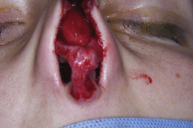
The osseous and cartilaginous frameworks are subject to many augmentation or reductive procedures in primary rhinoplasty. Therefore, excessive scarring, compromised vascularity, unpredictable healing, over-contoured and excessive reduction of native anatomy, grafts, sutures, and altered mucosa, perichondrium, and periosteum are among the changes seen in a previously operated nose. In particular, a clear path of dissection is not as easily identified in the sub-perichondrial plane at the nasal tip area. Figure 104-1 illustrates open access to a nose previously operated on 5 years previously that demonstrates scarring and altered cartilage anatomy.

Because autografts and alloplastic material are commonly used in primary rhinoplasty, the esthetic surgeon should be ready to confront anatomic changes that are not seen in a normal nose; each of these materials has its own characteristics and frequently provokes unpredictable tissue behavior.
The lower lateral cartilage is often significantly altered by reductive techniques. The continuity of this cartilage may be interrupted in several places. The presence of cartilage and bone grafts will also modify the anatomy. The upper lateral cartilage is usually weakened because of over-resection of the cartilaginous dorsum, which can result in severe esthetic and functional problems. The nasal septum is commonly manipulated in the first surgery to correct septal deviation or to harvest cartilage and is rarely intact in revision cases.
Pathologic Anatomy
The anatomy of an operated nose differs completely from its normal counterpart. For this reason, revision surgery requires special attention to anatomy. After the primary operation the skin redrapes over the new framework, followed by a dynamic healing process that extends well beyond the first year and affects the aging nose indefinitely. The result is shrinkage and contracture of skin, which sometimes limits its potential to undergo extensive dissection and major changes in volume. Subcutaneous scars are also inevitably seen in an operated nose. The extent of the scarring depends on the amount of previous surgical manipulations, the presence of dead space, and finally, the biologic behavior of the patient’s wound healing.
The osseous and cartilaginous frameworks are subject to many augmentation or reductive procedures in primary rhinoplasty. Therefore, excessive scarring, compromised vascularity, unpredictable healing, over-contoured and excessive reduction of native anatomy, grafts, sutures, and altered mucosa, perichondrium, and periosteum are among the changes seen in a previously operated nose. In particular, a clear path of dissection is not as easily identified in the sub-perichondrial plane at the nasal tip area. Figure 104-1 illustrates open access to a nose previously operated on 5 years previously that demonstrates scarring and altered cartilage anatomy.
Because autografts and alloplastic material are commonly used in primary rhinoplasty, the esthetic surgeon should be ready to confront anatomic changes that are not seen in a normal nose; each of these materials has its own characteristics and frequently provokes unpredictable tissue behavior.
The lower lateral cartilage is often significantly altered by reductive techniques. The continuity of this cartilage may be interrupted in several places. The presence of cartilage and bone grafts will also modify the anatomy. The upper lateral cartilage is usually weakened because of over-resection of the cartilaginous dorsum, which can result in severe esthetic and functional problems. The nasal septum is commonly manipulated in the first surgery to correct septal deviation or to harvest cartilage and is rarely intact in revision cases.
Diagnostic Studies
Proper diagnosis and treatment planning are key to successful revision rhinoplasty and are usually based on three important criteria: psychological evaluation, esthetic analysis, and functional examination. Each of these assessments should be performed before any corrective (or primary) surgery.
Psychological Evaluation
Apart from unsatisfactory esthetic and functional outcomes, patients desiring revision rhinoplasty can be emotionally affected by the unfavorable results of their primary elective surgery. Many revision patients will have a negative memory of their previous surgical experience, which can diminish their confidence and trust in any further esthetic surgery.
Psychological evaluation usually starts with a comprehensive history and a thorough interview. Revision patients seek to fulfill their unmet expectations in their second surgeries. Therefore, all expectations should be listed during the primary interview and any possible unrealistic expectations should be detected. Patients should be gradually prepared for possible outcomes of a well-planned revision surgery that are usually less than ideal. Consideration should be given to preoperative psychological consultation. Extensive preoperative consultation will reduce the frequency of postoperative problems related to inadequate communication and misdiagnosis.
Esthetic Analysis
Esthetic analysis of patients seeking revision rhinoplasty closely resembles that for primary rhinoplasty (see Chapter 103 ). Life-size photographs are helpful for rhinometric measurements and analysis of the actual size of the deformities. The use of predictive software will sometimes contribute to the perception of patients and can help esthetic surgeons make correct diagnoses and formulate proper treatment plans, although it can also affect patients’ postoperative expectations of the acceptable margin of error related to rhinoplasty surgery.
Functional Examination
Functional examination begins with visual inspection of the patient while making a deep inspiration. Any possible external valve collapse should be noted. To examine the inner valve, the nasal passages should be visualized with a headlight and nasal speculum. A Cottle test would be informative of the strength of the inner nasal valves and can help detect any valve stenosis.
Ancillary Diagnostic Studies
Computed Tomography And Plain Radiographs
Most structural abnormalities of the nose can be explored by careful physical examination. Computed tomography (CT) and plain radiographs are sometimes used to assess the condition of the paranasal sinuses or to document septal deformities.
Nasal Endoscopic Examination
Nasal endoscopy (rhinoscopic examination) can be used to further assess the nasal mucosa and visualize the nasal septum and associated structures. In the event of any uncertainty, nasal endoscopy will easily reveal even minor septal malformations or can be used to document and assess severe soft tissue complications such as septal perforation.
Rhinomanometry And Acoustic Rhinometry
These techniques are safe, non-invasive diagnostic modalities that are generally used for quantitative measurements of airway patency. Though not routine, they can be used to analyze and compare the effects of surgical corrective procedures with the preoperative parameters.
Treatment/Reconstructive Goals
The main goal of revision rhinoplasty is to restore both esthetic and functional damage caused by the primary surgery or to correct possible deformities that were left untreated in previous surgeries.
To achieve this objective the following minor goals should be taken into consideration: (1) proper diagnosis and treatment planning, (2) determination of the best graft donor site, (3) re-establishment of major and minor tip support, and (4) restoration of form and function of the external and internal nasal valves.
The modern trend in performing revision rhinoplasty is based on augmenting lost tissue, strengthening weakened structures, and precise refining of any possible excess tissue. This can be achieved by an endonasal (closed approach) or open approach (see Chapter 103 ). The approach depends on the complexity of the corrective surgery and the surgeon’s experience; however, minor corrections are usually done via a closed approach, and more complicated cases necessitate an open approach.
Augmentation Material
Many materials have been introduced for use in primary and revision rhinoplasty and are generally divided into three groups: autogenous grafts, allogeneic grafts, and alloplastic material. Table 104-1 lists the general characteristics and advantages and disadvantages of each augmentation material.
| Advantages | Disadvantages | ||
|---|---|---|---|
| Autogenous grafts | |||
| Fascia | Resistant to resorption Camouflage the sharp edge of grafts such as crushed cartilage |
Donor site morbidity Patient compliance |
|
| Cartilage | Septal cartilage | The longitudinal characteristic of this cartilage makes it a favorable choice for spreader grafts | Usually consumed or scarred during primary rhinoplasty, especially the caudal portion |
| Auricular cartilage | Excellent alternative choice of autogenous cartilage; the curved nature of auricular contours could be used for alar grafts and composite grafts The remaining subcutaneous portion assists in fixation of the graft |
Risk for hematoma Inherent anatomic curvatures Not suitable for crushing and morselizing |
|
| Costochondral cartilage | First choice for massive reconstructions; an abundant amount of cartilage is available | Extensive surgery needed Risk of warping of cartilage and complications at the donor site |
|
| Allograft | Available in large quantity Lack of donor site morbidity |
Risk of disease transmission Host immune reactions to the graft Resorption Warping |
|
| Alloplastic grafts | Available in every size, volume, and hardness Lack of donor site morbidity Easily contorted More favorable in dorsal augmentations |
Foreign body reaction or chronic inflammation Infection Extrusion Expensive Unfavorable result in the lower third of the nose and functional structures (e.g., strut, alar grafts, columella) Wound healing complications with subsequent nasal trauma Increased complications in younger patients |
|
Autogenous Grafts
Autogenous material consists of cartilage, fascia (temporalis fascia, fascia lata), skin, and bone (e.g., calvarial bone and iliac crest). Autogenous cartilage is the most common augmentation material and can be harvested from the nasal septum and auricular and costal cartilage.
Septal Cartilage
Septal cartilage is the donor material of choice for most augmentation procedures. The ease of access within the same surgical field, especially with concomitant septoplasty procedures, is a significant advantage. The cartilage is easily cut and tailored into different shapes and forms, and it can be crushed or morselized to achieve a smooth surface along with elimination of cartilage memory.
The major drawback of septal cartilage is its limited volume, particularly in a previously operated septum. In such cases the risk for septal perforation and inadequate volume of cartilage available make this donor site unacceptable.
Stay updated, free dental videos. Join our Telegram channel
VIDEdental - Online dental courses


