Fig. 3.1
(a) A patient presented to the clinic complaining of an unusual “bump on her gum”. Intraoral examination revealed localized swelling on the labial mucosa, buccal of left maxillary central incisor. Probing around the tooth was within normal limits and the patient was asymptomatic. Percussion was inconclusive on all anterior teeth. The patient could recall a history of dental trauma a year prior to the examination. This periapical radiograph of the left maxillary central incisor shows a periapical radiolucency with what seems to be a communicating internal-external inflammatory resorption. The tooth tested non-vital to pulpal testing. (b) Endodontic treatment was initiated and an attempt to induce calcification by using calcium hydroxide alone was made. (c) Root canal therapy was completed and adjunct surgical procedure was performed to apply ProRoot MTA (Dentsply Tulsa Dental Specialties) on the buccal aspect of the root, where the resorption communicated with the periodontal ligament space. A fiber post was bonded into the root canal and a provisional crown was fabricated
3.2.4 Periodontal Considerations
There are many factors to be evaluated when assessing the periodontal condition. The age of the patient, the initial bone loss, the probing depths, the clinical attachment loss, the mobility, the root form, the furcation involvement, and whether or not the patient is a smoker are to be considered when determining the prognosis of a tooth. In a retrospective study of 102 patients (816 molars) undergoing regular periodontal therapy, Miller et al. assigned scores to all teeth on the basis of periodontal prognostic factors. They determined that of all the factors evaluated, smoking had the most negative impact (246 % greater chance of losing their teeth), far exceeding the impact of pocket depth, mobility, or furcation involvement. The authors also mentioned that 78.3 % of the molars treated were never extracted and survived for an average of 24.2 years, which indicates that under preventive maintenance therapy, periodontal health can be maintained (Miller et al. 2014). One limitation of the study resides in the fact that the severity of the furcation involvement was not assessed; only its presence or absence was considered. However, it is understood by dental health professionals that the more severe the furcation involvement, the more difficult it is for the patient to maintain proper dental hygiene. The same could be said about pocket depths in excess of 3 mm. Surgical periodontal therapy could be indicated to reduce the pocket depth and increase the likelihood of dental hygiene to be effective.
Another prospective study on 100 treated periodontal patients under maintenance care (2509 teeth) was carried over a period of 16 years in an attempt to determine the effectiveness of commonly taught clinical parameters utilized in the assignment of prognosis to accurately predict tooth survival. The study concluded that initial probing depth, initial furcation involvement, initial mobility, initial crown-to-root ratio and parafunctional habit with no biteguard were all associated with tooth loss (McGuire and Nunn 1996). Teeth that were used as abutment for fixed partial dentures (FPDs) were lost at a lower rate than those who served the same function for removable partial dentures (RPDs). Interestingly, the authors suggested that the reasons that FPDs may have greater survival rate might be related to the initial choice of the tooth as an abutment, as only very healthy teeth would be used for a fixed abutment.
Multiple authors have reported that periodontal reasons are the most common cause for extraction of endodontically treated teeth with 59.4 % and 42.6 % of all extracted teeth (Fonzar et al. 2009). In esthetically challenging situations, with the presence of apical periodontitis, or when retreatment is needed, extraction of the tooth followed by implant placement has been recommended (Setzer and Kim 2014). However, as discussed above, if proper periodontal treatment is rendered, even on teeth with moderate vertical bone loss or furcation involvement, the prognosis could be good (Setzer and Kim 2014).
Any tooth is just as strong as its weakest link. If its foundation is compromised, its entire outcome also is. Besides a dental emergency, periodontal health has to be achieved and maintained before any treatment is to be initiated. As we will discuss later, when it comes to mechanical forces, a tooth is subjected to stresses that come from all directions. The weaker the support it has from its periodontium, the more likely the horizontal stresses on the entire system are to increase and the more strain the restoration and the periodontium have to absorb. Distribution of stress being a cornerstone concept in prosthodontics, the clinician must consider, when dealing with a less than ideal, yet healthy, periodontium to lighten the occlusion in eccentric movements. For example, when restoring a canine with a compromised, yet acceptable, crown-to-root ratio, the clinician should consider a group function rather than a canine guidance.
3.3 Tooth Restorability
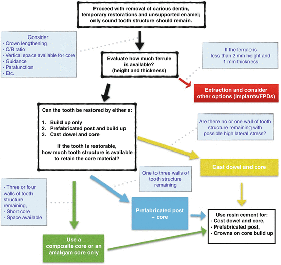
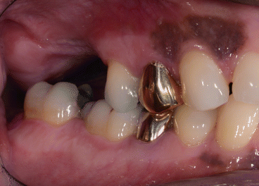
For a comprehensive treatment plan to be formulated and before using the treatment planning flowchart in Fig. 3.2, a complete evaluation of the mouth along with that of the particular tooth in question is necessary. The clinician must evaluate the overall periodontal support, the occlusal scheme, and the presence or absence of parafunction. With regard to the occlusal plane in a comprehensive treatment plan, a tooth that has extruded and is not in harmony with the occlusal plane might not allow enough vertical space for an antagonist. For this tooth to be restored, the plan might involve the possibilities of orthodontic intrusion or crown lengthening with or without root canal therapy (see Fig. 3.3). Also, when it comes to the tooth in question, one must evaluate the quality of the root canal treatment. The latter is still a major cause of failure, as reviewed in Chap. 1.


Fig. 3.2
This is the treatment plan flowchart that is referred to throughout this chapter

Fig. 3.3
This photograph shows the right mandibular second molar (mirror image) that has supra-erupted into the opposing edentulous space. Several years after its extraction, this patient is considering replacing his maxillary missing right maxillary first molar. An interdisciplinary approach, possibly involving orthodontic movement and/or crown lengthening and/or endodontic therapy, has to be considered to bring this tooth into the normal occlusal plane
A tooth serving as an abutment for an FPD or an RPD is subject to different stresses than if it were to support a single restoration. Lastly, if the possibility of crown lengthening is considered, the clinician must keep in mind some considerations for the crown-to-root ratio, the taper of the root, and the location of any furcation.
3.3.1 Evaluation of the Remaining Tooth Structure
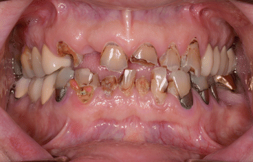
Some clinical conditions (e.g., a vertical root fracture or an infraction that extends far apically into the periodontium) could justify the extraction of the affected tooth, particularly if the patient is not keen on the clinician performing an explorative surgery to determine its restorability. Other, less dramatic scenarios require the removal of carious dentin and/or defective restorations in order to properly assess the overall condition of the tooth (Fig. 3.4). It is following this step that we would determine if the margin placement of the intended prosthesis is violating biologic principles (see the concept of biological width in Chap. 1, Fig. 1.2) and if the remaining sound tooth structure is sufficient in order to provide a strong support that will confer the restoration longevity and function. It is also at this stage that we assess the crown-to-root ratio and the occlusal forces the tooth is subject to in the dentition and determine the necessity of crown lengthening (Fig. 3.2).


Fig. 3.4
Several of these teeth would be deemed unrestorable even without removing the carious dentin and defective restorations. If crown lengthening is performed to obtain adequate ferrule on sound tooth structure, with or without endodontic therapy, the crown-to-root ratio becomes compromised. Considering the taper of the roots, the tooth preparation for any type of complete-coverage crown might result in very thin residual dentin walls
3.3.2 Ferrule
Following excavation of carious dentin and removal of defective restorations, it is essential that sound tooth structure remains circumferentially to produce a cervical ferrule. Please refer to Chap. 1 for more detailed explanation about the ferrule concept.
If the condition of the tooth is such that even adjunct surgical and/or orthodontic procedures cannot provide a 2 mm-high ferrule, without compromising significantly the prognosis of the tooth, extraction might be the solution (Fig. 3.4). When extensive tooth structure is lost after carious dentin or a faulty restoration are removed, or following trauma or endodontic access, but that the ferrule is at least 2 mm high and 1 mm thick, the clinician must consider the fabrication of a foundation prior to tooth preparation for complete-coverage restoration. In some cases, the tooth breakdown is so extensive that it could be in proximity to the pulp. These scenarios might necessitate an elective root canal treatment and the buildup of a core. The latter will increase the retention and resistance form of the future restoration.
According to Hempton and Dominici (2010), most of the retention and resistance to dislodgment of the restoration occurs at the apical third of the preparation. Therefore, the positioning of the margin remains of crucial importance. The clinician must avoid placing the margin if it is to be seated partially or completely on the core buildup. This precaution must be taken in order to avoid the stresses from occlusion to be transmitted to the foundation restoration or, in the case of a post and core, to the internal aspect of the post and the root. That interface is usually filled with cement, and, under occlusal stress, the fatigue of the cement could lead to dislodgment of the post and core or to the fracture of the tooth. In an in vitro study, Pilo et al. (2008) suggested that having a minimum thickness and length of ferrule is very important to prevent fractures. They explained that, in case fractures occur, they do so in the tooth structure, not in core material. Also, the potential of the teeth to fracture is directly related to the amount of dentin removed.
3.3.3 Dentin and Enamel Integrity
Worthy of mention, careful attention must be taken when instrumenting the canal during endodontic therapy as well as when preparing a post space. Over-instrumentation will contribute to over-enlargement of the root canal and unnecessary dentin removal. It is well accepted that a minimum of 1 mm of dentinal thickness wall is necessary to prevent its fracture and properly support the core foundation, if any is planned (Ouzounian and Schilder 1991).
As it was discussed in the previous chapter, mechanical properties of endodontically treated teeth could confer the dentin of the tooth different mechanical properties. However, it has been suggested that the type of cavity preparation could play an even more significant impact on cuspal deflection (separation of the cusps). In one study, it has been determined that intact mandibular molars had a cuspal deflection of up to 1.0 μm. As for MO cavity preparations, the deflection was noted to less than 2 μm of movement. MOD preparations showed a movement of 3–5 μm. Endodontic access preparations were responsible for a movement of 7.0–8.0 μm for the MO group and 12.0–17.0 μm for the MOD group (Panitvisai and Messer 1995). It has been advocated that maintaining the continuity of enamel maintains the tooth rigidity; henceforth, consideration should be given to some sort of cuspal protection, particularly when there is an increase of twofold or threefold from the MO to the MOD group with endodontic access preparation.
It seems that the mechanical properties of the endodontically treated tooth’s dentin might not be as critical as the initial appearance of the tooth that lead to the endodontic treatment. The integrity of the enamel seems to play a more important role than whether or not it has been treated endodontically. In the next section, we will have a closer look at the literature when it comes to the impact of such results on the overall outcome of the tooth.
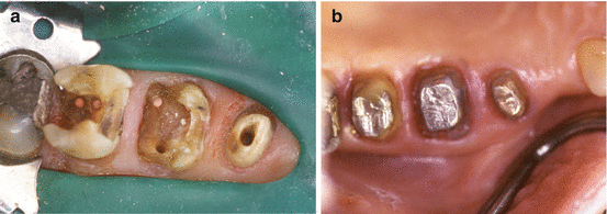
Let us go back to the treatment planning flowchart (Fig. 3.2). After determining the stress that will be applied to that tooth, we need to answer two questions: (1) Is there enough remaining sound tooth structure to retain a core? and (2) Is the remaining tooth structure strong enough to resist crown fracture at the neck of the tooth? If the answer to both these questions is “no,” then a cast dowel and core or a prefabricated post and core buildup are to be considered. If the answer to both questions is “yes,” then a (composite resin or amalgam) core restoration buildup should be considered (Figs. 3.2 and 3.5).


Fig. 3.5
(a) After removal of the carious dentin and defective margins, the remaining tooth structure on the maxillary right second premolar is insufficient to retain a core and is not strong enough to resist crown fracture at the neck of the tooth. As for the maxillary right first molar, it has fewer than three walls left and would need a post to retain a core. On the other hand, the maxillary right second molar has sufficient walls left to retain a core and resist crown fracture at its neck. (b) A cast dowel and core was placed on the maxillary right second premolar, a prefabricated metal post was inserted into the palatal root of the maxillary right first molar, and an amalgam core was built. As for the maxillary right second molar, an amalgam core was used to rebuild the missing tooth structure
In a randomized clinical trial on 360 premolars followed up for 3 years, Cagidiaco et al. (2008) divided the teeth in six groups of 60 premolars based on the amount of the dentin left at the coronal level after endodontic treatment and before abutment buildup. They then randomly assigned them into subgroups with or without fiber posts. They determined that a (fiber) post might not be needed in four coronal walls situation but that as soon as we lost one wall, we started seeing failures in the groups with no post. In the post group, failures increased in the group with one coronal wall and the groups with no wall with or without a 2 mm ferrule. Two studies by Ferrari et al. (2007, 2012) also confirmed that the placement of a fiber post reduced significantly the failures on endodontically treated premolars. The preservation of one coronal wall significantly reduced the failure risk.
In situations of three or four coronal walls remaining, one must choose between different core materials. In a fatigue study, Kovarik et al. (1992) tested glass ionomer cement (GIC), composite resin, and amalgam as a core material under crowns. It took 20,000 cycles to bring all GIC core crowns to failure. At 50,000 cycles, 80 % of composite core crowns experienced failures. As for amalgam core crowns, 30 % experienced failures at 70,000 cycles. Gateau et al. (2001) reported that two GIC-based materials used as core materials showed a higher number of defects than amalgam, suggesting that fatigue resistance of GIC-based materials may be inadequate for post and core applications. Some clinicians have used silver-reinforced GIC-based materials (known as cermets) for core buildups. As stated by Combe et al. (1999), cermet GIC materials are one of the weakest materials in terms of tensile, flexure, and modulus values, despite being similar to some resin materials in terms of compressive strength. Cermet GIC materials are not suitable for core buildup procedures in the posterior teeth.
3.4 Prognosis of Endodontically Treated Teeth
Restorative considerations put aside, endodontic therapy has been demonstrated to be a predictable procedure provided that the quality of the canal disinfection and that of the root canal obturation are good. In the absence of preoperative apical periodontitis, primary root canal therapy has shown success rates above 90 % (Marquis et al. 2006). However, when preoperative apical periodontitis was present, this number dropped to approximately 80 % (Sjogren et al. 1990; de Chevigny et al. 2008).
In their systematic review, Gillen et al. (2011) suggest that all aspect of the treatment, from the periodontal condition to the root canal therapy to the restoration, have an impact on the overall outcome. When coronal restorations are inadequate, the odds of maintaining the healed status of an apical periodontitis decrease as microbes ingress through the defective margins of the restorations.
3.4.1 Survival Rate of the Endodontically Treated Anterior Teeth
There is a belief that endodontically treated anterior teeth without crowns are not susceptible to as much fracture as posterior ones. However, a recent study on 1.4 million teeth (Salehrabi and Rotstein 2004).demonstrated that 83 % of teeth that were extracted had not received a crown, while 9.7 % of the extracted teeth had a crown and a post and 7.3 % of the extracted teeth had a crown without post. The result of this 2004 large-scale study contrasts the findings reported by Sorensen and Martinoff in 1984, where it was suggested that endodontically treated anterior teeth do not have a significantly better prognosis if they are crowned, compared if there were not (Sorensen and Martinoff 1984). Please refer to Chap. 6 for more evidence about the positive impact of fiber posts on endodontically treated anterior teeth.
3.4.2 Survival Rate of the Endodontically Treated Posterior Teeth
When endodontically treated posterior teeth are not restored with a crown, they are more likely to fracture than vital teeth (Aquilino and Caplan 2002). In another study, it was demonstrated that teeth without crowns failed after an average period of 50 months, while pulpless teeth restored with a full coverage restoration were lost after an average of 87 months following the placement of the restoration (Vire 1991). In a retrospective cohort study, it was demonstrated that endodontically treated molars that were intact, except for the endodontic access opening, were successfully restored using composite resin restorations. It is interesting to know that composite resin restorations had better clinical performance than dental amalgam restorations. On a 2-year basis, the survival of molars restored with composite resin restorations was 90 % vs. 77 % for amalgam restorations. At 5 years, the survival declined significantly for both restorative materials, to 38 % for composite resin and 17 % for dental amalgam restorations (Nagasiri and Chitmongkolsuk 2005). Similarly, a 3-year investigation found comparable success rates between endodontically treated premolars restored with only a post and direct class II composite resin and premolars restored with complete-coverage crowns (Mannocci et al. 2002).
Amalgam may be used provided all cusps adjacent to teeth with missing marginal ridges are covered and sufficient thickness of amalgam is present, as seen in Chap. 1. It has been recommended that a thickness of 4.0 mm of amalgam protects the functional cusp and 3.0 mm over the nonfunctional cusp (Liberman et al. 1987).
Once we have determined the restorability of the tooth as a unit, it is important to assess the stress that will be applied to that tooth. As we will discuss in the next section, there is scientific evidence that suggests that an endodontically treated tooth’s position in the arch as well as its future function has an impact on its survival rate. Also Chap. 6 covers the impact of placing fiber posts on the longevity and prognosis of posterior endodontically treated teeth.
3.4.3 The Importance of Proximal Contacts
It has been determined that the presence of two proximal contacts had a significant positive impact on the survival rates of endodontically treated teeth (Aquilino and Caplan 2002; Caplan et al. 2002). Caplan et al. (2002) performed a retrospective study in which they reviewed charts and radiographs of 400 teeth from 280 patients. They suggested that endodontically treated teeth with less than two proximal contacts underperform the ones with two proximal contacts. To reinforce that point, a meta-analysis of 14 clinical studies also pointed out that observation. In descending order of influence, the conditions increasing the survival rate of endodontically treated teeth were as follows: (1) a crown restoration after RCT, (2) tooth having both mesial and distal proximal contacts, (3) tooth not functioning as an abutment for removable or fixed prosthesis, and (4) tooth type or specifically non-molar teeth (Balto 2011).
Another 4-year prospective study involving 759 primary root canal-treated teeth and 858 endodontically retreated teeth demonstrated that teeth with two proximal contacts had a 50 % lower risk of being lost than those with less than two proximal contacts. It was demonstrated that terminal teeth were associated with almost 96 % more tooth loss than those that were not located distal most in the arch (Ng et al. 2011). In a 10-year follow-up study, Aquilino and Caplan demonstrated that second molars had a significantly lower survival rate than any other type of teeth. The greater than fivefold decrease in the endodontically treated second molar’s survival rate could be explained by the result of increased occlusal stresses and difficult endodontic treatment due to a compromised access and restricted visibility (Aquilino and Caplan 2002).
It is well documented that the position of the endodontically treated tooth in the arch and the presence or absence of proximal contact have a significant impact on its survival rate. This could be explained by the unfavorable distribution of occlusal forces and higher non-axial stress on these teeth. Also, regardless of the chewing forces of the patient, an endodontically treated tooth is better off, in terms of stress, to oppose an acrylic tooth from a complete conventional denture than it is to oppose a single implant crown. It has more to do resiliency of the opposing prosthesis than it is about the material.
3.4.4 FPDs and RPDs
Multiple clinical studies have suggested that FPDs supported by endodontically treated abutment teeth fail more often than those supported by vital abutment teeth (Reuter and Brose 1984; Karlsson 1986; Palmqvist and Swartz 1993; Sundh and Odman 1997). Over 20 years ago, Sorensen and Martinoff (1985) reviewed over 6000 patients’ records, and based on 1273 teeth endodontically treated that served as an abutment for either an FPD or an RPD, they concluded that abutments for FPDs and RPDs that were endodontically treated had significantly higher failure rates than single crowns. Respectively, the success rate of all endodontically treated RPD abutments was 77.4 % compared with FPDs, which was 89.2 %. Interestingly, they also found that post placement was associated with significantly decreased success rate for single crown, produced no significant change for FPD abutments, and significantly improved the success of RPD abutment teeth. The nature of the study being retrospective, variables that were not recorded and may have affected that function of the RPD, and stresses on the endodontically treated teeth include the rest and retainer design, the quality of the adaptation of the extension bases, and the occlusion. Also, the span of the distal extension was not recorded, and no distinction was made between a tooth-borne and a distal extension RPD.
A more recent clinical study compared FPDs and single crowns for up to 20 years. The authors reported that the survival rate of three-unit FPDs with at least one endodontically treated abutment was comparable to FPDs on vital teeth. More failures were associated with FPDs with cantilevered units and those with more than three units (De Backer et al. 2007
Stay updated, free dental videos. Join our Telegram channel

VIDEdental - Online dental courses


