Fig. 13.1
(a–c) Patients (middle to late-aged) presenting with moderate/severe tooth wear. (a) Anterior tooth wear mainly erosion/abrasion. (b) Tooth wear mainly attrition. (c) Generalised tooth wear multifactorial
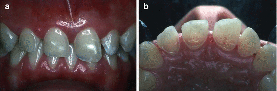
Fig. 13.2
(a, b) Young patient (age 17 years) exhibiting severe localised tooth wear mainly erosive in nature. (a) Labial view. (b) Palatal view
-
The patient feels that the appearance of the teeth is unacceptable.
-
Normal function is disrupted due to the loss of tooth tissue and/or ongoing pulp sensitivity which does not respond to conservative measures.
-
There is progressive tooth wear which may result in pulp necrosis and/or teeth becoming difficult to restore.
A broad range of restorative treatment options are possible with today’s materials and techniques and usually range between:
-
Conventional fixed restorations
-
Removable onlay/overlay prostheses
-
Minimal preparation adhesive restorations
This chapter will illustrate and discuss some of the conventional methods used to restore the worn dentition as well as the newer, less invasive and minimal preparation adhesive techniques now available to the dental surgeon.
13.2 Conventional Fixed Restorations
Conventional fixed restorations, usually in the form of porcelain-fused-to-metal (PFM) and all-metal crowns, have been used for many years for restoring worn and broken down teeth. The clinical performance of conventional crown and fixed bridge restorations have been well documented, and in general tolerable survival times have been demonstrated [9–12]. Unfortunately, studies have not assessed risk factors specifically associated with the various forms of tooth wear, although there is general agreement that restorations in patients exhibiting distinct parafunctional clenching and grinding habits have a higher risk of failure compared with other types of tooth wear.
Tooth preparation for conventional crown restorations is an invasive procedure, especially if porcelain is required to satisfy aesthetic demands, and as a consequence may further compromise an already damaged dentition [13]. Pulp necrosis, tooth fracture, loss of cementation and marginal caries can all occur following the placement of crown restorations, which will often complicate any subsequent repair or replacement, resulting in possible premature tooth loss [14, 15]. This is particularly the case if crowns are prescribed for the younger patient. Notwithstanding some of the risk factors described, these type of restorations are extensively used and offer an effective way of restoring worn and missing teeth, allowing a degree of versatility with regard to appearance and occlusal form. Additionally, one of the potential advantages of this treatment approach is the ability to use provisional crowns for a period of time, thus providing the opportunity for clinician and patient to assess appearance and function and make any modifications as necessary.
Often complicating the restoration of worn teeth is the need to recreate inter-occlusal space lost as a result of compensatory eruption of opposing teeth during the process of tooth wear [16]. This is often the case with anterior teeth and if not addressed will result in a compromised finish (Fig. 13.3). In these circumstances, there are a number of well-established conventional restorative techniques available to overcome the difficulties of reduced crown height and lack of inter-occlusal space [17]. The main options either individually or in combination are:
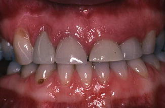
Fig. 13.3
Anterior crowns constructed to conform to the existing worn teeth without recreation of lost inter-incisal space resulting in poor aesthetics and retention form
-
Opposing tooth reduction
-
Elective endodontic treatment and post retention
-
Occlusal adjustment (retruded arc of mandibular closure)
-
Periodontal surgical crown lengthening
-
Localized orthodontic tooth movement (Conventional Fixed Appliance or ‘Dahl’ appliance)
-
Overall increase in occlusal vertical dimension
For an adequate restoration of good appearance and durable function, the role of opposing tooth reduction, elective pulp extirpation and post retention, occlusal adjustment and periodontal surgical crown lengthening alone may be of limited help and can compromise remaining tooth structure and periodontal support. Periodontal surgical crown lengthening is capable of dramatically improving available coronal tooth structure for adequate crown preparation (Fig. 13.4) and is often required when restoring more severe forms of tooth wear. However, it is an invasive procedure with postoperative sensitivity and can create a number of subsequent restorative difficulties, such as interproximal spacing and placement of crown margins on root surfaces [18, 19].
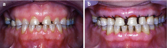
Fig. 13.4
(a, b) Periodontal surgical crown lengthening on worn upper anterior teeth prior to the construction of conventional crown restorations. (a) Before surgery. (b) Immediately after surgery
One of the more satisfactory and conservative ways of recreating space, particularly in situations of localized anterior tooth wear, is by orthodontic tooth movement. A number of techniques can be used to achieve this [20], with a fixed or removable metal-based anterior bite plane being an established method or more often today the use of direct composite resin techniques. The so-called Dahl appliance, described after the author, achieves space recreation by a combination of anterior tooth intrusion and posterior tooth extrusion [21, 22]. This localized orthodontic treatment provides the opportunity to maximize the appearance and function of the subsequent crowns and preserves tooth tissue (Fig. 13.5).
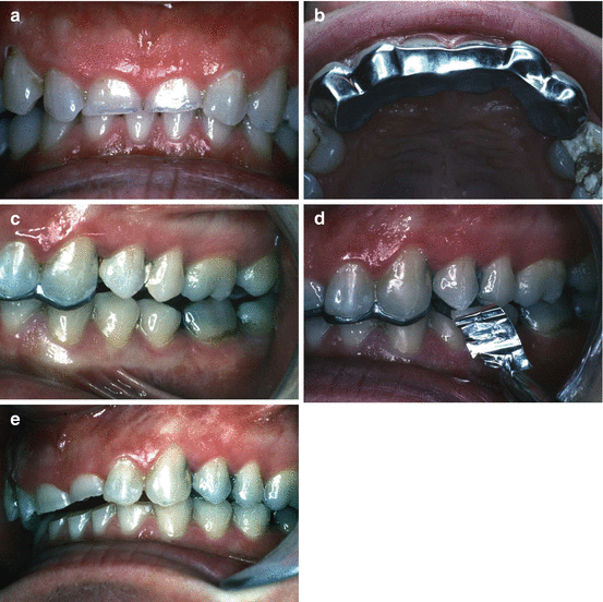
Fig. 13.5
(a–e) The use of an orthodontic ‘Dahl appliance’ to recreate lost inter-incisal space prior to the restoration of worn anterior teeth. (a) Localised anterior tooth wear. (b) ‘Dahl appliance’ cemented in place. (c) Initial space in posterior quadrants. (d) Regained posterior tooth contacts after 6 months. (e) Inter-incisal space recreated following the removal of the ‘Dahl appliance’ prior to anterior restorations
In certain situations where there is generalized tooth wear and sufficient indications to consider crown restorations for the posterior teeth, a full mouth crown reconstruction at an overall increase in occlusal vertical dimension will usually provide adequate space for anterior restorations (Fig. 13.6). This situation will avoid the need for an anterior orthodontic appliance as well as preserving valuable incisal and occlusal tooth tissue. Although excellent and predictable results can be achieved by this method, the process is relatively complex and requires a number of carefully planned stages, appropriate operator skills and knowledge, time and good technical laboratory support [23, 24]. As a consequence, this form of treatment may not be accessible for many patients.
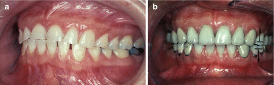
Fig. 13.6
(a, b) Generalised tooth wear restored with conventional crown restorations in the anterior and posterior segments at an overall increase in occlusal vertical dimension. (a) Before restoration. (b) After restoration
It is possible to achieve an increase in occlusal vertical dimension with the use of a removable posterior onlay prosthesis in combination with anterior fixed crown restorations. This approach relies heavily on patient compliance and may result in an unpredictable outcome for the anterior restorations due to adverse occlusal loads if the removable prosthesis is not used periodically. If a removable prosthesis is being considered to provide posterior occlusal support in this situation, then it may be more sensible and predictable to restore the worn anterior teeth with the same removable prosthesis.
13.3 Removable Onlay/Overlay Prostheses
The use of a removable onlay/overlay prosthesis can be a valuable means of rehabilitating patients with moderate/severe tooth wear, particularly when there are also missing strategic teeth to be replaced. This form of treatment has been advocated by a number of clinicians for patients with more severe forms of tooth wear [25–29]. This approach provides a relatively simple, non-invasive and cost-effective way to achieve improvements in appearance and function of the dentition.
The construction of a provisional acrylic resin removable prosthesis is recommended initially, allowing the opportunity to carry out modifications to the shape, position and occlusal relationship of the prosthetic teeth and soft tissues, as well as assessing the patient’s tolerance of a removable prosthesis (Fig. 13.7). It is advisable to avoid any significant tooth preparation at this stage, but if this treatment approach is to be continued in the longer term then subsequent prostheses, often incorporating a cobalt-chromium framework, will usually require some tooth preparation in order to optimize appearance, fit and retention. Space demands are usually greatest in the anterior region, both in the vertical and labio-lingual dimensions, and will be influenced by the amount of sound tooth structure remaining and changes to the occlusal vertical dimension. Unless modified by tooth reduction or extraction, the available space will determine whether or not an anterior labial flange can be used, or alternatively, gingival fitting and/or butt-fitting tooth facings (Fig. 13.8). The final decision may to some extent depend on the patient’s aesthetic demands and desire to avoid or limit any necessary tooth reduction.
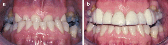
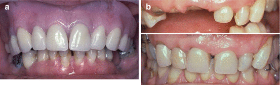
Fig. 13.7
(a, b) Moderate/severe tooth wear with an unfavourable occlusal relationship initially restored with a provisional onlay/overlay removable prosthesis to assess appearance and function. (a) Before restoration. (b) After restoration with removable prosthesis
Fig. 13.8
(a, b) Examples of overlay removable prostheses: (a) Full labial flange, (b) gingival-fitting anterior tooth facings
There are some well-established advantages in retaining teeth as overdenture abutments, such as maintenance of alveolar bone and support, improved sensory feedback, masticatory performance and reduced psychological trauma of tooth extraction [28]. Some disadvantages also exist, particularly if patients are unable to establish and maintain adequate oral and denture plaque control. In this situation, the abutment teeth will be at an increased risk of primary dental disease (Fig. 13.9), although the daily application of a non-acidulated fluoride gel may reduce the risk of root surface caries [30].
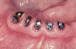
Fig. 13.9
Root surface caries affecting grossly worn teeth restored with an overlay removable prosthesis
Maintenance demands are relatively high for this form of prosthesis [31], with material wear and fracture being common, particularly in patients exhibiting parafunctional clenching/grinding habits (Fig. 13.10). The use of extensive metal frameworks, including incisal and occlusal coverage (Fig. 13.11), may reduce the regularity of repair but increase the clinical and technical complexity when repair or replacement eventually becomes necessary. This complexity may be further exacerbated if copings and precision attachments have been utilized on abutment teeth to increase retention and stability of the prosthesis [28].
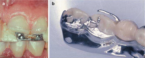
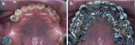
Fig. 13.10
(a, b) Fracture and wear of acrylic resin facings on removable prostheses often demonstrated in patients with parafunctional clenching/grinding habits. (a) Anterior tooth facings. (b) Posterior onlay components
Fig. 13.11
(a, b) The use of a metal framework incorporating incisal and occlusal coverage used to strengthen removable onlay/overlay prostheses for patients demonstrating significant parafunctional clenching/grinding habits. (a) Before restoration. (b) After restoration with removable onlay/overlay prosthesis
While for certain situations this form of treatment offers a satisfactory way of restoring the worn dentition, for many patients difficulties arise in adapting both functionally and psychologically to a removable prosthesis and this approach is often seen as a last resort.
13.4 Minimal Preparation Adhesive Restorations
Since the introduction to the dental profession of the acid-etch technique using phosphoric acid by Buonocore in 1955 [32], and an early form of Bis-GMA-based composite resin by Bowen in the early 1960s [33], there has been significant progression in the development of adhesive materials and techniques within dentistry. With the additional development of glass ionomer cements, dentin bonding agents, silane primers and composite resin luting cements, it is now possible to produce acceptable adhesive bonds between enamel and dentin tooth tissue with a variety of materials such as composite resins, ceramics and metal alloys suitable for use in situations where restoration of the worn dentition is necessary [34–42].
The final section highlights the range of adhesive materials available to the dental surgeon and discuss how they may be applied when restoring the dentition of patients with moderate to severe tooth wear. The following areas are discussed:
-
Cervical tooth wear
-
Anterior tooth wear
-
Palatal tooth wear
-
Incisal/palatal tooth wear
-
Labial/incisal/palatal tooth wear
-
-
Posterior and generalised tooth wear
13.4.1 Cervical Tooth Wear
Cervical tooth wear lesions are common and present in a variety of forms depending on the type and severity of the causative factors. Not all lesions require restoration, but if aesthetics, sensitivity, or prevention of further tooth wear dictates then some form of adhesive restoration will usually be most suitable.
A plethora of tooth-coloured restorative materials are now available. Materials can either be composite resin or glass ionomer-based, or a combination of both; either in a layered technique with the individual materials or with formulated resin-modified glass-ionomer cements. The choice of materials can be bewildering, with new materials and techniques seemingly introduced to the market on a monthly basis [38, 43–45].
There are a number of approaches to bonding restorations to cervical tooth tissue. For lesions with margins that are still confined to enamel, the use of a microfine or polishable densified composite resin, in conjunction with acid-etched enamel, will usually produce good aesthetic and durable results. Unfortunately, most cervical lesion margins are not confined to enamel and usually involve root cementum and dentin. In this situation, some form of dentin bonding agent is required, in combination with a composite resin or a self-adhesive composite resin formulation. Alternatively, a glass-ionomer cement restoration with inherent bonding properties to both dentin and enamel may be considered.
In situations where aesthetics are paramount, then a polishable composite resin combined with some form of adhesive bonding agent is often the material of choice. However, despite ongoing improvements, questions remain as to the longer term durability of dentin bonding agents, which may result in micro-leakage and characteristic marginal discolouration of the restoration [46].
Where lesions are not as visually prominent and involve more of the root surface, often partly below the gingival margin, then a glass-ionomer material may prove to be more durable. The dynamic bond of glass-ionomer cements to both dentin and enamel through an ionic exchange provides the opportunity for continual repair of the adhesive bond at the tooth and cement interface. There is also the possible additional benefit of fluoride ion release from the glass-ionomer cement reducing the possibility of marginal caries in susceptible individuals [47].
Although much improved over recent years, the colour properties of conventional glass-ionomer cements are not ideal. However, in deeper cervical lesions, it is possible to consider a layered technique combining the adhesive properties of the glass-ionomer cement with the superior colour properties of a polishable composite resin. This can be carried out in one visit or preferably following a minimum set time of 24 h; the superficial portion of the glass-ionomer cement restoration can be reduced and a layer of composite resin added (Fig. 13.12). The new generation of light-activated resin-modified glass-ionomer materials attempt to combine some of the better properties of composite resin and conventional glass-ionomer cements. Certainly the command set, improved colour and easier finishing of some of these materials allow the opportunity to provide very acceptable conservative restorations for cervical tooth wear lesions [48, 49].
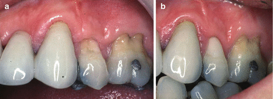
Fig. 13.12
(a, b) Restoration of a cervical abrasion/erosion lesion using a layered glass-ionomer and direct composite resin technique. (a) Before restoration. (b) After restoration
Only longer term observation and assessment will determine how durable the variety of materials and techniques available for non-carious cervical lesions will ultimately prove to be [50].
13.4.2 Anterior Tooth Wear
Although tooth wear can generally affect the whole dentition, it is often localized to the anterior teeth and the maxillary anterior teeth in particular.
13.4.2.1 Palatal Tooth Wear
This pattern of tooth wear is usually characteristic of acid erosion, possibly combined with a degree of attrition, and is the type more commonly seen in the younger age groups. Often, the labial and incisal surfaces are relatively intact and the main indications for restorative treatment are to offer some resistance to further palatal tooth wear which will reduce the risk of significant enamel fractures to the weakened incisal edges and pulp tissue inflammation. The use of resin-bonded palatal metal alloy veneers is an acceptable method to manage this form of tooth wear [51, 52] and has been shown to be a relatively durable technique [39, 53]. Either heat-treated gold alloys or nickel-chromium alloys, as used in resin-bonded bridge frameworks, are currently the cast metal alloys of choice. The decision as to which of the two materials to use is based on the improved bond strength of resin to nickel-chromium alloys versus the easier working properties and wear characteristics of the gold alloys.
Tooth preparation is minimal, usually restricted to smoothing the incisal and palatal peripheral enamel margins. Laboratory fabrication of the metal alloy veneers is either directly on a refractory working cast or by a wax/resin ‘lift-off’ technique. When restoring worn anterior teeth, creation of inter-occlusal space is usually required in order to accommodate the thickness of the restoring material. As the tooth structure is already compromised, avoiding further tooth reduction to create space is obviously advantageous. Orthodontic tooth movement using an anterior ‘Dahl’ appliance has been described as a method of achieving inter-occlusal space. Although this is a predictable method, there are some disadvantages, not least the increased treatment time and extra laboratory procedures. An alternative approach, based on similar principles to the Dahl appliance, is to deliberately design and construct the palatal veneers in such a manner that they are cemented initially high in occlusion. Expected tooth movement is enhanced if a positive cingulum contact can be achieved with the occluding lower incisor teeth in an attempt to direct orthodontic forces along the long axis of the contacting teeth. This method would appear to contradict traditional occlusal teaching but to date has proved to work well in these particular circumstances where tooth preparation is minimal [54].
Luting cements are usually resin-based and used in combination with the manufacturer’s dentin bonding agent where appropriate. The use of opaque resin-based cement will overcome any potential greying of the incisal third caused by the underlying palatal metal veneer. Rubber dam isolation is used when necessary and occasionally gingival retraction cord in situations where there has been excessive tooth wear in the cervical region. By including metal coverage of the palatal veneers onto the incisal edges, location during cementation is made somewhat easier and this design will also offer some increased resistance to shearing loads. An example of the use of resin-bonded metal alloy palatal veneers is illustrated in Fig. 13.13
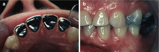
Fig. 13.13
(a, b) The use of nickel/chromium alloy resin-bonded palatal veneers used to restore localised palatal tooth wear for maxillary incisor teeth. (a) Palatal view of veneers. (b) Labial view demonstrating re-establishment of posterior occlusal contacts
13.4.2.2 Incisal/Palatal Tooth Wear
Although the use of metal alloy palatal veneers is an excellent conservative method of managing localized anterior tooth wear, it is not possible to improve the appearance of lost incisal and labial tooth tissue. In these circumstances, it is feasible to build up the incisal portion of the tooth with direct acid-etch retained composite resin and then construct a resin-bonded metal alloy palatal veneer to cover both the palatal tooth tissue and composite resin [55, 56]. However, potential difficulties arise with this technique, in that it is difficult to know where to finish the composite resin build-up palatally and the adhesive bond of the metal alloy palatal veneer will be somewhat compromised because of the reduction in available tooth enamel.
Stay updated, free dental videos. Join our Telegram channel
VIDEdental - Online dental courses


