Introduction
Pharynx dimensions may vary according to the position and morphology of the mandible. The aim of this study was to test the hypothesis that craniofacial morphology affects pharyngeal volume and its cross-sectional areas.
Methods
Seventy-four subjects (38 men, 36 women) aged 18 to 56 years (mean ± SD 32.8 ± 1.8 years) were scanned with a cone-beam computed tomography unit and had facial profile photographs taken. All participants were classified according to skeletal anteroposterior (Class II and Class III) and vertical facial patterns (brachyfacial, mesofacial, and dolichofacial). Facial profile analyses and pharyngeal volume and cross-sectional area determinations were performed.
Results
The soft palate cross-sectional area of the oropharynx was significantly greater in the Class III patients. The upper facial third (trichion to soft tissue glabella/facial height) correlated negatively with pharyngeal volume and with soft palate cross-sectional area in the oropharynx. Alternatively, the midfacial third (soft tissue glabella to subnasale/facial height) correlated positively with soft palate cross-sectional area of the oropharynx. No significant differences across sexes and facial patterns regarding pharyngeal volume were found ( P >0.05).
Conclusions
The soft palate cross-sectional area is larger in skeletal Class III subjects. It seems that analysis of the facial profile focusing on the proportions of the facial thirds allows for inferences on pharyngeal dimensions. However, anteroposterior skeletal facial type and vertical facial pattern do not seem to affect pharyngeal volume.
Highlights
- •
Volumes and 5 cross-sectional areas of the pharynx were measured.
- •
The subjects were classified by anteroposterior skeletal types and vertical patterns.
- •
Soft palate cross-sectional area of the oropharynx was greater in the Class III skeletal type.
- •
Facial profiles show trends of correlations between facial thirds and the pharynx.
- •
Facial patterns do not influence pharynx volumes.
Craniofacial growth has been the subject of several studies, some claiming that breathing dysfunctions may disrupt facial growth in a morphologic and functional fashion by producing skeletal changes and malocclusions. Other authors believe that facial appearance is influenced by breathing and other craniofacial factors.
Among other modifiers of craniofacial growth, nasopharyngeal airway narrowing (usually associated with hypertrophic tonsils) may hinder the passage of air and cause a respiratory disorder characterized by sleep apnea. Such changes may induce mouth breathing, which affects mandibular positioning and, consequently, the mandibular growth vector. It is suggested that changes in facial hard and soft tissues and malocclusion indicate breathing dysfunction.
Lateral cephalometric projection can be used for the analysis of hard and soft facial structures and allows for the study of upper airway morphology. However, it is limited because it provides a 2-dimensional view of complex, 3-dimensional (3D) anatomic structures.
On the other hand, cone-beam computed tomography (CBCT) is appropriate for the analysis of 3D structures such as the upper airway. CBCT allows for a low radiation dose during image acquisition and offers capabilities such as multiplanar image manipulation and 3D image reconstruction; altogether, it provides for a more reliable characterization of the airways. Therefore, several authors have studied the association of craniofacial morphology with the upper airway using CBCT scans. Thus far, however, studies assessing the correlations between facial profile and the volume and the area of the pharynx are still missing.
Because early examination of surgical and conventional orthodontic patients includes (among other studies) CBCT images and facial photographs, and because orthodontic or orthognathic interventions may affect the airway, information regarding the influence of facial patterns and facial profiles on the pharyngeal morphology would improve the diagnosis and treatment of surgical and conventional orthodontic patients. Thus, the aim of this study was to examine the hypothesis that craniofacial morphology influences the pharyngeal volume and the cross-sectional area of the pharynx.
Material and methods
The research ethics committee of Piracicaba Dental School–UNICAMP approved this study under protocol number 071/2013.
Data of patients who had CBCT head scans and facial profile photographs taken were obtained from the records of the oral radiology clinic. Six hundred eighty images with different fields of view were acquired from March 2011 to February 2014. One hundred three images with extended fields of view were selected. Those with signs of orthognathic surgery, radiographic signs of upper airway pathology, cervical spine that was not upright, and CBCT images without nasion or the base of the third cervical vertebra region were excluded. Therefore, 74 subjects (38 men, 36 women) from 18 to 56 years old (mean ± SD, 32.8 ± 1.8 years) were selected as the convenience sample.
CBCT images were acquired with an i-CAT unit (Imaging Sciences, Hatfield, Pa) with the following settings: 80 kVp, 4.8 mA, acquisition time of 40 seconds, reconstruction time of 62 seconds, voxel size of 0.4 mm, and field of view of 23 × 17 cm. The facial profile photographs were taken with the patients facing laterally away from the camera in natural head position, at maximal intercuspation and with the lips at rest.
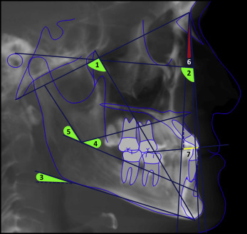
The sample was classified according to vertical facial patterns (brachyfacial, mesofacial, and dolichofacial) and anteroposterior facial skeletal types (Class II and Class III) by cephalometric analysis. A study of the facial profile was performed for each subject.
Lateral cephalograms from images acquired with the CBCT scans were used for the analysis, which was performed using Nemotec software (Nemotec, Madrid, Spain). For all patients, the vertical facial patterns were labeled according to the VERT index in Ricketts’ analysis. Moreover, Steiner’s and Jacobson’s measurements were used to determine the anteroposterior facial skeletal type ( Fig 1 ). The corresponding medians and interquartile deviations to the measurements obtained with Ricketts’, Steiner’s, and Jacobson’s analyses are shown in the Supplementary Table .

Facial analysis was performed using facial profile photographs analyzed with the Facial Analysis Sideview (photo) functionality feature of the Cef X software (CDT Software, Bauru, São Paulo, Brazil). Facial thirds were measured according to the following reference points.
- 1.
The proportion of the upper facial third (trichion to soft tissue glabella) in relation to facial height (trichion to soft tissue menton).
- 2.
The proportion of the midfacial third (soft tissue glabella to subnasale) in relation to facial height.
- 3.
To the lower facial third: the proportion of stomion to soft tissue menton (Stm-Me′) and subnasale to stomion (Sn-Stm) in relation to lower facial third (subnasale to soft tissue menton). Moreover, the proportion of lower facial third in relation to facial height.
To assess the pharyngeal dimensions, 3D reconstructions were created from the CBCT data with Insight ITK-SNAP version 2.4.0 ( www.itksnap.org ). One examiner (D.M.B.), who has solid experience with reconstruction software, performed all measurements. For the initial calibration, the examiner assessed 10 pictures repeatedly until consistent measurement replications were achieved. After that, all images were assessed, and measurements were taken. To determine intraexaminer agreement, 15% of the sample was reassessed a second time.
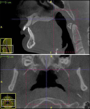
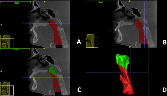
The 3D pharyngeal reconstruction was achieved with semiautomatic segmentation software. Limits for the area of interest are shown in Figure 2 . Values for total pharyngeal, nasopharyngeal, and oropharyngeal volume calculations were obtained ( Fig 3 ).


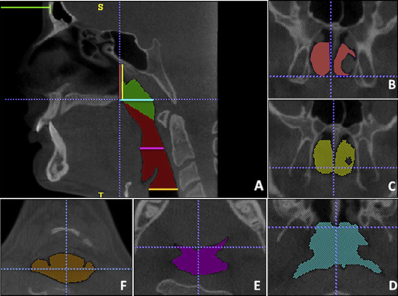
To determine the cross-sectional pharyngeal areas, 5 spatial planes were set and identified with different colors in the 3D model. The areas were calculated using the manual segmentation tool of the Insight ITK-SNAP software, as shown in Table I and Figure 4 .
| Area | Reference | Color | Figure |
|---|---|---|---|
| PNS-coronal area | Coronal plane through the PNS | Pink | Figure 4 , B |
| PNS-posterior area | Coronal plane through the point on the anterior pharyngeal wall extending from the PNS | Yellow | Figure 4 , C |
| PNS-axial area | Axial plane that passes through the PNS (perpendicular to the PNS-coronal) | Blue | Figure 4 , D |
| Soft palate area | Axial plane through the most inferior point of the soft palate against the soft palate with the tongue dorsum | Purple | Figure 4 , E |
| Epiglottis area | Axial plane through the base of the epiglottis at the height of the medial caudal projection of third cervical vertebra | Orange | Figure 4 , F |

Statistical analysis
The statistical significance level was set at 5% for all tests. The Fisher exact test was used to assess the distributions of anteroposterior facial skeletal types and vertical facial patterns. The Mann-Whitney test was used to determine the influence of sex and anteroposterior facial skeletal types on the dependent variables; the Kruskal-Wallis test was used to evaluate any correlations among vertical facial patterns and other variables; and the Spearman correlation test was used to analyze possible correlations among the measurements in a given vertical facial pattern, in the same anteroposterior facial skeletal type, and in other variables of the facial analysis and measurements proposed.
Results
Distributions regarding anteroposterior facial skeletal types and vertical facial patterns were evaluated with the Fisher exact test. There were no statistically significant differences between the Class II and the Class III distributions ( Table II ).
| Class II | Class III | Total | |
|---|---|---|---|
| Brachyfacial | 22 (44%) | 17 (70.8%) | 39 (52.7%) |
| Mesofacial | 15 (30%) | 4 (16.7%) | 19 (25.7%) |
| Dolichofacial | 13 (26%) | 3 (12.5%) | 16 (21.6%) |
| Total | 50 (100%) | 24 (100%) | 74 (100%) |
The results of the reproducibility were considered excellent, with intraclass correlation coefficients greater than 0.9 and P <0.001.
Among the anteroposterior facial skeletal types, the soft palate cross-sectional area ( P <0.05) was greater for the Class III subjects. There was no statistically significant difference between this group and the others for pharynx measures ( Table III ).
| Class II (n = 50) | Class III (n = 24) | P | |||
|---|---|---|---|---|---|
| Median | ID | Median | ID | ||
| Pharyngeal volumes | |||||
| Total | 21649.8 | 10044.4 | 24031 | 8682.6 | 0.221 |
| Nasopharynx | 10868.8 | 3734 | 8948.6 | 3307.1 | 0.467 |
| Oropharynx | 10619.6 | 7377.8 | 13132.6 | 5491.4 | 0.094 |
| Pharyngeal cross-sectional areas | |||||
| PNS-coronal | 218.2 | 121.0 | 236.3 | 123.9 | 0.446 |
| PNS-posterior | 299.4 | 149.5 | 341.0 | 108.3 | 0.347 |
| PNS-axial | 526.3 | 211.4 | 523.4 | 178.4 | 0.648 |
| SP | 160.0 | 220.7 | 270.6 | 132.3 | 0.017 ∗ |
| EP | 175.2 | 135 | 231.5 | 178.9 | 0.077 |
| Facial analysis indexes | |||||
| Stm-Me′ | 66.6 | 3.6 | 69.3 | 5.1 | 0.001 † |
| Sn-Stm | 33.3 | 3.6 | 30.7 | 5.0 | 0.001 † |
| Lower third | 36.6 | 3.1 | 36.9 | 3.1 | 0.383 |
| Middle third | 36.1 | 2.9 | 35.6 | 2.4 | 0.234 |
| Upper third | 27.1 | 4.4 | 27.2 | 4.9 | 0.953 |
No significant differences among the vertical facial patterns concerning the pharyngeal dimensions were detected ( Table IV ).
| Brachyfacial (n = 39) | Mesofacial (n = 19) | Dolichofacial (n = 16) | P | ||||
|---|---|---|---|---|---|---|---|
| Median | ID | Median | ID | Median | ID | ||
| Pharyngeal volumes | |||||||
| Total | 23282.7 | 13335.1 | 21291.1 | 7779.3 | 22237.1 | 7633.9 | 0.966 |
| Nasopharynx | 9207.9 | 4001.6 | 9756.6 | 3953.6 | 11061.4 | 4300.5 | 0.320 |
| Oropharynx | 12987.8 | 9695.3 | 10872.9 | 4854.6 | 11513.6 | 4825.3 | 0.632 |
| Pharyngeal cross-sectional areas | |||||||
| PNS-coronal | 213.8 | 141.7 | 214.1 | 103.7 | 265.9 | 105.5 | 0.162 |
| PNS-posterior | 334.7 | 187.3 | 297.4 | 96.3 | 356.6 | 109.4 | 0.435 |
| PNS-axial | 523.2 | 246.1 | 506.2 | 197.0 | 558.5 | 147.5 | 0.389 |
| SP | 207.4 | 211.1 | 216.6 | 197.1 | 154.0 | 112.4 | 0.733 |
| EP | 193.1 | 188.3 | 166.9 | 147.6 | 199.7 | 125.9 | 0.712 |
| Facial analysis indexes | |||||||
| Stm-Me′ | 67.8 | 4.7 | 66.9 | 3.3 | 68.0 | 3.5 | 0.600 |
| Sn-Stm | 32.1 | 5.0 | 33.0 | 3.3 | 32.0 | 3.5 | 0.500 |
| Lower third | 36.1 B | 2.9 | 36.6 A | 2.2 | 38.6 A | 3.0 | 0.035 ∗ |
| Middle third | 35.7 | 2.7 | 36.2 | 2.8 | 36.4 | 2.8 | 0.550 |
| Upper third | 28.3 A | 4.8 | 27.3 B | 3.0 | 24.6 C | 3.1 | 0.006 ∗ |
No statistically significant sex differences were found for the pharyngeal volume and cross-sectional areas, except for postnasal spine (PNS)-coronal ( P <0.01) and PNS-posterior ( P <0.01), which were significantly greater for the men ( Table V ).
| Female (n = 36) | Male (n = 38) | P | |||
|---|---|---|---|---|---|
| Median | ID | Median | ID | ||
| Pharyngeal volumes | |||||
| Total | 23032.1 | 9923.2 | 22510.6 | 10508.2 | 0.991 |
| Nasopharynx | 9640.7 | 3620.0 | 10132.6 | 4020.3 | 0.489 |
| Oropharynx | 10748.2 | 6964.6 | 12712.7 | 7374.0 | 0.315 |
| Pharyngeal cross-sectional areas | |||||
| PNS-coronal | 192.3 | 101.8 | 255.4 | 136.8 | 0.009 † |
| PNS-posterior | 298.0 | 130.1 | 360.1 | 164.9 | 0.007 † |
| PNS-axial | 525.0 | 214.6 | 524.7 | 208.5 | 0.485 |
| SP | 188.3 | 211.4 | 228.0 | 210.4 | 0.299 |
| EP | 179.9 | 148.4 | 206.2 | 164.9 | 0.118 |
| Facial analysis indexes | |||||
| Stm-Me′ | 67.5 | 5.2 | 67.8 | 3.0 | 0.638 |
| Sn-Stm | 32.5 | 5.6 | 32.2 | 3.0 | 0.520 |
| Lower third | 36.4 | 2.8 | 37.2 | 3.2 | 0.022 ∗ |
| Middle third | 36.4 | 2.8 | 35.5 | 3.1 | 0.052 |
| Upper third | 27.3 | 3.3 | 26.1 | 5.1 | 0.530 |
A positive correlation between the midfacial third and the soft palate cross-sectional area of the oropharynx was found, as well as negative correlations between the upper facial third and total pharyngeal volume, oropharyngeal volume, and soft palate cross-sectional area ( Table VI ).
| Facial analysis indexes | Pharyngeal volumes | Pharyngeal cross-sectional areas | ||||||
|---|---|---|---|---|---|---|---|---|
| Total pharynx | Nasopharynx | Oropharynx | PNS-coronal | PNS-posterior | PNS-axial | SP | EP | |
| Stm-Me′ | 0.016 (0.890) | −0.096 (0.416) | 0.066 (0.574) | 0.144 (0.220) | 0.178 (0.130) | −0.035 (0.765) | 0.024 (0.838) | −0.070 (0.552) |
| Sn-Stm | 0.010 (0.934) | 0.102 (0.386) | −0.037 (0.756) | −0.135 (0.250) | −0.155 (0.187) | 0.045 (0.706) | 0.003 (0.977) | 0.066 (0.577) |
| Lower third | 0.090 (0.443) | 0.064 (0.590) | 0.104 (0.379) | 0.043 (0.716) | −0.022 (0.851) | 0.063 (0.596) | 0.061 (0.606) | 0.127 (0.281) |
| Middle third | 0.086 (0.467) | −0.045 (0.701) | 0.104 (0.376) | −0.073 (0.538) | −0.021 (0.858) | 0.114 (0.334) | 0.245 ∗ (0.036) | 0.109 (0.353) |
| Upper third | −0.239 ∗ (0.040) | −0.117 (0.320) | −0.243 ∗ (0.037) | −0.069 (0.560) | −0.028 (0.813) | −0.176 (0.133) | −0.289 ∗ (0.012) | −0.200 (0.087) |
Moreover, correlations seemed more frequent in the Class II subjects when compared with the Class III group, and in the brachyfacial and dolichofacial subjects compared with the mesofacial group. There were correlations between pharynx volumes and soft palate cross-sectional areas of the oropharynx in the anteroposterior facial skeletal patterns and vertical patterns ( Tables VII and VIII ).
| Anteroposterior skeletal facial types | Pharyngeal volumes | Pharyngeal cross-sectional areas | |||||
|---|---|---|---|---|---|---|---|
| Total pharynx | Nasopharynx | Oropharynx | PNS-coronal | PNS-posterior | PNS-axial | SP | |
| Class II (n = 50) | |||||||
| Nasopharynx | 0.831 † (0) | ||||||
| Oropharynx | 0.927 † (0) | 0.624 † (0) | |||||
| PNS-coronal | 0.317 ∗ (0.025) | 0.394 † (0.005) | 0.255 (0.073) | ||||
| PNS-posterior | 0.112 (0.438) | 0.269 ∗ (0.036) | 0.046 (0.753) | 0.797 † (0) | |||
| PNS-axial | 0.695 † (0) | 0.751 † (0) | 0.585 † (0) | 0.500 † (0) | 0.353 ∗ (0.012) | ||
| SP | 0.744 † (0) | 0.436 † (0.001) | 0.815 † (0) | 0.287 ∗ (0.043) | 0.126 (0.383) | 0.504 † (0) | |
| EP | 0.526 † (0) | 0.273 ∗ (0.021) | 0.546 † (0) | 0.262 (0.066) | 0.177 (0.218) | 0.370 ∗ (0.008) | 0.642 † (0) |
| Class III (n = 24) | |||||||
| Nasopharynx | 0.583 † (0.003) | ||||||
| Oropharynx | 0.820 † (0) | 0.214 (0.315) | |||||
| PNS-coronal | 0.396 † (0.003) | 0.575 † (0.003) | 0.096 (0.657) | ||||
| PNS-posterior | 0.199 (0.351) | 0.341 (0.103) | −0.003 (0.990) | 0.807 † (0) | |||
| PNS-axial | 0.561 † (0.004) | 0.657 † (0) | 0.311 (0.139) | 0.605 † (0.002) | 0.361 (0.083) | ||
| SP | 0.789 † (0) | 0.227 (0.286) | 0.752 † (0) | 0.122 (0.571) | −0.001 (0.997) | 0.393 (0.057) | |
| EP | 0.709 † (0) | 0.172 (0.421) | 0.664 † (0) | 0.170 (0.426) | 0.176 (0.412) | 0.326 (0.120) | 0.587 † (0.003) |
Stay updated, free dental videos. Join our Telegram channel

VIDEdental - Online dental courses


