Gastroesophageal reflux disease, or GERD, is a common disease
10–20 % of the general population suffers from GERD
GERD affects adults and children
GERD can cause dental erosion
24 % of patients with GERD have dental erosion
32 % of adults with dental erosion have GERD
17 % of children with dental erosion have GERD
Patients with GERD are not only more likely to have dental erosion but also more likely to have severe erosion
The prevalence of GERD in the United States appears to be rising. It accounts for at least nine million physician office visits in the United States each year, and annual costs for managing GERD are estimated to be greater than $9 million [8]. In Western populations, 25 % of the people report having heartburn at least once a month, 12 % at least once a week, and 5 % describe having symptoms daily (Table 12.2). There appears to be no gender predominance of heartburn symptoms; men and women are affected equally. The relationship of age and reflux is unclear. One study has suggested an association between advancing age and fewer reflux symptoms; however, there is a higher presence of more severe esophagitis, or inflammation of the esophagus due to acid reflux [9].
Table 12.2
Frequency of heartburn in Western populations
|
Symptoms once a month
|
25 %
|
|
Symptoms once a week
|
12 %
|
|
Symptoms daily
|
5 %
|
Studies have shown that up to 60 % of patients with dental erosions had pathological levels of acid reflux when assessed by ambulatory esophageal pH monitoring [3]. Dental erosions as a manifestation of GERD have not only been seen in adults but also in children. A recent study of 249 children and adult, of whom 91 had molar erosion and/or symptoms of acid reflux and had undergone endoscopy, esophageal manometry, and 24-h esophageal pH monitoring, found a significant association between diagnosed GERD and dental erosion [10]. Studies have shown that children diagnosed with GERD according to esophageal pH monitoring had more dental erosions compared to healthy children. A study of 38 children with a diagnosis of GERD according to esophageal pH recordings was evaluated for the prevalence of dental erosions. Children with confirmed GERD had more severe dental erosions compared to children with no GERD [11]. Another study evaluated 52 children with GERD and 52 healthy siblings. In this study, the severity of dental erosion was greater in the study group; 43 % of the affected teeth had grade 3 erosions and 9 % in the control group [12]. In a study in the United States, dental erosion was measured using Aine index in 24 children diagnosed with GERD. Twenty children had dental erosion, ten with mild erosion (grade 1), six with moderate (grade 2), and four with severe erosion (grade 3) [13]. The presence of dental erosions, especially in posterior primary teeth in children, is associated with GERD [14].
12.3 Etiology
Gastroesophageal reflux is the retrograde flow or reflux of gastric contents other than air into or through the esophagus. GERD refers to reflux that produces frequent symptoms or results in damage to the esophageal mucosa or contiguous organs of the upper respiratory tract and occasionally the lower respiratory tract and oral cavity.
The pathogenesis of gastroesophageal reflux is complicated and multifactorial. The antireflux barriers of the gastroesophageal junction are anatomically and physiologically complex and vulnerable to a number of potential mechanisms of reflux. The basic cause of GERD is incompetent antireflux barriers at the gastroesophageal junction, which normally prevents backflow of gastric acid into the esophagus. Normally, a ring of muscle tissue called the lower esophageal sphincter (LES), which is located in the lower portion of the esophagus where it joins the stomach (gastroesophageal junction), prevents reflux (or backing up) of acid from the stomach. Normally, this sphincter relaxes during swallowing to allow food to pass and then tightens to prevent flow in the opposite direction (i.e., reflux). With GERD, however, the sphincter relaxes between swallows or is weakened, allowing stomach contents and corrosive acid to reflux up the esophagus and damage the lining of the esophagus. The two main patterns of LES dysfunction are (1) hypotensive LES and (2) pathologic transient lower esophageal sphincter relaxations (TLESRs). Anatomic disruption of the gastroesophageal junction is commonly associated with a hiatal hernia, which is a partial herniation of the stomach through the diaphragm and into the thorax. Hiatal hernias contribute to the pathogenesis of reflux disease by impairing LES function [15, 16].
Gastric factors can play a significant role in producing GERD. Gastric factors that promote GERD include poor acid clearance from the esophagus; diminished salivary flow; increased gastric acid production; increased gastric volume after meals; increase gastric pressure due to obesity, recumbency, or lying down after meals; and delayed gastric emptying or gastroparesis. Increased gastric distention can cause an increase in transient LES relaxation and the volume of reflux, particularly in GERD patients with large hiatal hernias. Delayed gastric emptying may be present in approximately 15% of patients with GERD and is frequently underdiagnosed [17]. There is a well-known association between body mass index (BMI) and reflux symptoms. Evidence has shown that inappropriate relaxation of the LES can be exacerbated by obesity [18, 19]. Even moderate weight gain among people with normal weight is thought to cause or exacerbate reflux symptoms [20] (Table 12.3, Fig. 12.1).
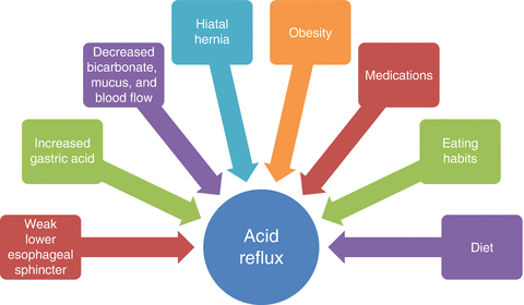
Table 12.3
Etiologic factors of gastroesophageal reflux disease (GERD)
|
Motility disorders
|
Damaging factors
|
Resistance factors
|
Others
|
|---|---|---|---|
|
Transient lower esophageal sphincter relaxations (TLESRs)
|
Increased gastric acid production
|
Reduced saliva and bicarbonate production
|
Hiatal hernia
|
|
Weak lower esophageal sphincter (LES)
|
Reflux of bile and pancreatic juice from small intestines
|
Diminished mucosal blood flow
|
Diet
|
|
Weak esophageal peristalsis or motility
|
Decreased protective mucus
|
Eating habits
|
|
|
Delayed gastric emptying or gastroparesis
|
Increased intra-abdominal pressure
|
||
|
Scleroderma and CREST syndromea
|
Obesity
|
||
|
Medications
|
|||
|
Obstructive sleep apnea
|
Fig. 12.1
Factors contributing to gastroesophageal reflux disease (GERD)
Other factors that decrease LES pressure and contribute to GERD are medications, lifestyle behaviors, and the ingestion of certain foods. Certain medications can exacerbate GERD by lowering LES pressure, like calcium channel blockers and nitrates; others can cause esophagitis, or damage to the lining of the esophagus, by direct mucosal injury, like nonsteroidal anti-inflammatory drugs. Behaviors like binge eating; bulimia, which is binge eating followed by regurgitation of food in order to lose weight; and recumbency after eating all create gastric distension and weaken the LES leading to acid reflux. Certain foods like fatty foods, peppermint, chocolate, caffeinated beverages, and alcohol can all decrease LES pressure and contribute to GERD. Smoking tobacco products can also lead to GERD by decreasing LES pressure. The contribution of these various factors varies from patient to patient [9] (Table 12.4, Fig. 12.1).
Table 12.4
Factors that precipitate or exacerbate GERD symptoms
|
Medications
|
Foods
|
Behaviors or conditions that increase intra-abdominal pressure
|
|---|---|---|
|
α-Adrenergic antagonists
(e.g., doxazosin, prazosin, tamsulosin, terazosin)
|
Alcohol
|
Binge eating
|
|
Anticholinergics
(e.g., atropine, benztropine, buproprion, dextromethorphan, ipratropium, oxybutynin, tolterodine, tiotropium)
|
Caffeine
|
Bulimia
(binge eating followed by regurgitation in order to lose weight)
|
|
β-Adrenergic agonists
(e.g., albuterol, formoterol)
|
Carbonated beverages
|
Eating large-volume meals
|
|
Calcium channel blockers
(e.g., amlodipine, diltiazem, nifedipine, verapamil)
|
Chocolate
|
Eating prior to recumbency (lying down)
|
|
Diazepam
|
Citrus fruits
|
Pregnancy
|
|
Estrogens
|
Fatty foods
|
Sleep apnea
(pause in breathing during sleep)
|
|
Narcotics
|
Peppermint
|
Smoking
|
|
Nitrates
(e.g., isosorbide mononitrate or dinitrate, nitroglycerin)
|
Spicy foods
|
Weight gain
|
|
Nonsteroidal anti-inflammatory drugs (NSAIDs)
(e.g., aspirin, diclofenac, ibuprofen, naproxen, meloxicam, piroxicam)
|
Tomato-based products
|
|
|
Progesterone
|
Vinegar
|
|
|
Theophylline
|
||
|
Tricyclic antidepressants
(e.g., amitriptyline, desipramine, imipramine, nortriptyline)
|
12.4 Pathogenesis
Esophageal clearance is important to prevent gastric content from reaching the oral cavity and causing dental erosion. Esophageal clearance is achieved by two mechanisms: esophageal peristalsis, or sequential esophageal contractions causing propulsion of food to stomach, and the buffering action of saliva, which neutralizes the acid. Peristalsis empties the esophagus of its contents, and this process is followed by the neutralization of the acid of the esophageal lumen by the saliva [21]. Saliva has a buffering and protective role against the demineralizing effects of acids in the oral cavity. The saliva in patients with GERD has a greater buffering capacity compared to patients without GERD. This difference is due to modifications in the saliva composition resulting from an increased concentration of inorganic phosphates [22].
The two possible mechanisms by which acid reflux damages extraesophageal tissue may include (1) direct damage from mucosal contact (reflux theory) and (2) vagal nerve-mediated reflex from distal esophageal acid exposure (reflex theory) [23]. In either case, the refluxed acid can damage extraesophageal tissues including the oral tissues, particularly its hard and soft tissues. In these cases, the quality and amount of saliva play an important role in hard and soft oral tissues changes [24]. The hydroxyapatite crystals constituting the dental inorganic material may be dissolved by acid having a pH under the critical pH (5.5) for dental enamel dissolution [25]. The gastric reflux has a pH less than 2.0, so it can erode dental tissues [26].
The surfaces of the teeth during active endogenous acid erosion are largely devoid of protective biofilm and saliva due to gastric acid and also possible proteolytic pepsin. The raw products resulting from hard tooth tissue demineralization are lost and are not available to be reused when the oral pH increases back to neutral levels [27]. The chemical action causes rapid dissolution of exposed tooth surfaces that is distinctly different from the subsurface dissolution seen with plaque acids [28]. Under magnification, the eroded tooth surfaces will show damage to the ends of the enamel rods, which will only remineralize after the endogenous acid has been cleared from the oral cavity and after salivary pellicle has been reestablished on the tooth surfaces.
The addition of remineralizing ions to the eroded surfaces will only result in the repair of the ends of the enamel rods as the “gross” surface damage is irreversible. Even when fluorapatite is present in high concentrations, the remineralized surfaces provide little or no extra protection to further sustained demineralization as the endogenous acid has a pH well below 4.5, which is the approximate critical pH for fluorapatite dissolution [29]. These findings are supported by observations that fluoride-based and casein-based (amorphous calcium phosphate stabilized by casein phosphopeptide) remineralizing agents provide some protection against erosion at pH 3.0 [30–33], but not at a highly erosive environment of pH less than 2 [34–36].
12.5 Clinical Manifestations
Typical manifestations of GERD are heartburn, regurgitation, and dysphagia (difficulty swallowing). Other symptoms associated with GERD include water brash (regurgitation of excessive saliva), a globus sensation (lump in the throat), odynophagia (pain with swallowing), and nausea (Table 12.5). Heartburn, or pyrosis, is defined as a retrosternal burning discomfort located in the midepigastric area that may radiate up toward the neck or throat and typically occurs in the postprandial (post-meal) period [37]. Patients who present with typical symptoms with a minimum frequency of twice a week for 4–8 weeks or more should be considered as having GERD. At initial presentation, it is important to consider a patient’s age and the presence of “alarm signs” (Table 12.6). Alarm signs should be investigated in every patient with GERD symptoms as they may indicate complications from GERD, including esophageal stricture (i.e., scar tissue resulting in narrowing of the esophageal lumen), Barrett’s esophagus (i.e., a precancerous condition with changes in the cells lining the esophagus), or malignancy (i.e., esophageal or gastric cancer). The presence of any alarm signs necessitates the evaluation of GERD symptoms with further testing such as an upper endoscopy and/or imaging [9].
Table 12.5
Typical GERD symptoms
|
Heartburn
|
|
Regurgitation
|
|
Dysphagia (i.e., difficulty swallowing)
|
|
Water brash (i.e., regurgitation of excessive saliva)
|
|
Globus (i.e., sensation of lump in the throat)
|
|
Odynophagia (i.e., pain with swallowing)
|
|
Nausea
|
Table 12.6
“Alarm signs” that necessitate further evaluation of GERD
|
Dysphagia (i.e., difficulty swallowing)
|
|
Odynophagia (i.e., pain with swallowing)
|
|
Weight loss
|
|
Gastrointestinal bleeding (i.e., vomiting blood, blood in stools, or black stools)
|
|
Anemia
|
|
Advanced age (>50 years old)
|
|
Chest pain
|
|
Family history of upper GI tract cancer
|
Atypical manifestations of GERD refer to symptoms that are extraesophageal, including pulmonary, ear, nose, and throat manifestations, as well as noncardiac chest pain. Studies have shown that GERD can be found in 30–80% of adults with asthma. A new, more frequently recognized atypical manifestation of GERD is otitis media in children [38]. It is recommended that in patients who present with atypical respiratory, or ear, nose, or throat (ENT) symptoms, GERD needs to be ruled out (Table 12.7).
Table 12.7
Extraesophageal manifestations of GERD
|
Otitis media
|
|
Chronic sinusitis
|
|
Dental erosions
|
|
Aphthous ulcers
|
|
Halitosis
|
|
Pharyngitis
|
|
Laryngitis
|
|
Hoarseness of voice
|
|
Subglottic stenosis
|
|
Laryngospasm
|
|
Postnasal drip
|
|
Frequent throat clearing
|
|
Globus
|
|
Tracheobronchitis
|
|
Chronic cough
|
|
Asthma
|
|
Aspiration pneumonia
|
|
Pulmonary fibrosis
|
|
Chronic bronchitis
|
|
Bronchiectasis
|
|
Noncardiac chest pain
|
|
Sleep apnea
|
Patients with gastroparesis (i.e., delayed gastric emptying) and GERD may present with concomitant nausea, vomiting, or early satiety (i.e., sensation of feeling full early after eating a small amount). Gastroparesis should be suspected in patients with acute or subacute onset of GERD, particularly after an episode of viral respiratory infection or gastroenteritis [9].
12.5.1 Oral Manifestation
With respect to dental erosions, the location of the dental erosive zones is specific to each etiologic factor. The diagnostic criteria for dental erosions are loss of tooth structure of non-carious etiology, outside the areas of contact or occlusal guidance, and a glossy, smooth, rounded shape. In addition, negative gap areas at the edges of amalgam or composite fillings can also be seen [39]. If lesions occur due to extrinsic factors, then they are more frequently situated on the vestibular areas of the teeth. When the etiology is intrinsic in nature (gastric acids), the areas of erosion have characteristic distribution. Refluxed acid first attacks the palatal surface of the upper incisors (Figs. 12.2 and 12.3). The palatal surfaces of the maxillary teeth are also affected early due to the fact that they are not protected by the major salivary glands (Fig. 12.4). In the early stages, the tongue protects the lower teeth, but in the later stages, if the exposure to acid continues, erosion of the posterior mandibular teeth occurs, starting with the lingual, then occlusal, and buccal surfaces (Fig. 12.5). The labial or the buccal surfaces are affected by erosion only if acid reflux persists for an extended period of time. In chronic GERD, the occlusal surfaces of maxillary and mandibular teeth are affected. In the early stages, lesions are difficult to identify during normal examination, but advanced erosions are easily noticeable and have characteristic appearances (Table 12.8) [3, 40].
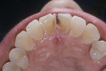
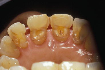
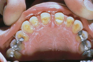
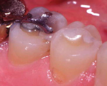
Fig. 12.2
Dental erosion on the palatal surface of upper incisors (Courtesy of Dr. Bennett T. Amaechi)
Fig. 12.3
Severe dental erosions on the palatal surface of upper incisors (Courtesy of Dr. Bennett T. Amaechi)
Fig. 12.4
Severe dental erosions on the palatal surface of upper incisors and maxillary premolars (Courtesy of Dr. Bennett T. Amaechi)
Fig. 12.5
Early stage erosion on lingual surface of mandibular molar and premolar (Courtesy of Dr. Bennett T. Amaechi)
Table 12.8
Pattern of dental erosion due to gastroesophageal reflux disease (GERD)
|
Refluxed acid first damages the palatal surface of the upper incisors then the palatal surfaces of the maxillary teeth
|
|
In the early stages, the tongue protects the lower teeth, but in the later stages, erosion of the posterior mandibular teeth occurs, starting with the lingual, then occlusal, and buccal surfaces
|
|
The labial or the buccal surfaces are affected by erosion only if acid reflux persists for an extended period of time
|
|
In chronic GERD, the occlusal surfaces of maxillary and mandibular teeth are affected
|
12.6 Diagnosis and Workup
The diagnosis of GERD may be:
-
Clinical (presentation with typical symptoms, such as heartburn)
-
Physiologic (evidence of abnormal esophageal pH levels in the distal esophagus)
-
Anatomic (evidence of esophagitis on endoscopy)
-
Functional (clinical response to antacid medications)
However, the correlation between these diagnostic approaches is relatively poor. For example, many asymptomatic patients have esophagitis, abnormal pH values, or even Barrett’s esophagus.
There are several different methods for diagnosing GERD. Classic GERD can also be diagnosed by taking a thorough symptom history and confirmed by a complete response to medical therapy (a “PPI [proton pump inhibitor] test”). Acid-suppressive medications like proton pump inhibitors (PPIs) can be used not only as therapy for GERD but also as a diagnostic test. A meta-analysis that assessed the accuracy of normal to high-dose PPI for 1–4 weeks in the diagnosis of GERD found a sensitivity of 78 % and a specificity of 54 % when ambulatory esophageal pH was used as a gold standard. In the absence of serious symptoms and signs, PPIs administered over 1–4 weeks are a cost-effective initial diagnostic test and treatment therapy for GERD [41]. Patients who have no symptom response to PPI therapy are unlikely to have GERD [9].
In addition to a simple PPI test, several different questionnaires have been developed to facilitate the diagnosis of GERD, but many of them lack proper validation or lack the simplicity required to be an integrated part of routine care [42–46]. Recently, the Gastroesophageal Reflux Disease Questionnaire (GerdQ) has been developed as a tool to improve and standardize symptom-based diagnosis and evaluation of treatment response in patients with GERD. Studies have shown that family practitioners and gastroenterologists who use the GerdQ have moderate and similar accuracy for diagnosing GERD, approximately 70 % [47]. In this patient symptom-centered questionnaire, a total score of 0–2 points indicates a 0 % likelihood of GERD, a score of 3–7 points indicates a 50 % likelihood of GERD, a score of 8–10 points indicates a 79 % likelihood of GERD, and a score of 11–18 indicates an 89 % likelihood of GERD (Fig. 12.6).
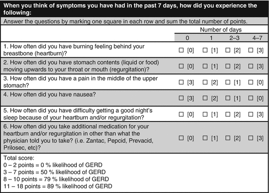
Fig. 12.6
GerdQ (gastroesophageal reflux disease questionnaire)
Studies indicate that symptom-based questionnaires have been demonstrated to be useful research tools, but they are either of insufficient validity or are too complicated to use to be useful in clinical practice. They may however have potential in directing the appropriate diagnostic workup and for tailoring of subsequent medical treatment. Therefore, its value in the evaluation of patients with possible GERD in clinical practice may be in aiding primary care physicians in identifying patients with low likelihood of GERD who may benefit more from further testing and in providing the typical GERD patient with better treatment and follow-up [48–50]. However, at this time, current guidelines do not recommend routine use of symptom questionnaires for the diagnosis or management of GERD.
In general, diagnostic testing is reserved for patients who fail to respond to a trial of adequate medical therapy, like histamine-2 receptor blocker (H2RB) or PPI, or for patients who have alarm symptoms with GERD. Available tests include upper GI series, upper endoscopy (esophagogastroduodenoscopy [EGD]), 24-h esophageal pH study, wireless capsule pH study, and esophageal impedance testing [8, 9] (Table 12.9
Stay updated, free dental videos. Join our Telegram channel
VIDEdental - Online dental courses


