Introduction
The purpose of this study was to evaluate the efficacy of the Schwarz appliance with a new method of superimposing detailed cone-beam computed tomography (CBCT) images.
Methods
The subjects were 28 patients with Angle Class I molar relationships and crowding; they were randomly divided into 2 groups: 14 expanded and 14 nonexpanded patients. Three-dimensional Rugle CBCT software (Medic Engineering, Kyoto, Japan) was used to measure 10 reference points before treatment (T0) and during the retention period of approximately 9 months after 6 to 12 months of expansion (T1). Cephalometric and cast measurements were used to evaluate the treatments in both groups. Also, the mandibular widths of both groups were measured along an axial plane at 2 levels below the cementoenamel junction from a CBCT scan. Differences between the 2 groups at T0 and T1 were analyzed by using the Mann-Whitney U test.
Results
The dental arch (including tooth root apices) had expanded; however, alveolar bone expansion was only up to 2 mm below the cementoenamel junction. There was a statistically significant ( P <0.05) difference between the groups in terms of crown, cementoenamel junction, root, and upper alveolar process. However, no significant ( P >0.05) differences were observed in the interwidths of the mandibular body, zygomatic bones, condylar heads, or mandibular antegonial notches. In the mandibular cast measurements, arch crowding and arch perimeter showed statistically significant changes in the expanded group. The buccal mandibular width and lingual mandibular width values had significant changes as measured from a point 2 mm below the cementoenamel junction.
Conclusions
The findings suggest that the Schwarz appliance primarily affected the dentoalveolar complex, but it had little effect on either the mandibular body or any associated structures. In addition, the molar center of rotation was observed to be below the root apex.
The maxilla has a midpalatal suture, but the mandible does not. Therefore, rapid maxillary expansion increases the transverse dimension of the maxillary arch by separating the suture, whereas the effects of mandibular expansion are localized to alveolar bones and mainly induce tooth inclination. For lateral expansion of the mandible during mixed dentition, appliances with expansion screws, such as the Schwarz appliance, have been widely used. Although expansion of the mandible has theoretically never been successful, several studies have reported good clinical results with the technique.
However, gaining space in the mandibular arch has been a limiting factor because of the belief that the expansion is not stable. Reidel stated that arch form, particularly in the mandibular arch, could not be altered by appliance therapy. Intercanine and intermolar widths tend to decrease during the postretention period, especially if they were expanded during treatment. Average mandibular intercanine widths have been reported to be 24 to 26 mm, suggesting possibly a biologically optimal range for achieving stability. Several reports contend that moderate increases in arch width are possible, especially in the anterior regions of the arches, until the permanent canines erupt. After this, arch width usually decreases in both anterior and posterior regions. In contrast, Walter concluded that mandibular arch width could be expanded permanently. Lutz and Poulton also reported that, during the early mixed dentition, if the dental arches are expanded in patients with crowding, the path of eruption of the mandibular permanent canines and premolars can be altered to the increased width. Furthermore, they found that the increased maxillary and mandibular deciduous second molar width can be maintained. However, the increased maxillary and mandibular deciduous canine widths tend to return to the widths before treatment without retention appliances. Motoyoshi et al also found that retention duration is the most important factor for maintaining the expansion effect. Although the literature has provided little guidance for predicting which patients can tolerate greater expansion, certain malocclusions seem to have greater potential for retaining some increase in arch width. In addition, Schulhof derived a formula indicating that a patient with a brachyfacial pattern will have a wider arch than the dolichofacial type.
However, a method to objectively evaluate mandibular shape change has not yet been developed. Currently, analysis methods have been with cast models or cephalometric radiographs. Therefore, we have devised a new method of superimposing images from cone-beam computed tomography (CBCT) to evaluate the changes in mandibular shape. The purpose of this research was to analyze the efficacy of the Schwarz appliance with this method of CBCT imaging.
Material and methods
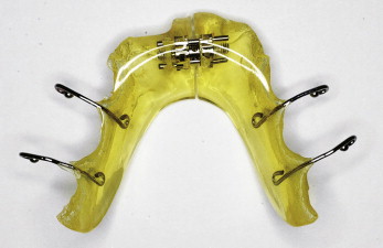
The subjects comprised 28 patients from a private orthodontic office who were diagnosed with Angle Class I malocclusions with crowding and normal vertical dimensions and no posterior crossbites. The study was approved by the Kansai Hospital ethics committee, and informed consent was obtained from the participants. After initial recording of the data, the patients were randomized to 2 groups: those treated with Schwarz appliances (expanded group) and a nonexpanded control group (nonexpanded group) ( Fig 1 ). Records were gathered at 2 times from both groups. For the expanded group, the first records were gathered before treatment (T0); the second set was obtained during a retention period of approximately 9 months after 6 to 12 months of expansion (T1). In the control group, 2 sets of records were obtained approximately 13 to 21 months apart. The expanded group included 14 patients (6 boys, 8 girls) with average ages of 7 years 11 months at T0 and 9 years 8 months at T1. The nonexpanded group received no treatment and included 14 patients (6 boys, 8 girls) with average ages of 8 years at T0 and 9 years 8 months at T1. The expanded group used a Schwarz expansion appliance on the maxillary and the mandibular dentitions to relieve anterior crowding. The maxillary dental arches were also expanded with a Schwarz appliance in all expanded group patients to maintain the buccolingual relationships of occlusal contact in the posterior teeth during expansion. The patients wore the expansion appliances at night. The treatment period was approximately 1 year. The appliance was activated by rotating its screws once a week (90° for 0.175 mm). The expansion plates were adjusted when the appliance disturbed an erupting tooth or did not fit in the dental arch. A new appliance was fabricated when the screw reached its limit.
After 12 months of expansion, the screw of the Schwarz appliance was fixed with composite (cured) and used as a retainer. The patients wore Schwarz appliances only to prevent any effects from other appliances.
Lateral cephalometric radiographs were traced by 1 investigator (K.T.) to minimize error in measurement. Sixteen points were digitized on each cephalometric radiograph, and 12 cephalometric measurements were made.
Three measurements were made on the mandibular casts: arch crowding, arch perimeter, and arch length. The dental cast measurements were made with Digimatic calipers (NTD 12-15PMX, Mitsutoyo, Kanagawa, Japan) accurate to 0.01 mm.
The CBCT images at T0 were taken at average ages of 7 years 11 months (expanded group) and 8 years 0 months (nonexpanded group), and, in both groups, the CBCT images at T1 were taken at an average age of 9 years 8 months.
The head position was oriented so that the Frankfort plane was parallel to the floor in a seated position, and an image was taken at the intercuspal position using CB MercuRay (Hitachi Medical, Tokyo, Japan).
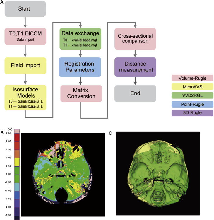
To transform the digital imaging and communication in medicine (DICOM) data from the CBCT images into polygon data, 5 software programs were used—Volume-Rugle (Medic Engineering, Kyoto, Japan), MicroAVS (KGT, Tokyo, Japan), VVD2RGL, Point-Rugle, and 3D-Rugle (Medic Engineering) ( Fig 2 , A ). The iterative closest point (ICP) method can superimpose precisely with repeatability because numerous corresponding points are used to compare with point-based registrations. To superimpose two 3-dimensional (3D) images at T0 and T1, the ICP method was used ( Fig 2 , B and C ). Furthermore, to superimpose 2 separate multi-planar reconstruction (MPR) images accurately, specific points of the cranial base were used as the reference points for superimposition, because the cranial base is not greatly influenced by growth ( Figs 2 , B and C ). As a result, accurate superimposition of 2 separate MPR images is possible. Furthermore, to enhance the accuracy of the ICP method, specific points of the cranial base were used as reference points for registration (superimposition), because the cranial base is not greatly influenced by growth ( Fig 2 , B and C ). Then the combined images were cut down an arbitrary plane and made into 2 units. The MPR images, which have excellent dimensional accuracy, were used to compare the data at T0 and T1.
A slice plane perpendicular to the occlusal plane, passing through both sides of the mesiobuccal cusp tips of the mandibular first molars, was prepared for the measurements. The 3D-Rugle software program was used for the measurements, and the following distances were measured at T0 and T1. Ten points of interest were measured including mandibular first-molar crowns, cementoenamel junctions (CEJs), roots, buccal and lingual alveolar processes, inner and outer surfaces of the mandibular bodies, zygomatic bones, condylar heads, and antegonial notches ( Table I ; Fig 3 , A and B ).
| Landmark for parameter | Measured landmarks |
|---|---|
| 1. M1Crown | Distance between crowns (lingual surfaces of the mandibular right and left first molar crowns 3 mm above the CEJ) |
| 2. M1CEJ | Distance between the CEJs |
| 3. M1LAP | Distance between lingual surfaces of the alveolar processes at the mandibular right and left first molar areas 2 mm below the CEJ |
| 4. M1BAP | Distance between buccal surfaces of the alveolar processes at the mandibular right and left first molar areas 2 mm below the CEJ |
| 5. M1Root | Distance between roots (the distance between the mandibular right and left first molar roots 7 mm below the CEJ) |
| 6. M1IMB | Distance between inner surface of the mandibular bodies 13 mm below the CEJ |
| 7. M1OMB | Distance between outer surface of the mandibular bodies 13 mm below the CEJ |
| 8. Zyg | Distance between zygomatic bones at the outermost part of the zygomatic buttresses |
| 9. CoO | Distance between the outermost part of the condylar heads |
| 10. Ag | Distance between antegonial notches |
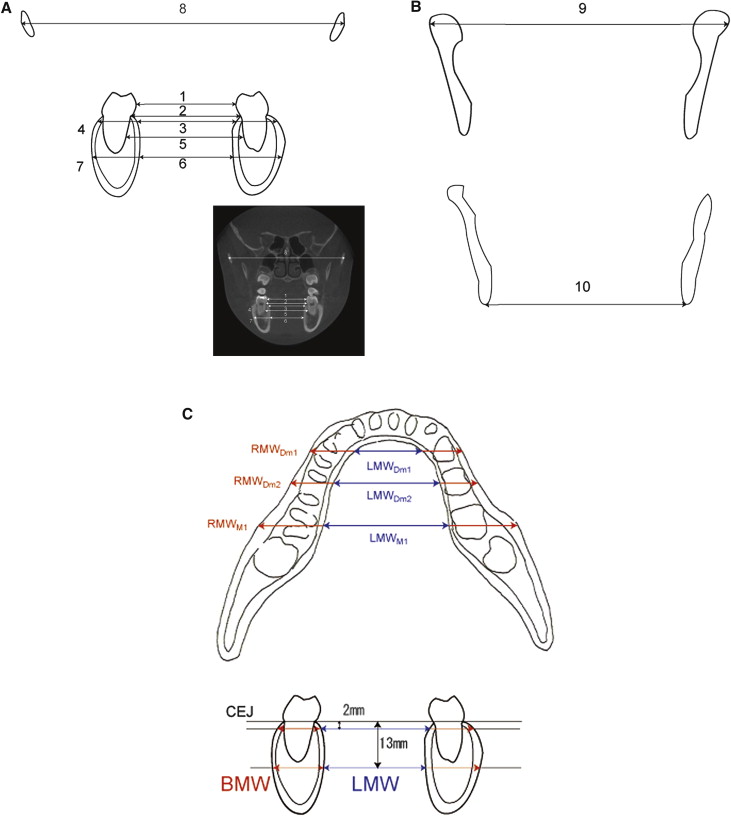
In addition, the distances of the crowns, CEJs, labial and lingual alveolar processes, and inner and outer surfaces of the mandibular bodies of the mandibular first deciduous molars were measured at a point 18 mm anterior from the mesial cusps of the mandibular first molars.
To determine whether there was a relationship between the amount of expansion or tipping and the change in cortical bone thickness at the mandibular first molars, the mandibular first deciduous molars, and the mandibular second deciduous molars, axial CBCT images were taken at 2 and 13 mm below the CEJ ( Fig 3 , C ).
One examiner (K.T.) made all measurements to eliminate interexaminer errors. To identify systematic errors and compare measurement accuracy, intraexaminer reliability was evaluated. Sources of error included landmark location, anatomic contours, tracing from the cephalograms, digitizing the cephalograms, data conversion from the different software programs, all linear and angular measurements from the CBCT, and all linear measurements from the dental casts. Also, to prevent bias in the measurement of the expanded and nonexpanded groups, the investigator was blinded. Tracing and digitizing errors of cephalograms, data conversion errors from the different software programs, linear and angular measurement errors of the CBCT, and all linear measurements from the dental casts were determined by remeasurement at least twice on 2 separate occasions, 2 weeks apart. Five randomly selected subjects from each group were measured at least twice on 2 separate occasions, 2 weeks apart, by the same investigator. No statistically significant difference was found for any measurement by using intraclass correlation coefficients.
Statistical analysis
Descriptive statistics were calculated for each measurement. The data were analyzed by using a statistical software package (version 16.0, SPSS, Chicago, Ill). Treatment changes between the 2 groups and between T0 and T1 were analyzed with the Mann-Whitney U test. A value of P <0.05 was considered significant.
Results
Table II shows the measurements at T0 and T1 of both groups. There were no significant skeletal, dental, and soft-tissue lip profile changes from T0 to T1. No mandibular rotation or displacement was seen between T0 and T1. However, in the mandibular cast measurements of arch crowding and arch perimeter, there were statistically significant changes from T0 to T1 in the expanded group.
| Group | Nonexpanded group | Expanded group | Mann-Whitney U test (significance) | ||||||||||
|---|---|---|---|---|---|---|---|---|---|---|---|---|---|
| T0 | T1 | Changes with growth | T0 | T1 | Changes with treatment | ||||||||
| Mean | SD | Mean | SD | Mean | SD | Mean | SD | Mean | SD | Mean | SD | ||
| Angular skeletal (°) | |||||||||||||
| Facial angle | 84.39 | 2.64 | 83.80 | 2.59 | −0.59 | 0.39 | 83.57 | 2.63 | 83.71 | 2.61 | 0.14 | 0.12 | NS |
| Angle of convexity | 4.21 | 1.89 | 3.80 | 1.79 | −0.41 | 0.77 | 4.17 | 1.32 | 3.61 | 1.29 | −0.56 | 0.40 | NS |
| FMA | 29.79 | 2.64 | 30.26 | 2.54 | 0.47 | 0.93 | 28.90 | 2.28 | 29.49 | 2.25 | 0.59 | 0.51 | NS |
| SNA | 82.16 | 3.73 | 82.23 | 3.66 | 0.07 | 0.05 | 81.56 | 4.05 | 82.11 | 4.02 | 0.55 | 0.11 | NS |
| SNB | 80.01 | 2.43 | 80.12 | 2.32 | 0.11 | 0.09 | 79.59 | 3.45 | 80.01 | 3.39 | 0.43 | 0.14 | NS |
| ANB | 2.15 | 0.73 | 2.11 | 0.64 | −0.04 | 0.04 | 1.97 | 1.55 | 2.10 | 1.49 | 0.13 | 0.10 | NS |
| Angular and linear dental | |||||||||||||
| U1 to SN (mm) | 100.90 | 11.60 | 104.52 | 10.50 | 3.62 | 3.59 | 101.19 | 7.82 | 105.14 | 7.01 | 3.95 | 3.39 | NS |
| Interincisal angle (°) | 132.03 | 9.04 | 125.54 | 10.52 | −6.49 | 3.02 | 133.37 | 7.54 | 124.86 | 7.74 | −8.51 | 5.53 | NS |
| IMPA (°) | 92.60 | 7.11 | 93.89 | 7.52 | 1.29 | 2.10 | 93.59 | 5.45 | 96.36 | 4.85 | 2.78 | 1.91 | NS |
| L1-AP o (°) | 25.19 | 2.84 | 25.73 | 2.63 | 0.54 | 0.45 | 24.29 | 2.36 | 25.28 | 2.28 | 0.99 | 0.50 | NS |
| L1-AP o (mm) | 3.06 | 1.50 | 3.39 | 1.40 | 0.34 | 0.29 | 2.97 | 1.81 | 3.70 | 1.52 | 0.73 | 0.45 | NS |
| Linear soft tissues (mm) | |||||||||||||
| Upper lip E-line | 2.00 | 1.71 | 1.37 | 1.68 | −0.63 | 0.50 | 2.08 | 1.90 | 1.60 | 1.75 | −0.48 | 0.35 | NS |
| Lower lip E-line | 2.61 | 2.04 | 1.91 | 1.67 | −0.71 | 0.63 | 2.95 | 1.73 | 2.39 | 1.57 | −0.56 | 0.50 | NS |
| Mandibular cast measurements (mm) | |||||||||||||
| Arch crowding | −3.83 | 0.87 | −3.17 | 1.27 | 0.66 | 0.50 | −3.59 | 1.21 | −0.78 | 0.70 | 2.80 | 1.14 | ∗ |
| Arch perimeter | 66.26 | 4.77 | 67.04 | 4.95 | 0.78 | 0.65 | 66.35 | 3.36 | 70.11 | 3.58 | 3.76 | 1.62 | ∗ |
| Arch length | 23.06 | 2.28 | 23.87 | 2.56 | 0.81 | 0.75 | 24.92 | 2.11 | 25.84 | 1.95 | 0.92 | 0.80 | NS |
The Schwarz expanded group showed marked expansion compared with the nonexpanded group. The intermandibular first molar lengths increased by 5.41 mm at the crown level, 4.39 mm at the CEJ, 2.40 mm at the root, 3.75 mm at the mandibular alveolar lingual point, and 3.84 mm at the mandibular alveolar buccal point. There were significant ( P <0.05) differences between the groups for teeth and alveolar bone. However, there were no significant differences for the mandibular bodies ( P = 0.695), zygomatic bones ( P = 0.893), condylar heads ( P = 0.613), and antegonial notches ( P = 0.724) ( Table III ).
| Nonexpanded group | Expanded group | Mann-Whitney U test (significance) | |||
|---|---|---|---|---|---|
| Mean | SD | Mean | SD | ||
| M1Crown (mm) | 0.83 | 0.28 | 5.41 | 1.61 | ∗ |
| M1CEJ (mm) | 0.80 | 0.39 | 4.39 | 1.46 | ∗ |
| M1LAP (mm) | 0.81 | 0.43 | 3.75 | 1.58 | ∗ |
| M1BAP (mm) | 0.76 | 0.39 | 3.84 | 1.74 | ∗ |
| M1Root (mm) | 0.77 | 0.30 | 2.40 | 1.22 | ∗ |
| M1IMB (mm) | 0.69 | 0.30 | 0.66 | 0.39 | NS |
| M1OMB (mm) | 0.82 | 0.52 | 0.73 | 0.36 | NS |
| Dm1Crown (mm) | 1.18 | 0.50 | 5.90 | 1.61 | ∗ |
| Dm1CEJ (mm) | 1.12 | 0.71 | 4.69 | 1.19 | ∗ |
| Dm1LAP (mm) | 0.99 | 0.32 | 4.20 | 1.74 | ∗ |
| Dm1BAP (mm) | 1.02 | 0.54 | 4.25 | 1.54 | ∗ |
| Dm1IMB (mm) | 0.89 | 0.62 | 0.88 | 0.49 | NS |
| Dm1OMB (mm) | 0.85 | 0.36 | 0.91 | 0.61 | NS |
| Zyg (mm) | 3.68 | 1.02 | 3.70 | 1.04 | NS |
| CoO (mm) | 3.64 | 1.06 | 3.68 | 1.27 | NS |
| Ag (mm) | 3.59 | 0.79 | 3.64 | 1.74 | NS |
Stay updated, free dental videos. Join our Telegram channel
VIDEdental - Online dental courses


