Fig. 5.1
Explorer window
Operations that can be done at this level allow for improving the quality of 2D and 3D images in order to optimize their use.
Scroll bars can be used to browse through images, each linked to a type of section. The visual reference is shown by lines on complementary planes.
Focus On
-
Coronal section: vertical plane that extends from left side to right side (or vice versa) and that divides the skull in front and rear portions.
-
Axial section: sections of the axial plane. These sections correspond to horizontal cut of the reconstructed volume.
-
Sagittal section: vertical plane which extends from the front part to the rear one (or vice versa) and that divides the skull in right and left portions Fig. 5.2.
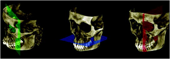 Fig. 5.2Anatomical planes
The operator can move and rotate the image plane clockwise/counterclockwise; the software records these shifts and changes the images of the other two planes in order to have a proper centering in space. The operator can also scroll through each single image that compose the exam.
The operator can adjust the properties of planar images by modifying saturation, contrast, and, brightness; it is also possible to change slice thickness and crop images to highlight a specific area.
The volume cutout applied to 3D rendering is applied on planar images but affects only the three-dimensional reconstruction; the result is a 3D volume that recreates the area of interest of the patient, thus promoting its clinical focus.
Bone density can be measured in a circumscribed area using a circular tool that creates a region of interest (ROI), that can be placed on any projection.
The measurement is given in Hounsfield Units. However, due to the intrinsic property of CBCT, the densitometric result may not be as precise as what would be obtained by CT (Fig. 5.3).
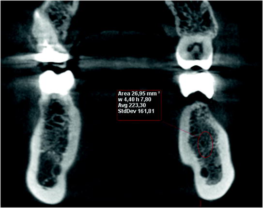
Fig. 5.3
Bone density measurement
Focus On Different systems of analysis of the densitometric data have been proposed in order to choose the best screws for implants: weak bone requires thin but long screw, while a bone in good condition can be implanted with shorter screws.
The most widely used classification is by Misch, which includes four density classes (D1–D4) that indicate a decreasing bone demineralization:
-
D1: thick cortical and dense cancellous bone
-
D2: thick cortical and fenestrated cancellous bone
-
D3: thin cortical and dense cancellous bone
-
D4: thin cortical and fenestrated cancellous bone
Currently, the Misch classification is highly debated. Some authors proposed a correspondence with the values of Hounsfield Units, while others have questioned the validity of the classification.
The 3D reconstruction can be navigated separately from the planar representations, in order to permit a full exploration of the three-dimensional model without changing the axial images. The exploration of the rendering happens through rotations and enlargements.
A number of 3D image adjustments are possible, which allow for creating high-quality models in a few steps. Adjustments can be made in respect to image parameters (contrast, brightness) and to series of advanced tools, such as the choice of lighting source (to eliminate shadows in areas of interest), the transformation from isometric model to model in perspective, or anti-aliasing to smooth the edges (Fig. 5.4).
Fig. 5.4
Advanced graphics settings
Finally, it is possible to highlight on the model the axial planes (Fig. 5.5).
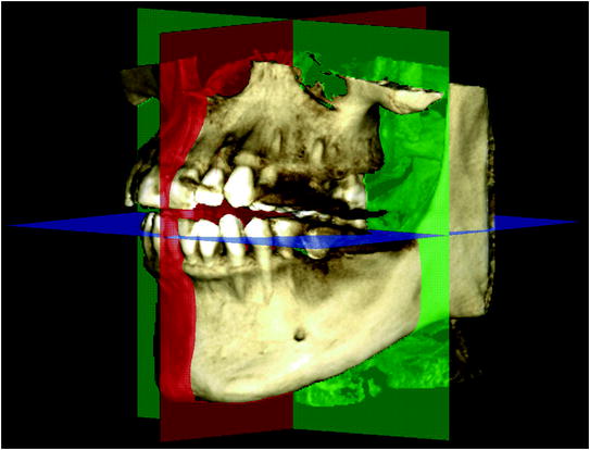
Fig. 5.5
Anatomical planes on the 3D model
5.2 Dental Arch and Panoramic Images
The second phase of the study is concerned with the analysis of panoramic images.
Panoramic images are sections perpendicular to the axial plane, typically designed to represent the entire dental arch (preferably to include the temporomandibular joints).
Stay updated, free dental videos. Join our Telegram channel
VIDEdental - Online dental courses


