Fig. 4.1
Removal of metal objects
Patient must wear radiation protective equipment (Fig. 4.2).
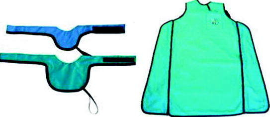
Fig. 4.2
Personal protective equipment
The patient should also wear surgical template that is prescribed by the dentist. They are acrylic radiotransparent masks that fit perfectly to the teeth and residual alveolar processes.
Radio-opaque markers are applied in specific places of the masks to facilitate information transfer from the images to the patient, in respect to implant orientation (Fig. 4.3).
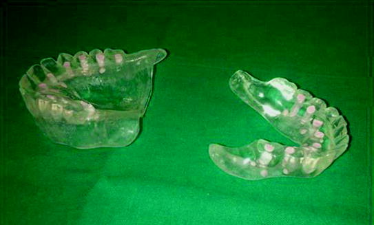
Fig. 4.3
Surgical template for dental implant
For some procedures, scanning of the surgical template alone should be performed. This allows for digitally subtracting the images of the implant from the whole examination of the patient at later stages.
Step 3
Patient Positioning
Patient positioning is crucial to obtain adequate images. As acquisition time may be long, the patient should be positioned in a comfortable position, to avoid motion artifacts.
Patient has to:
-
Maintain the position according to the relevant imaging system in use (Fig. 4.4);
 Fig. 4.4Correct position of the patient
 -
Grip both handles;
-
Relax shoulders to allow for a complete movement of the swivel arm;
-
Place the chin on the support, according to the relevant imaging system in use (Fig. 4.5).
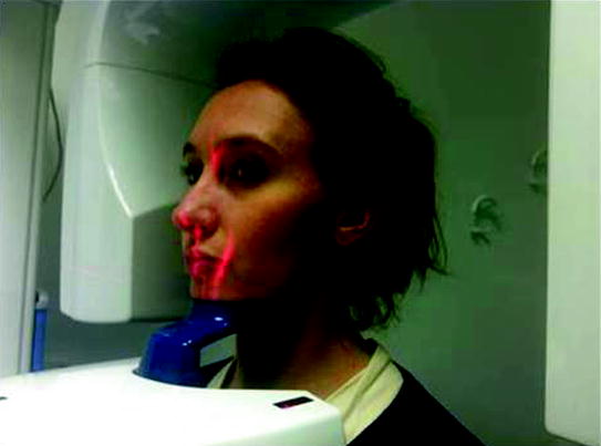 Fig. 4.5Position of the face
The same operation can also be done with the patient sitting on a chair/wheelchair, placing feet slightly forward (Fig. 4.6) to reduce motion artifacts.
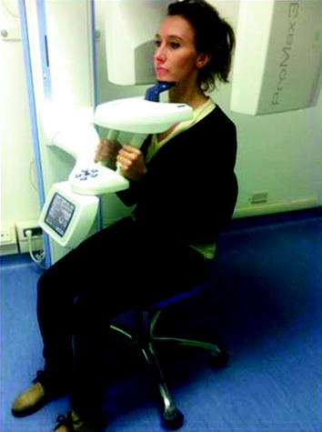
Fig. 4.6
Correct position of the patient sitting on a chair/wheelchair
Check the correct position of the patient using laser markers (Fig. 4.7).
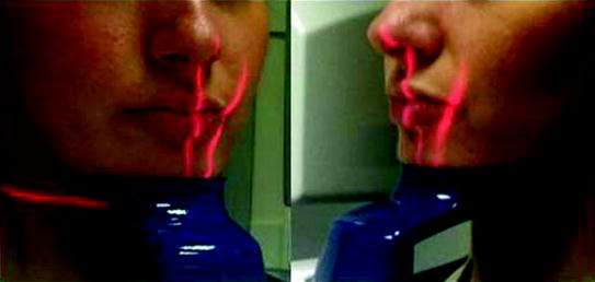
Fig. 4.7
Detail on the centering laser
Children undergoing this type of examination deserve a particular attention in terms of radioprotection. It has been estimated that radiation exposure during the first 10 years of life produce an overall risk of health detriment two to three times higher than that incurred between 30 and 40 years, and five to seven times higher than that incurred over 50. Thus, it is important to protect not only the gonads but also other radiosensitive tissues, such as bone marrow, breast, and thyroid. Approximately 35 % of red marrow is found in long bones and 40 % in the skull.
To reduce the dose, particular attention should be paid to acquisition data. Also, all cares should be taken to obtain a good scan at first attempt. As far as possible, cooperation of the young patient is crucial. The operators should speak to the child and to parents, explaining the procedure. The waiting room may be equipped with color furniture and toys suitable for different ages. Also, drawings and cartoons on the walls of the examination room may reduce fear and make the process easier. The examination should be performed as quick as possible. Also, remember how useful the so-called “positive reinforcement” is, i.e., continuous encouragement and confirmation that he is doing well during the examination. In some cases, presence of parents during examination may help.
4.2 Scanning Parameters
Examination duration, image quality, and radiation dose depend on the parameters of the scan. Thus, it is extremely important to take advantage of the features and benefits they bring in terms of quality and radiation protection.
Scanning parameters include:
1.
exposure parameters
2.
geometric parameters
Stay updated, free dental videos. Join our Telegram channel
VIDEdental - Online dental courses


