and Jasdeep Kaur1
(1)
Earth and Life Sciences Vrije Universiteit Amsterdam and ILEWG, Amsterdam, The Netherlands
10.1 Introduction
10.5.1 Identification
10.5.2 Ethnic Groups
10.5.3 Sex Determination
10.6 Conclusions
Abstract
Palatal rugae are anatomical wrinkles or folds called “plica palatine,” the asymmetrical connective tissue located on the anterior third of the palate behind the incisive papilla. This chapter focuses on the role of palatal rugoscopy in forensic odontology.
10.1 Introduction
Establishing a person’s identity can be a very difficult process in forensic identification. Dental, fingerprint, and DNA comparisons are the most common techniques used in this context, allowing fast and secure identification. However, since they cannot always be used, sometimes simple techniques can be used successfully in human identification, such as palatal rugoscopy, which is the study of the palatal rugae (Caldas et al. 2007). Palatal rugoscopy was first proposed in 1932 by the Spanish investigator Trobo Hermosa. Palatoscopy, or palatal rugoscopy, is the name given to the study of the palatal rugae in order to establish a person’s identity. Palatal rugae have been compared with fingerprints and are unique to an individual (English et al. 1988). They can be of particular significance in edentulous cases and also in certain conditions where there are no fingers to be studied, such as burned bodies or bodies that underwent severe decomposition (Caldas et al. 2007). The uniqueness, postmortem resistance, overall stability, and, in addition, low utilization cost make palatal rugae an ideal forensic identification parameter (Kapali et al. 1997).
10.2 Classification of Palatal Rugae
The first system of classification was developed by C. Goria in 1911 and was rudimentary. The rugae pattern was categorized in two ways: specifying the number of rugae and specifying the extent of the rugal zone relative to the teeth. In this system, compound rugae of two or more branches were counted as one, whether they were V- or Y-shaped. Goria further distinguished two types: simple or primitive and more developed. L. Lysell’s (English et al. 1988) classification in 1955 is the most important, and it has been used widely in research involving rugae. It is comprehensive and includes the inter rugae (IR). Rugae are measured in a straight line between the origin and termination and are grouped into three categories:
1.
Primary: ≥5 mm
2.
Secondary: 3–5 mm
3.
Fragmentary: 2–3 mm
Rugae smaller than 2 mm are disregarded. This is a rather simplified picture of the intricate form that rugae usually present. Therefore, Thomas and Kotze (1983) further detailed the various patterns of primary rugae. These include branched, unified, cross-linked, annular, and papillary, among others. Their classification is based on the length of rugae:
1.
Primary rugae: >5 mm.
2.
Secondary rugae: 3–5 mm.
3.
The shapes of individual rugae were classified into four major types: curved, wavy, straight, and circular (Fig. 10.1).
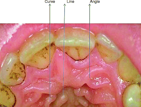
Fig. 10.1
Different types of rugae
4.
Unification occurs when two rugae are joined at their origin or termination. Rugae are considered diverging if two rugae had the same origin but immediately branched, whereas rugae with different origins that joined on their lateral portions are considered converging.
Stay updated, free dental videos. Join our Telegram channel
VIDEdental - Online dental courses


