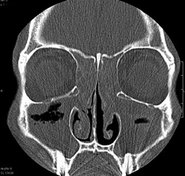
Fig. 4.1
Right nasal fossa showing acute maxillary sinusitis
4.3 Management of Rhinosinus Dysfunction
The diagnostic approach to sinonasal disease, based on the medical history, endonasal endoscopy, and reading of the sinus CT images, will enable the ENT to restore the maxillary sinus prior to the implant through medical and/or surgical treatment of the following infectious or inflammatory diseases (Pignataro et al. 2008; Torretta et al. 2011):
-
Anatomical and functional changes in the ostiomeatal complex (high and posterior septal deviation, concha bullosa, reverse curvature of the middle turbinate, large Haller cell, anatomical change in the uncinate, synechia of the middle turbinate), responsible for anterior localized sinusitis (Fig. 4.2)
 Fig. 4.2Coronal CT scan. Anatomical and functional abnormalities in the ostiomeatal complex
 -
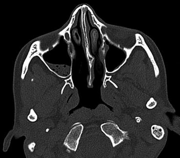
Killian’s antrochoanal polyp, foreign bodies destabilizing the maxillary sinus, and endosinus mycotic diseases (Fig. 4.3)
 Fig. 4.3Coronal CT scan showing an Aspergillosis of the maxillary sinus
 -
Sinonasal polyposis outside of stages 3 and 4 of the Rouviere classification (Fig. 4.6)
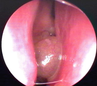 Fig. 4.6Presence of a nasal polyposis in the right nasal fossa
Endoscopic and radiological control in the third week will ensure the physiological restoration of the sinus in the case of medical treatment. Endoscopic endonasal surgery supplements, if needed for the medical treatment, will allow continuation of the implant plan in the case of normalization of the mucosa of the maxillary sinus after the 6th week following surgery.
4.4 Treatment of Endosinus Abnormalities During the Preimplant Radiological Assessment
They are detectable when reading the panoramic or DentaScan images and are represented by the mucosal abnormalities of the maxillary sinus and bone abnormalities of the floor. They require us to take them into account, even in the absence of a sinonasal history, in asymptomatic patients.
4.4.1 Mucosal Abnormalities of the Sinus Floor
They are represented by polypoid or hypertrophic opacities.
4.4.1.1 Polypoid Opacities
-
Non-cystic polypoid opacities (usually associated with hypertrophy of the endosinus mucosa) are indolent and are represented by antrochoanal polyps in their incipient form, and polypoid opacities reacting to submucosal foreign bodies or prior tooth root disease (Fig. 4.7).
 Fig. 4.7Cone beam CT. Non-cystic polypoid opacity of the maxillary sinus
Stay updated, free dental videos. Join our Telegram channel
VIDEdental - Online dental courses


