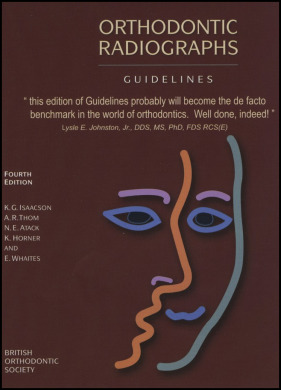
A concise and precise book, Orthodontic Radiographs provides effective and meaningful insight into the use of radiographs in clinical orthodontics. Now in its fourth edition, the book is published by the British Orthodontic Society. As argued by its authors, it is not intended to represent absolute rules but, rather, be a guide in an area in which attitudes are changing rapidly.
This evidence-based book is written in 16 chapters, including sections on the damaging effects of ionizing radiation, the aims of radiation protection, imaging equipment and techniques available for orthodontic radiography, methods for radiation protection including quality assurance, selection criteria for the use of radiographs, applications of cone-beam computed tomography (CBCT) in orthodontics, and others. Throughout the text, high-quality color photographs, radiographs, and tables are included. Indications for radiographs according to a patient’s age and type of encounter are presented in engaging charts.
Whereas no similar text has been provided by the American Association of Orthodontists, the American Dental Association (ADA) offers general guidelines for patient selection and limiting radiation exposure during dental radiograph examinations. For example, the ADA recommends that dentists should conduct a clinical examination before a radiographic examination. The ADA also recommends that CBCT should be prescribed only when the diagnostic yield will benefit the patient or improve the clinical outcomes signficantly. The American Academy of Oral and Maxillofacial Radiology also recommends that the use of CBCT in orthodontics should be justified on an individual basis according to the clinical presentation.
Several guidelines provided by the ADA and the American Academy of Oral and Maxillofacial Radiology on the use of CBCT and other radiographic examinations echo the central ideas of the British Orthodontic Society. However, one senses that the British guidelines are overall more conservative. Taking radiographs only when justified clinically is a legal requirement in the United Kingdom, and the use of ionizing radiation in clinical practice is governed by criminal law. Some practitioners in the United Kingdom have had their licenses to practice withdrawn as a result of inappropriate use of x-rays, and others received a custodial sentence.
Such strict regulations are in the best interest of orthodontic patients, especially because children who are sensitive to radiation comprise most of these patients. The authors stressed that since radiographic examinations are invasive procedures, they put children at the greatest risk. Nevertheless, they also acknowledged that the benefits of diagnostic radiology generally outweigh the risks; consequently, they provided robust yet flexible guidelines for the appropriate acquisition of radiographs in clinical orthodontics, including common-sense rationales for orthodontic practice, clinical audit, and research.
The authors advised that there is no indication for the following practices: radiographs before a clinical examination, radiographs when only minimal tooth movement is planned, full-mouth periapical views before treatment, a cephalometric lateral radiograph for the prediction of facial growth, radiographs of the hand and wrist to predict the onset of the pubertal growth spurt, radiographs to investigate temporomandibular disorders associated with temporomandibular joint pain and dysfunction, prospective radiographs only for medical or legal reasons (ie, “defensive dentistry”), posttreatment radiographs for professional examinations and clinical presentations, and routine CBCT images for all orthodontic patients.
Some orthodontists take radiographs in these clinical situations, either to follow the status quo or to maximize risk management or practice management. You will find that this book is a valuable resource by which to change some of these practices, which can ultimately reduce the collective effective dose for orthodontic patients.
Stay updated, free dental videos. Join our Telegram channel

VIDEdental - Online dental courses


