Oral ecosystem and dental plaque
Publisher Summary
The human mouth is lined by a stratified squamous epithelium, which is modified in different areas according to function. The oral mucous membrane is interrupted by salivary ducts and by teeth, if present. The gingival tissues are bound to the tooth surface by the epithelial attachment, and crevicular fluid passes into the mouth from the gingival crevice. A thin layer of saliva bathes the surface of the mucosa and contained in this layer are epithelial squames, polymorphonuclear leucocytes, and members of the oral commensal microflora. However, within the general environment of the mouth, there exist a number of different microenvironments or niches, each of which supports its own peculiar microflora. The microbiological differences between some sites are qualitative and quantitative, while quantitative differences exist between other surfaces. These variations are because of the complex interactions of a wide range of ecological factors.
The oral environment
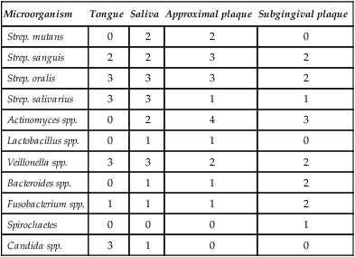
However, within the general environment of the mouth there exists a number of different microenvironments or niches, each of which support their own peculiar microflora (Table 3.1). The microbiological differences between some sites are both qualitative and quantitative, e.g. the flora of the tongue and subgingival plaque, while mainly quantitative differences exist between other surfaces, e.g. tongue and saliva. These variations are due to the complex interactions of a wide range of ecological factors, many of which are poorly understood.
Table 3.1
Relative proportions of oral microorganisms in health
| Microorganism | Tongue | Saliva | Approximal plaque | Subgingival plaque |
| Strep. mutans | 0 | 2 | 2 | 0 |
| Strep. sanguis | 2 | 2 | 3 | 2 |
| Strep. oralis | 3 | 3 | 3 | 2 |
| Strep. salivarius | 3 | 3 | 1 | 1 |
| Actinomyces spp. | 0 | 2 | 4 | 3 |
| Lactobacillus spp. | 0 | 1 | 1 | 0 |
| Veillonella spp. | 3 | 3 | 2 | 2 |
| Bacteroides spp. | 0 | 1 | 1 | 2 |
| Fusobacterium spp. | 1 | 1 | 1 | 2 |
| Spirochaetes | 0 | 0 | 0 | 1 |
| Candida spp. | 3 | 1 | 0 | 0 |
The main factors involved are the following.
Salivary factors
Mixed saliva plays a central role in the microbial ecology of the mouth, receiving secretions from the parotid, submandibular, sublingual and minor glands. The composition of saliva from these glands is different and further variation can be produced by other factors, e.g. increase or decrease in salivary flow rate. The main factors and their effect on the oral microflora are shown in Table 3.2. Although the precise role of these different factors in oral ecology is uncertain, there is good evidence that, overall, saliva is important. Patients with severe xerostomia due to the effect of radiotherapy or degenerative disease of the salivary glands (e.g. Sjögren’s syndrome), can develop rapidly progressive dental caries, candidosis and acute sialadenitis (see Chapter 12).
Table 3.2
Salivary factors involved in oral ecology
| Factor | Mechanism | Effect on oral microflora |
| Mechanical washing action and swallowing | Powerful mechanism which results in microorganisms either in aggregates or bound to epithelial cells being swallowed and killed in gastric acid | Prevents overgrowth of microflora |
| Glycoproteins | Important in the formation of acquired pellicle on enamel and in coating epithelial cells | May promote or inhibit microbial adherence to oral and dental surfaces. Nutrient source for microbial growth |
| Lysozyme, lactoferrin, lactoperoxidase | Inhibit the growth of or kill microorganisms | Most effective against exogenous bacteria but also have some effect on members of the commensal flora |
| Buffering capacity and pH | Bicarbonate system most important; tends to stabilize pH to about 6.7 | Tends to prevent overgrowth of potentially pathogenic miroorganisms which require low or high pH environments for maximal growth |
| Immunoglobulins | Mainly secretory IgA which coats oral and dental surfaces and interacts with specific antigens if present | May inhibit or in some cases promote microbial adherence to surfaces. Also may alter microbial metabolism |
Microbial factors
The complex mixture of microorganisms which make up the oral microflora can interact both with each other and with the host factors described earlier. A number of the most important of these interactions are shown in Table 3.3.
Table 3.3
Microbial factors involved in oral ecology
| Factor | Effect |
| Cell wall components, extracellular polymers and surface fibrils | Adherence of microorganisms to oral and dental surfaces. Aggregation of organisms of the same species (homotypic) and of different species (heterotypic). Important mechanisms in plaque formation |
| Bacteriocins and similar products | Chemicals produced by one microbial species which are antagonistic to cells of the same or related species |
Stay updated, free dental videos. Join our Telegram channel
VIDEdental - Online dental courses


